Explodable 3D Dog Skull for Veterinary Education
3D models of a Sheep and Goat Skull and Inner ear
3D models of Miocene vertebrates from Tavers
3D GM dataset of bird skeletal variation
Skeletal embryonic development in the catshark
Bony connexions of the petrosal bone of extant hippos
bony labyrinth (11) , inner ear (10) , Eocene (8) , South America (8) , Paleobiogeography (7) , skull (7) , phylogeny (6)
Lionel Hautier (23) , Maëva Judith Orliac (21) , Laurent Marivaux (16) , Rodolphe Tabuce (14) , Bastien Mennecart (13) , Pierre-Olivier Antoine (12) , Renaud Lebrun (11)

|
3D atlas and comparative osteology of the middle ear ossicles among Eulipotyphla (Mammalia, Placentalia).Daisuke Koyabu
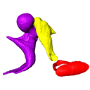
Published online: 03/05/2017 Keywords: aquatic adaptation; convergence; Eulipotyphla; fossorial adaptation; hearing https://doi.org/10.18563/m3.3.2.e3 Cite this article: Daisuke Koyabu, 2017. 3D atlas and comparative osteology of the middle ear ossicles among Eulipotyphla (Mammalia, Placentalia). MorphoMuseuM 3 (2)-e3. doi: 10.18563/m3.3.2.e3 Export citationAbstract Considerable morphological variations are found in the middle ear among mammals. Here I present a three-dimensional atlas of the middle ear ossicles of eulipotyphlan mammals. This group has radiated into various environments as terrestrial, aquatic, and subterranean habitats independently in multiple lineages. Therefore, eulipotyphlans are an ideal group to explore the form-function relationship of the middle ear ossicles. This comparative atlas of hedgehogs, true shrews, water shrews, mole shrews, true moles, and shrew moles encourages future studies of the middle ear morphology of this diverse group. M3 article infos Published in Volume 03, Issue 02 (2017) |
|

|
M3#166Left middle ear ossicles Type: "3D_surfaces"doi: 10.18563/m3.sf.166 Data citation: Daisuke Koyabu, 2017. M3#166. doi: 10.18563/m3.sf.166 state:published |
Download 3D surface file |