
Abstract book of the 18th Conference of the EAVP
Pterosaurs from Coahuila
Pliocene-Pleistocene large mammals from Le Riège and Saint-Palais
Les sélaciens du Miocène de la région de Montpellier
Muridae du Pliocène supérieur d'Espagne et du midi de la France.
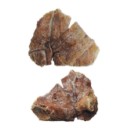
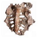
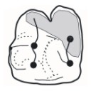
Contribution à l'étude des genres Gliravus et Microparamys.
Eocene (57) , Quercy Phosphorites (38) , Systematics (32) , Rodents (29) , Mammalia (27) , Rodentia (25) , Miocene (24)
|
Dortokid turtle remains from the Upper Cretaceous of Cruzy (Hérault, southern France) and phylogenetic implicationsHaiyan Tong, Eric Buffetaut
Published online: 14/11/2022 |
|

|
The Late Cretaceous nesting site of Auca Mahuevo (Patagonia, Argentina): eggs, nests, and embryos of titanosaurian sauropods.Luis M. Chiappe
Published online: 15/12/2003 |
|

|
Physogaleus hemmooriensis (Carcharhinidae, Elasmobranchii), a new shark species from the early to middle Miocene of the north sea basin.Thomas Reinecke and Kristiaan HoedemakersPublished online: 15/10/2006Keywords: Carcharhinidae; Early Miocene; Elasmobranchii; Hemmoorian; new species; North Sea Basin; Physogaleus https://doi.org/10.18563/pv.34.e14 Abstract A new carcharhinid shark species, Physogaleus hemmooriensis sp. nov., is described from the Lower Hemmoorian (Behrendorfian, late Burdigalian, early Miocene) of Werder, Lower Saxony, Germany. P. hemmooriensis also occurs in the Edegem and Antwerpen Sands Members of the Berchem Formation, Belgium, and in the Miste Bed, Aalten Member of the Breda Formation, The Netherlands, which have an early to middle Miocene age. In the Western Atlantic region, the taxon is present in the early Miocene Calvert Formation of Delaware, U.S.A, which is largely contemporaneous with the Hemmoorian. PV article infos Published in Vol. 34, Fasc. 1-2 (2006) |
|

|
La palichnofaune de vertébrés tétrapodes du permien supérieur du Bassin de Lodève (Languedoc-France).Georges Gand, Jacques Garric, Georges Demathieu and Paul EllenbergerPublished online: 15/09/2000Keywords: Footprints; France; Languedoc; Lodève basin; new ichnotypus; Saxonian; Upper Permian Abstract Near "la Lieude", in the Lodève basin, more than a thousand of footprints are distributed in a twenty of trackways which amounts to 220 m length. They have been found on calcareous siltstone level in the B site named also "Réserve Naturelle Volontaire". This last is located in the Saxonian summit dated Upper Permian. "La Lieude" tracks are described by using statistical methods then they are compared with others from the world Permian. What allows to distinguish 4 following ichnotaxa: Lunaepes ollierorum nov.ichnosp., Merifontichnus thalerius nov. ichnogen. and nov. ichnosp., Planipes brachydactylus nov.ichnosp. and Brontopus circagiganteus nov. ichnosp. All these traces are attributed with possibility or probability to Therapsida or to Therosauria, except Brontopus circagiganteus nov. ichnosp. that could be due to Caseamorpha. All these animals whose sizes have been estimated between l and 5 m lived probably in a playa environment.The biological and sedimentological data from "la Lieude" footprints levels compared with informations provided by the tracks orientations, suggest the following scenario. Animals coming from the North have crossed a sandy channel bank with plants zones by directing to the South for the majority. Maybe, they were going to the lacustrine part of the playa, close to "la Lieude" footprints that they have just trampled on. PV article infos Published in Vol. 29, Fasc. 1 (2000) |
|
|
New remains of the giant bird Gargantuavis philoinos from the Late Cretaceous of Provence (south-eastern France)Eric Buffetaut
Published online: 27/08/2015 |
|

|
Macroscelidea, Insectivora and Chiroptera from the Miocene of east Africa.Percy M. ButlerPublished online: 15/11/1984Keywords: Chiroptera; East Africa; Insectivora; Macroscelidea; Miocene; Systematics https://doi.org/10.18563/pv.14.3.117-198 Abstract The East African Miocene Macroscelidea, lnsectivora and Chiroptera are revised on the basis of new material. New taxa proposed are: Miorhynchocyon, .n. gen. (Macroscelididae): Míorhynchocyon meswae, n. sp.: Pronasílío ternanensis. n. gen.. n. sp. (Macroscelididae); Hiwegicyon juvenalis, n. gen. n. sp. (Macroscelididae); Parageogale, n. gen. (Tenrecidae): Prochrysochlorinae, n. subfam. (Chrysochloridae): Propottininae, n. subfam, (Pteropodidae); Chamtwaria pickfordi, n. gen., n. sp. (Vespertilionidae). Gymnurechnínus songhorensis is synonymised with G. camptolophus. The new material provides additional information on the dentition, especially of Myohyrax oswaldi. Galerix africanus. Amphechínus rusingensis, Protenrec tricuspis and Parageogale aletris. Partial skulls are described of Amphechinus rusingensis, Protenrec tricuspis, Prochrysochloris míocaenicus and Taphozous incognita. The oldest member of the Macroscelidinae (Pronasilio) is described from Fort Ternan. Galerix africanus is closely related to G. exilis from Europe. Amphechinus rusingenesis is compared with Asiatic Oligocene Erinaceinae. The Miocene age of Crocidura is rejected. On the evidence of humeri, the following families of Chiroptera are newly reported: Pteropodidae, Nycterididae, Vespertilionidae, Molossidae. Propotto is regarded as an offshoot from the Pteropodidae, not ancestral to modern forms. Chamtwaria is a primitive vespertilionoid, provisionally placed in the Kerivoulinae. Erinaceidae probably entered Africa at the beginning of the Miocene, before 20 Ma. Faunistic differences between deposits are largely to be ascribed to differences in local environment. PV article infos Published in Vol. 14, Fasc. 3 (1984) |
|

|
Analysis of changing diversity patterns in Cenozoic land mammal age faunas, South AmericaLarry G. Marshall and Richard L. CifelliPublished online: 30/03/1990Keywords: Cenozoic; Chronofaunas; diversity; Equilibrium theory; Extinction; Land mammal faunas; Origination; South America https://doi.org/10.18563/pv.19.4.169-210 Abstract Comparison of various measurements of taxonomic evolution using stratigraphic range data for orders, families and genera of land mammals indicates several means by which deficiencies of the South American fossil record (e.g., presence of hiatuses, unequal temporal and geographic representation of ages, unequal systematic treatment) may be normalized, thus permitting a less distorted appreciation of diversity pattern and trend. Initial radiation of native taxa resulted in a relative equilibrium by early Eocene time. Subsequent increases in absolute diversity were apparently induced by immigration at the family level and by environmental factors at the generic level. Miocene through Pleistocene phases of faunal stability, herein characterized as chronofaunas, are punctuated by rapid turnover events resulting from a complex of factors, including adaptive radiation of immigrant taxa into unoccupied eco-space; environmental and concomitant habitat change induced by orogenic events of the Andes; and biotic interactions between native and immigrant taxa, including competition and prey naivete. The first two factors account for major faunal transitions in the South American middle and late Tertiary; immigration-induced turnover may have been of greater importance in shaping the character of the fauna upon the Great American Interchange and the arrival of man in the Neotropics PV article infos Published in Vol. 19, Fasc. 4 (1990) |
|
|
A late Eocene palaeoamasiine embrithopod (Mammalia, Afrotheria) from the Adriatic realm (Island of Rab, Croatia)Fabrice Lihoreau
Published online: 14/12/2023 |
|

|
Archosauriform teeth from the upper Triassic of Saint Nicolas-de-Port (Northeastern France).Pascal Godefroit
Published online: 15/12/1997 |
|
|
Angolabatis nom. nov.,a replacement name for the Cretaceous genus Angolaia Antunes & Cappetta, 2002 (Chondrichthyes: Rajiformes), a preoccupied name.Miguel T. Antunes
Published online: 15/10/2006 |
|
|
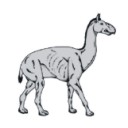
Reconstruction of the cervical skeleton posture of the recently-extinct litoptern mammal Macrauchenia patachonica Owen, 1838R. E. Blanco, Lara Yorio
Published online: 02/05/2023 |
|

|
Field trip guides of the 20th Annual Conference of the European Association of Vertebrate Palaeontologists, 26th June – 1st July 2023, Sabadell (Barcelona), Spain
|
|

|
Book of Abstracts of the 20th Annual Conference of the European Association of Vertebrate Palaeontologists, 26th June – 1st July 2023, Sabadell (Barcelona), SpainDavid M. Alba
Published online: 15/06/2023 |
|

|
Dating dinosaur oodiversity: chronostratigraphic control of LateCretaceous oospecies succession.Nieves Lopez-MartinezPublished online: 15/12/2003Keywords: Biostratigraphy; Chronology; dinosaur eggshells; Late Cretaceous Abstract An increasing fossil record of dinosaur eggs and eggshells allows putting ootaxa within a chronostratigraphic framework, in order to study their distribution pattern leading eventually to their use as biochronological markers. For these purposes, high-quality data exists in four major regions; North America, South America, Europe and Asia (Central Asia and India). Most of the highly diverse dinosaur egg record has been dated as Latest Cretaceous in age (Campanian-Maastrichtian), reaching the Cretaceous-Tertiary boundary closer than the dinosaur bone record. However, dating continental sections is problematic and need to be carefully verified, as it appears when comparing the European dinosaur eggshell record from two well-studied areas. Ootaxa distribution in both sides of the Pyrenees (Tremp and Aix basins) shows comparable oospecies successions, but different chronology. This disagreement probably indicates that one or both successions have a wrong chronostratigraphic calibration. PV article infos Published in Vol. 32, Fasc. 2-4 (2003) |
|

|
Les oiseaux aquatiques (Gaviiformes à Anseriformes) du gisement Aquitanien de Saint-Gerand-le-Puy (Allier, France): Révision systématique.Jacques ChenevalPublished online: 01/11/1984Keywords: Aves; Early Miocene; Osteology; Palaeoecology; Systematics https://doi.org/10.18563/pv.14.2.33-115 Abstract Six orders of birds adapted to aquatic life are represented among the numerous avifauna of "Saint-Gérand-le-Puy": Gaviiformes, Procellariiformes, Pelecaniformes, Ciconiiformes, Phoenicopteriformes, and Anseriformes. The present study of this avifauna proposes several changes in systematics:- Procellariiformes: Puffinus arvernensis does not belong in Procellariidae but in Diomodeidae, and it is transferred to the fossil genus Plotornis previously described in the Middle Miocene of France. - Pelecaniformes: Phalacrocorax littoralis remains in Phalacrocoracidae; P. míocaenus is different from the modern species, and is transferred to the new genus Nectornis. Empheresula arvernensis, described in the Oligocene deposits of Gannat, seems to be present in Saint-Gérand-le-Puy too. Pelecanus gracilis shows many differences from the modern species, and belongs to the new genus Miopelecanus, - Ciconiiformes: Ardea formosa nom. oblit. is a synonym of Proardeola walkeri. - Anseriformes: a new species closely related to swans is described, and belongs to the fossil genus Cygnopterus, of the Middle Oligocene of Europe; this species is called C. alphonsi. The ecology of each species is suggested by comparison with that of its nearest living relatives, and by study of osteological adaptations. PV article infos Published in Vol. 14, Fasc. 2 (1984) |
|

|
First record of dinosaur eggshells and teeth from the north-west african Maastrichtian (Morocco).Géraldine Garcia
Published online: 15/12/2003 |
|

|
Palaeotis weigelti restudied : a small middle Eocene Ostrich (Aves : Struthioniformes)Peter Houde
Published online: 20/06/1987 |
|

|
Diversity among north african dinosaur eggshells.Monique Vianey-Liaud
Published online: 15/12/2003 |
|
|
First record of the family Protocetidae in the Lutetian of Senegal (West Africa)Lionel Hautier
Published online: 05/12/2014 |
|

|
Mammals and stratigraphy : Geochronology of the continental mammal-bearing Tertiary of south America.Larry G. Marshall, Robert Hoffstetter and Rosendo PascualPublished online: 15/12/1983Keywords: Cenozoic; Geochronology; Mammalia; South America; Stratigraphy; Tertiary https://doi.org/10.18563/pv.13.ext Abstract The principles and practices employed in establishment and recognition of South American land mammal ages are reviewed along with previous and present concepts of distinguishing time, rock, and faunal units. Previous chronological arrangements of South American Tertiary land mammal faunas are appraised on the basis of recent geological and paleontological data. Twelve South American Tertiary land mammal ages are here recognized [from oldest to youngest, Riochican (middle to late Paleocene); Casamayoran (early Eocene); Mustersan (middle Eocene); Divisaderan (late Eocene); Deseadan (early [to middle?] Oligocene); Colhuehuapian (late Oligocene); Santacrucian (early Miocene); Friasan (middle Miocene); Chasicoan (late Miocene); Huayquerian (latest Miocene); Montehermosan (early to middle Pliocene); and Chapadmalalan (late Pliocene)]. As all except the Friasian were originally defined on the basis of Argentine faunas, these are discussed first and at length, and each is reviewed with discussion of type locality, stratigraphy, type fauna, and faunal correlations. Non-Argentine faunas are then discussed country by country in alphabetical order. PV article infos Published in Vol. 13, Ext (1983) |
|