|
Abstract book of the 18th Conference of the European Association of Vertebrate Palaeontologists (EAVP), 5-9 July 2021, Benevento, Italy
Published online: 12/07/2021
Keywords:
2021; Abstracts; Benevento; EAVP
https://doi.org/10.18563/pv.eavp2021
Abstract
Welcome to the 18th conference of the EAVP, the first online meeting of our association. The pandemic emergency made it impossible to organize the in-person meeting in Benevento as we all had hoped. However, we couldn’t miss another EAVP meeting. Therefore, this year we are meeting online, trying to make the experience the closest to the in-person meeting possible, in order to offer the delegates the opportunity to share knowledge, build new networks and reinforce the old ones. We have received 137 communications, with more than 150 delegates from 24 countries. All the abstracts have passed a peer review process and are part of this special volume of Palaeovertebrata, the official journal of the EAVP. This year we are also offering a variety of workshops, roundtables and symposia on different topics. These include the annual “Pride EAVP: An LGBTQ+ Roundtable” and “Women in Palaeontology Roundtable Discussion”, together with the workshops on “Gendered Perspective in Palaeontological Research: from Definition to Action”, “International Palaeontology Education: Virtual Teaching and Real-World Learning”, “Stepping out of Academia: Why, When and How?”, “Introduction to Hypothesis Testing in Statistics”, “The Early-Middle Pleistocene Transition: Marked Mammal Turnover and Ecosystem Dynamic” (included in the early event for the XXI INQUA Congress in Rome 2023, “A Mediterranean Perspective on Quaternary Sciences”). To conclude, we are hosting two symposia on “Palaeoart: Diversity on and behind the Canvas” and “3D fossils, Robotic and Experimental Palaeontology”. We wish you all a happy and productive meeting. And see you in Benevento next year!
PV article infos
Published in Special Volume 1-2021 (2021)
|
PDF
|
|
Pterosaurs (Pterosauria) from the Cerro del Pueblo Formation (Late Campanian) of Coahuila, Mexico
Published online: 21/11/2025
Keywords:
Azhdarchoidea; Coahuila; Mexico; Pterodactyloidea; Pterosauria
https://doi.org/10.18563/pv.48.2.e1
Abstract
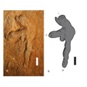
The Late Campanian Cerro del Pueblo Formation, located in southeastern Coahuila, Mexico, has produced a diverse array of vertebrate fossils. However, pterosaur remains from this unit are notably scarce. In this study, we describe new pterosaur material from the formation. The specimens include a fragmentary vertebra identified as belonging to an indeterminate, but derived pterodactyloid, along with the distal condyle of a left metacarpal, referable to an azhdarchoid pterosaur, and a left manus print. While these specimens provide additional evidence of pterosaur presence in the region during the Late Cretaceous, their fragmentary nature limits precise taxonomic and ichnotaxonomic identification. Nevertheless, they highlight the potential for future discoveries that could refine our understanding of the diversity and distribution of pterosaurs in Mexico.
PV article infos
Published in 48-2 (2025)
|
PDF
|
|
Rodent paleocommunities from the Oligocene of Ulantatal (Inner Mongolia, China)
Published online: 10/06/2014
Keywords:
late Paleogene; Mammalia; Mongolian Plateau; Rodentia; Systematics
https://doi.org/10.18563/pv.38.1.e3
Abstract
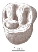
The Oligocene deposits of the Ulantatal area in Inner Mongolia (China) contain among the richest mammalian faunas from Asia. To date, only some parts of the rodent faunas have been described. Here, we propose to review the rodent faunal lists for each site, including the description of a few new rodent specimens. We describe three additional rodent species: the Cylindrodontidae Anomoemys lohiculus, the Eomyidae Asianeomys sp., and the Dipodidae Litodonomys huangheensis. This study allows us to constrain the stratigraphic range of Anomoemys lohiculus, which ranged from the late Early Oligocene to the early Late Oligocene in this area. Asianeomys sp. and Litodonomys huangheensis are dated from the latest Oligocene. These Oligocene deposits consist now of more than 70 species of mammals if we include the fauna from Kekeamu. This latter corresponds to the basal part of the Ulantatal Formation and could be dated biochronologically from the earliest Oligocene. When compared to the faunas from the Valley of Lakes in Central Mongolia, the Ulantatal faunas present a great majority of rodents, and this difference can be partly explained by sampling and description biases regarding macro-mammals. This study also shows that variations existed between Inner and Central Mongolia, especially regarding the composition of the rodent paleocommunities. However, the assessment of their evolutionary history in this part of Asia with respect to the important climate and environment changes, require further precisions and more material than current data allow.
PV article infos
Published in Vol.38-1 (2014)
|
PDF
|

|
A new hypothesis for the origin of African Anomaluridae and Graphiuridae (Rodentia)
Published online: 16/12/1996
Keywords:
Africa; Anomaluridae; Gliridae; Graphiuridae; Paleontology; PHYLOGENY; Rodentia
Abstract
A new hypothesis for the phylogenetic relationships of recent anomalurids and graphiurids is proposed, based on information from evolutionary lineages of Paleogene European rodents, particularly Gliridae, and Eocene Algerian Zegdoumyidae. Differences in first occurrences, in paleogeography, and in infraorbital structure in glirids (protrogomorphy and pseudomyomorphy) and graphiurids (hystricomorphy) separate Graphiuridae from Gliridae (Graphiurinae is here raised to family rank). Similar considerations, and dental morphology, suggest that Anomaluridae (appearing in the late Eocene) and Graphiuridae (appearing in the Pliocene) are related to early Eocene Zegdoumyidae.
PV article infos
Published in Vol. 25, Fasc. 2-4 (1996)
|
PDF
|

|
Révision des Chiroptères Lutériens de Messel (Hesse, Allemagne).
Published online: 04/04/1970
Keywords:
Chiroptera; Lutetian; Messel
https://doi.org/10.18563/pv.3.4.83-182
Abstract
The revision of the Lutetian chiropterans from Messel, first described by Revilliod in 1917, is based on the anatomy of the teeth and the skeleton. A figuration or refiguration of thematerial utilized accompanies the new description, which goes beyond that of the original monograph.
The study shows a certain variability of the dental structure within the genera Palaeochiropteryx Revilliod and Archaeonycteris Revilliod, as well as a general resemblance of the two forms. The morphology of the teeth permits, however, the verification of the validity of the different species: Palaeochiropteryx tupaiodon Revilliod, P. spiegeli Revilliod, Archaeonycterís trigonodon Revilliod, and Archaeonycteris revilliodi, n. sp.
Some differences of the skeletal and dental anatomy tend to indicate a stage of evolution less advanced for the genus Archaeonycteris.
The comparison of the chiropterans of Messel with the principal groups of living chiropterans, as well as with different Eocene fossíls (notably Cecílionycteris Heller and Icaronycteris Jepsen) leads to a more precise idea of the anatomy of primitive chiropterans. This comparison also permits the proposition that the oid forms so far described by integrated in a superfamily, the Palaeochiropterygoidea and allows a general phylogenetic hypothesis to be advanced for the order Chiroptera.
PV article infos
Published in Vol. 03, Fasc. 4 (1970)
|
PDF
|

|
Les rongeurs du site Pliocène à Hominidés de Hadar (Ethiope)
Published online: 15/02/1982
Keywords:
Ethiopia; hominids; Muridae; Pliocene
https://doi.org/10.18563/pv.12.1.1-56
Abstract
The intensive exploration of the Pliocene Hadar Formation, rich in hominid remains, led us to the discovery of several micromammals levels. ln some of them, rodents are very abundant. The stratigraphic repartition of these levels do not cover the whole fossiliferous series of the formation but takes place only in the sedimentary members from Sidi Hakoma and Denen-Dora (rancing from 3.1 - 3.2 MY to 2.8 - 2.9 MY, according to the recent geochronological data). During this gap of time, the species do not show morphological changes, what allowed us to gather, in the same taxa, forms of slighty different ages.
Two striking facts, giving a lot of indications, characterize these small rodents'faunas. First, we notice the domination of the Muridae, as well on a qualitative way (number of species) as on a quantitative one (number of individuals). Then, it appears that, until now, two genera of these murids were known only in the south-western asiatic regions. So, we can suppose continuous biotops between Africa and Indian Subcontinent before 3 MY. In this hypothesis, the hominids had already the possibility to leave their african « cradle ››. Finally, almost all studied genera are still represented at the present time. This fact, previously observed in Laetolil, Omo, Olduvai contributes to remove hope of establishing a biochronological scale based on rodents, in tropical zone. Nethertheless, that allows to try a reconstruction of the palaeoenvironnement, by using the principle of actualism.
PV article infos
Published in Vol. 12, Fasc. 1 (1982)
|
PDF
|

|
Muridae (Rodentia) du Pliocène supérieur d'Espagne et du midi de la France.
Published online: 20/09/1969
Keywords:
Anthracomys meini; Castillomys crusafonti; Pliocene; Rodents; Valerymys ellenbergeri
https://doi.org/10.18563/pv.3.1.1-25
Abstract
The murid fauna of the terminal Pliocene of southwest Europe is rich in at least eight genera and ten species. With the species belonging to the genera Apodemus, Rhagapodemus, and Stephanomys not being studied here, the study of the other murids resulted for one thing in the description of three new genera and three new species: Castillomys crusafonti n. g., n. sp., Occitanomys brailloni n. g., n. sp., Anthracomys meini n. sp., Valerymys ellenbergerí (THALER) n. g., and for another thing in the recognition of a form hitherto unknown in this region, Micromys praeminutus KRETZOI. Systematic study has shown that certain species of the terminal Pliocene fauna had their ancestors in the Turolian fauna presently known in Spain. The evolutionary lineages thereby recognized have been studied more in detail and a list of the evolutionary tendencies of the dendal characters has been given. A chart of the probable phyletic relationships between the different murids of the Pliocene faunas of southwest Europe (With the genus Rhagapodemus and Apodemus dominans being excluded) is given in conclusion of this work.
PV article infos
Published in Vol. 03, Fasc. 1 (1969)
|
PDF
|

|
Les sélaciens du Miocène de la région de Montpellier
Published online: 15/12/1970
Keywords:
Ichtyofauna; Miocene; Montpellier
https://doi.org/10.18563/pv.3.ext.1-139
Abstract
The utilization of screen-washing and attack by dilute acetic acid has permitted the collecting, in the Miocene of the department of Hérault (France), of a very rich ichthyofauna. This fauna is presently comprised of about 60 studied species, of which 11 are new, and represents, in the present state of knowledge, the most varied Miocene selachian fauna described in the world.
The abondance of material has allowed an overall revision to be made; it has thus been possible to complete the description and the figuration of species that were poorly known until now, and to synonymize species that were established on simple morphotypes. Paleo-ecologic study of the ichthyofauna has permitted conclusions to be drawn relative to climate and bathymetry; it was thus possible to show that the Miocene fauna of Hérault was a fauna of a subtropical sea, essentially neritic with rare pelagic contributions.
Knowing the stratigraphic position of the localities, it has been possible to distinguish three faunal assemblages based on associations of species. Some hypotheses on the evolution of certain lineages have been expressed.
The comparison of this fauna with that of other regions permitted the relationships of two diflerent faunal provinces to be specified: the first belongs to the northern domain, characterized by a fauna still subtropical but with numerous temperate water elements; the leoond belongs to the Mesogean domain characterized by warm water forms. It has also lhovm that contemporary faunas could be very different according to the bathymetric zone in which they lived, which furnishes valuable information for the paleogeographic reconstruction of sedimentary basins.
PV article infos
Published in Vol. 3, Ext (1970)
|
PDF
|
|
Comparative bone histology of rhabdodontid dinosaurs
Published online: 17/11/2014
Keywords:
bone histology-based ontogeny; Mochlodon; Rhabdodon; skeletal maturation; Zalmoxes
https://doi.org/10.18563/pv.38.2.e1
Abstract
A comparative bone histological study of the three known genera of the endemic European ornithopod dinosaur family, Rhabdodontidae, is presented here in an ontogenetic context. Investigated specimens were assigned to different ontogenetic stages based exclusively on the histological indicators of osteologic maturation during diametrical bone growth; an entirely size-independent method as opposed to most previous studies. Qualitative comparison of bone histology of corresponding ontogenetic stages and elements among the three valid rhabdodontid genera, Mochlodon, Zalmoxes, and Rhabdodon, revealed some consistent patterns. Genus specific histological differences within Rhabdodontidae are most expressed between Rhabdodon and the Mochlodon-Zalmoxes clade. These indicate a prolonged phase of fast growth and a less constrained cyclicity in the growth dynamics of Rhabdodon, as opposed to the slower and more regulated growth strategy reflected in the bones of Mochlodon and Zalmoxes. These genus specific differences are consistent with the phylogenetic interrelation of the genera and are most probably related to the pronounced differences in body size. However, when compared to other ornithopods, most detected histological features in rhabdodontids do not seem to reliably reflect either phylogenetic relations or body size. A notable common feature of all rhabdodontid genera irrespective of body size is the ontogenetically early onset of cyclical growth and secondary remodelling; a pattern that more resembles the condition found in derived ornithopods than that described in more basal taxa which are closer relatives of rhabdodontids. The recognition of taxon-specific histological patterns as well as patterns indicative of ecological and thereby functional traits clearly requires more accurate, preferably quantitative evaluations.
PV article infos
Published in Vol.38-2 (2014)
|
PDF
|
|
Revision of the historical collections of Pliocene-Pleistocene large mammals from Le Riège and Saint-Palais localities, near Pézenas (Southern France)
Published online: 23/06/2025
Keywords:
Hérault; Mammalia; Montpellier; Neogene; Quaternary
https://doi.org/10.18563/pv.48.1.e2
Abstract
Numerous “Quaternary” large-mammal fossils have been collected since the 1830s along the Le Riège stream, near Pézenas (Southern France). More than 120 specimens are stored in the collections of the Université de Montpellier (UM) under the name “Le Riège”. A major operation aiming at relocating the palaeontological collections of the University has made it possible to group together all the specimens of interest and launch their systematic revision for the first time. The fossils belong to the Reboul (1839; 51 samples) and de Christol (1865; 18 samples) Collections and 17 samples compose the Crochet & Ivorra Collection (1998). The remaining 38 samples have no mention about the exact time and location of their finding. We provide a critical inventory with literal transcription of inscriptions on specimens and historical labels. This revision confirms the presence of two distinct faunal assemblages under the name of “Le Riège”: Saint-Palais (Early Pliocene, MN14–15) and Le Riège sensu stricto (late Early Pleistocene, most likely MNQ19). The former assemblage, with coastal affinities, is composed of the ruminants Alephis sp. and Procapreolus cf. pyrenaicus, the rhinocerotid Pliorhinus megarhinus, the gomphotheriid Anancus arvernensis and marine mammals, all emblematic taxa for the Early Pliocene of Montpellier and Perpignan. The latter assemblage documents a late Early Pleistocene fluvio-volcanic sequence, yielding the bovid Bison (Eobison) spp., the cervid Eucladoceros cf. giulii, the hippopotamid Hippopotamus antiquus, the rhinocerotid Stephanorhinus etruscus, the equid Equus cf. altidens, and the elephantid Mammuthus cf. meridionalis, plus a few specimens of uncertain taxonomic affinities. This revision underscores the interest of revisiting historical collections and further provides a starting point for future research.
PV article infos
Published in 48-1 (2025)
|
PDF
S.I. Data
|
|
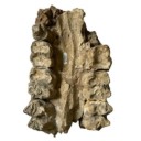
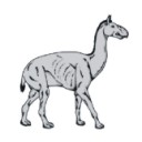
Reconstruction of the cervical skeleton posture of the recently-extinct litoptern mammal Macrauchenia patachonica Owen, 1838
Published online: 02/05/2023
Keywords:
biomechanics; cervical posture; functional anatomy; Litopterna; Macrauchenia
https://doi.org/10.18563/pv.46.1.e1
Abstract
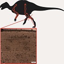
Macrauchenia patachonica was among the largest litopterns. It had a long neck with elongated cervical vertebrae, unique among endemic South American ungulates. We calculated the pattern of stress in the joints between the vertebral centra along the neck of the recently-extinct litoptern mammal M. patachonica for various hypothetical neck postures to determine which one is optimal. We also determined the zygapophyseal alignment positions for the neck, assuming a wide range of values for the thickness of the intervertebral discs. We concluded that a vertical posture is the one that best meets the requirements of nearly constant stress. This upright posture was probably a frequently adopted posture by M. patachonica while feeding or standing. It is also possible that occasionally it could adopt a gerenuk-like posture. In almost any other position, the standard deviations of stress values (SD) divided by mean stress (MS) have values between 0.4 and 0.5. Since it was a mixed feeder, M. patachonica probably used different postures to reach resources at different heights. However, an almost horizontal posture was required for the optimal articulation of the neck vertebrae. It probably represents the posture during fast locomotion, as suggested in a previous biomechanical study of locomotion.
PV article infos
Published in 46-1 (2023)
|
PDF
|

|
The late Miocene percrocutas (Carnivora,Mammalia) of Madedonia, Greece.
Published online: 14/06/1995
Keywords:
Biochronology; Carnivora; Comparisons; Dinocrocuta; Greece; Late Miocene; Mammalia
Abstract
Some new material of percrocutas from the late Miocene of Axios valley (Macedonia, Greece) is studied. They have been found in the locality of "Pentalophos 1" (PNT). The material has been described and compared with the known late Miocene percrocutas of Eurasia. This comparison indicates that it can be identified as Dinocrocuta gigantea (SCHLOSSER, 1903). A maxilla of a percrocuta, named ”Hyaena" salonicae, was found in the same area (Andrews, 1918). "Hyaena" salonicae is smaller than the PNT material. It is also compared with other material from Eurasia while its taxonomic and age problems are discussed. It belongs to Dinocrocuta and shows close relationships with D. robusta and D. senyureki; its age can be considered as late Vallesian-early Turolian. The age of the locality PNT is also discussed and a possible Vallesian age is proposed for it.
PV article infos
Published in Vol. 24, Fasc. 1-2 (1995)
|
PDF
|

|
Norselaspis glacialis n.g., n.sp, et les relations phylogénétiques entre les kiaeraspidiens (Osteostraci) du dévonien inférieur du Spitsberg.
Published online: 15/06/1981
Keywords:
Devonian; kiaeraspids; Osteostraci; Spitsbergen
https://doi.org/10.18563/pv.11.2-3.19-131
Abstract
The anatomy of Norselaspis glacialis n.g., n.sp., a primitive kiaeraspidian from the Lower Devonian of Spitsbergen, is described on the basis of spécimens studied by grinding sections or prepared with dilute formic acid. This study yielded some new anatomical details, including the presence of a canal prolonging posteromedially the canal alloted to the facial nerve by Stensiö. This posterior prolongation of the « facial canal ›› into the posterolateral part of the labyrinth cavity is consistent with the hypothesis put forward by Allis, Lindström, Jefferies and Whiting, that this canal housed the glossopharyngeus nerve. Furthermore, in N. glacialis, the foramen usually referred to as the foramen for the œsophagus opens posteriorly into a cavity in the postbranchial wall, referred to here as the intramural cavity, and which is interpreted as having housed the heart. Consequently, the œsophagus probably accompanied the dorsal aorta through the aortic canal. Finally, the foramen generally interpreted as having transmitted the ventral afferent arterial trunk is here considered as having housed the hepatic vein, which emptied into the venous sinus of the heart. The ventral afferent arterial trunk may thus have passed through the former «œsophageal ›› foramen.
The problem of the position of the dorsal nerves in the Osteostraci is discussed, and it is suggested that the three foremost nerve canals opening into the oralobranchial cavity housed the maxillary ramus of the trigeminus, the facial nerve and the glossopharyngeus nerve respectively. The mandibular ramus of the trigeminus must have accompanied one of the two foremost nerves, but for the moment it is impossible to decide which.
The problem of the nature of the interbranchial crests of the Osteostraci is briefly discussed. Comparison with the branchial apparatus of the Petromyzontida does not support the hypothesis that the interbranchial crests are part of the branchial arches, incorporated into the endoskeletal shield. A different hypothesis is proposed, that the branchial skeleton of the Osteostraci was situated entirely inside the oralobranchial cavity, and was attached to the endoskeletal shield only by the ventromedial processes. The grooves classically allotted to the efferent branchial arteries would thus have housed extrabranchial arteries, branching off from the dorsal aorta, and irrigating the ventral branchial musculature.
A phylogeny and a classification of the kiaeraspidians are proposed. The evolution of this monophyletic group is characterized by, e.g., reduction of cornual processes, shortening of the abdominal division of the shield, subdivision of the lateral fields, and enlargement of the supraoral fossae.
The phylogenetic position of the kiaeraspidians within the Osteostraci remains uncertain. Their sister-group may be either the benneviaspidiens or the thyestidians, or Thyestes alone (in which case they would have to be included within the thyestidians).
PV article infos
Published in Vol. 11, Fasc. 2-3 (1981)
|
PDF
|
|
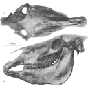
Old world hemiones and new world slender species (Mammalia, Equidae)
Published online: 16/12/2008
Keywords:
Amerhippus; biometry; Equus; Holocene; New World; Old World; Osteology; Pleistocene; Pliocene
https://doi.org/10.18563/pv.36.1-4.159-233
Abstract
Morphological and biometrical description of skulls, teeth, and limb bones of extant and fossil Old World herniones (including E. hydruntinus) and of New World 'stilt-Iegged' and other slender species from Blancan to Holocene. An Appendix presents ways in which the approximate size of some missing bones or dimensions may be deduced from available ones.
The discussed and/or illustrated fossils were found in Bolivia (Tarija), Canada (Yukon), China (Choukoutien, Gulongshan, Jiling, Loufangzi), Ecuador (Oil Fields), Ethiopia (Melka Kunturé), France (Lunel-Viel), Germany (Süssenborn), Greece (Agios Georgios, Petralona), Hungary (Dorog), Italy (Romanelli), Mexico (Cedazo, San Josecito), Mongolia (Sjara-osso-gol), Spain (Venta Micena), ex-Soviet Union (Akhalkalaki, Binagady, Chokurcha, Chukochya, Kabazi, Kolyma, Krestovka, Kurtak, Staroselie, Tologoj), USA (Alaska, Arkalon, Cedar Meadow, Channing, Conkling, Dry Mountains, Hay Springs, Leisey Shell Pit A, Lissie Formation, Natural Trap, Pool Branch, Powers Ranch, Rock Creek, San Diego, Santo Domingo, Seymour Formation, Shelter, Slaton, Trinity River). Numerous raw or statistically elaborated data are given in Tables.
There is no evidence for the existence of Old World hemiones in the New World nor of 'stilt-Iegged' equids in the Old World. The first 'stilt-Iegged' equid was found at Santo Domingo, New Mexico, and is believed to be Late Blancan. It was probably at the origin of E. calobatus (Arkalon, Rock Creek) and of the smaller E. semiplicatus (Channing, Rock Creek). Slender, but not 'stilt-Iegged', equids found at Natural Trap, Wyoming, ca. 12 ky ago belong to Amerhippus. AlI these species share with Oid World Sussemiones (and some hemiones) peculiar patterns on the lower cheek teeth.
The slender Equus sp. B of Leisey Pit A, Florida, ca. 1.2 Ma, as weIl as Amerhippus francisci and E. tau (probably a senior synonym of E. quinni) share conventional lower cheek teeth patterns. The skulls of A. francisci and E. tau, however, are quite different.
Paleontological data suggest a common origin of Amerhippus, Sussemiones, and 'stilt-Iegged' equids during the late Blancan. Old World hemiones seem to have differentiated later.
PV article infos
Published in Vol. 36, Fasc. 1-4 (2008)
|
PDF
|

|
Les Dipodidae (Mammalia, Rodentia) d'Europe occidentale au Paléogène et au Néogène inférieur: origine et évolution.
Published online: 01/10/1980
Keywords:
Dipodidae; Late Oligocene; Quercy Phosphorites
https://doi.org/10.18563/pv.9.ext.302-342
Abstract
The study of three new populations of Plesiosminthuspromyarion from the "phosphorites du Quercy" and of material from "Auvergne" brings new data on european oligocene Dipodidae. They appear in Western Europe at the beginning of late Oligocene. Evolutionary trends of the group are drawn and particularly the emergence of morphotypes announcing P. schaubi, from the Coderet level, is revealed among the most recent populations of P. promyarion. Differences are attempted to be drawn between the three species : P. promyarion, P. myarion and P. schaubi by restudying the type-population of P. myarion from the aquitanian deposits of Chavroches (Allier) in comparison with two other populations from the same age and the same region. Relationships between early european, american and asiatic Dipodidae are discussed.
PV article infos
Published in Vol. 9, Ext (1980)
|
PDF
|
|
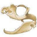
The skull of Tetraceratops insignis (Synapsida, Sphenacodontia)
Published online: 09/01/2020
Keywords:
cranium; pelycosaur; Permian; therapsid origins
https://doi.org/10.18563/pv.43.1.e1
Abstract
Tetraceratops insignis is known from a single, crushed skull from the Lower Permian of Texas. Its unique proportions and osteological details gained central meaning in the question of the origins of Therapsida since this early synapsid has been determined as the oldest and less derived therapsid. Apart from Tetraceratops, the ‘mammal-like’ Therapsida and their sister, the pelycosaur-grade Sphenacodontidae, are separated by one of the longest ghost lineages in tetrapod fossil record. However, the minor, though well justified critique faced insistent publication regarding the therapsid hypothesis. A carefull re-evaluation of the holotypic skull reveals that therapsid traits cannot be supported, including a rejection of the formerly supposed adductor shelf in the temporal fenestra. Increased understanding of ‘pelycosaur’ character variation underlines a haptodontine-grade or, less likely, sphenacodontid position for Tetraceratops.
PV article infos
Published in Vol 43-1 (2020)
|
PDF
|

|
Mammals of the Eocene locality Toru Ajgyr (Kyrgyzstan)
Published online: 15/12/2006
Keywords:
Eocene; Kyrgyzstan; Mammalia; Olsenia; Palaeoecology; Stratigraphy; taxonomy
https://doi.org/10.18563/pv.34.e12
Abstract
Morphological descriptions are given of Eocene mammals from the locality Toru Ajgyr (NEKyrgyzstan) that were excavated in 1997 and 1998 in a cooperation between the Martin-Luther-University Halle (Germany), the Zoological Institute in St. Petersburg (Russia) and the Seismological Institute in Bishkek (Kyrgyzstan). The species found belong mostly to perissodactyls, as Lophialetes sp., Teleolophus sp. and brontotheres. The primitive ungulate family Olseniidae is represented by a complete foot skeleton of cf. Olsenia sp. In addition, postcranial materials of Gobiatherium mirificum (Dinocerata) and of artiodactyls have been collected and are described herein. Based on mammals, the locality is part of the Asian Land Mammal Age Arshantan and is stratigraphically equivalent with the Bridgerian Land Mammal Age in North America and with the lower and middle Geiseltalian of the European Middle Eocene.
PV article infos
Published in Vol. 34, Fasc. 3-4 (2006)
|
PDF
|

|
Les gisements de Robiac (Eocène supérieur) et leurs faunes de Mammifères.
Published online: 05/04/1969
Keywords:
Fauna; Late Eocene; Mammalia; Robiac
https://doi.org/10.18563/pv.2.3.95-156
Abstract
Designated the type-locality of a late Eocene paleomammal zone, Robiac has recently been the object of important excavations. The first results of the new collecting, as well as a revision of the material in old collections, are given in this work.
Two stratigraphic section, cutting through the two sites presently distinguished (Robiac-Nord and Robiac-Sud) reveal the lithologic variation at the base of the Fons Limestone and the localization of three fossiliferous beds at Robiac-Sud.
The molluscan fauna and the flora (charophytes), which have already been described, as well as that of the lower vertebrates, have been listed.
A list of 46 mammalian species (only 16 species were known previous to 1964) has been established. The micro-mammals, nearly all new in this fauna (marsupials, insectivores, bats, rodents, primates, and some smallsized artiodactyls), Were obtained only after screen-washing of the matrix; about 4 tons of sediment were thus treated.
The artiodactyls have been the most extensively analyzed; 6 genera, of which one is new, have been recognized. The latter is described as Robiacina minum n.g., n.sp., and represents a very small artiodactyl of the family Anoplotheriidae. The taxonomie status of certain species formerly described has been clarified by the designation of lectotypes (Cebochoerus robiucensis, Catodonerium robiacense, Xiphodon castrense).
The paleontologic corrélations at present possible between the late Eocene faunas have allowed the relative positions of te different French localities of this age to be established; the Guépelle locality, it seems, could define in the future a new paleomammal zone.
PV article infos
Published in Vol. 02, Fasc. 3 (1969)
|
PDF
|

|
Artiodactyla aus den Eozänen Braunkohlen des Geiseltales bei Halle (DDR)
Published online: 04/12/1989
Keywords:
Artiodactyles; Eocene; Europe; Paleoecology; Stratigraphy; taxonomy
https://doi.org/10.18563/pv.19.3.131-160
Abstract
The present study of Artiodactyla from the Middle Eocene of the Geiseltal lignite beds concems systematics, biostratigraphy, and palaeoecology on the basis of 174 specimens: isolated remains to more complete skeletons. Instead of the formerly known five species of two families are now recognized 14 species of the Diacodexeidae, Dichobunidae, Cebochoeridae, and Haplobunodontidae. New species are Aumelasia maniai, Anthracobunodon neumarkensis, Masillabune franzeni. Four species of the Geiseltalfauna are definitely known from elswere, and five species are closely related to those from other European localities. Evidently the faunal situation of artiodactyls during the Middle Eocene of Europe was largely uniform. The distribution of artiodactyls within the sequence of the Geiseltal strata corroborates the biostratigraphical concept of the land mammal age Geiseltalian (Franzen & Haubold l986b) as well as the mammalian reference levels MP 11-13 (Franzen 1987). Reconstructions of the skulls and skeletons allow conclusions on the functional morphology and palaeoecology of the artiodactyls of the European Middle Eocene
PV article infos
Published in Vol. 19, Fasc. 3 (1989)
|
PDF
|
|
Les Périssodactyles (Mammalia) du gisement Bartonien supérieur de Robiac (Éocène moyen du Gard, Sud de la France)
Published online: 04/05/2015
Keywords:
Chasmotherium; new species; Palaeotheriidae; paleoenvironments
https://doi.org/10.18563/pv.39.1.e3
Abstract
We present here a new updated counting of the perissodactyls of Robiac, the type locality of the MP 16 level of the biochronological scale of paleogene mammals and that of the Robiacian stage of Eocene Land Mammals Ages in Western Europe.
The outcrop of Robiac consists actually of two 500m apart loci, Robiac-Nord and Robiac-Sud, considered of the same age according to the current discriminating power, and is dated from -38,7 MA after the last faunal, magnetostratigraphic and climatic calibrations.
It has yielded a very abundant and rich of 21 taxa perissodactyl fauna, topped by the giant Lophiodon lautricense, last representative of the family Lophiodontidae, of which it is the last proved deposit. The Palaeotheriidae are much diversified with 5 genera and 9 species of "Pachynolophinae", 3 genera and 10 species of Palaeotheriinae. Nine taxa have been defined from Robiac: Chasmotherium depereti n. sp., Palaeotherium castrense robiacense Franzen, 1968, the genus Leptolophus Remy, 1965 with the species L. stehlini Remy, 1965 and L. magnus Remy, 1998, Anchilophus (Paranchilophus) jeanteti Remy, 2012, Metanchilophus chaubeti Remy, 2012, Lophiotherium robiacense Depéret, 1917 and Pachynolophus gaytei n. sp.
The faunal Robiac cenogram with the associated flora testify to a hot, wet and forestal environment, likely corresponding to a short warming climatic phase; the broken up fossil bones should have been carried away and then gathered in swamp areas along the banks of a meandering river.
The swarm of mammals of Robiac, the richest of contemporaneous deposits, has been followed by a drastic drop in perissodactyl diversity at the MP 17A level; a crisis which could have originated in a renewal of the global Eocene cooling. Fons 4, the type-locality of this level, is largely scarcer in perissodactyls and its cenogram testifies to a less diversified fauna, with on the whole smaller species, that likely means a cooler and drier climatic environment; a new perissodactyl diversification occurred but later.
PV article infos
Published in Vol.39-1 (2015)
|
PDF
|
