|
Abstract book of the 18th Conference of the European Association of Vertebrate Palaeontologists (EAVP), 5-9 July 2021, Benevento, Italy
Published online: 12/07/2021
Keywords:
2021; Abstracts; Benevento; EAVP
https://doi.org/10.18563/pv.eavp2021
Abstract
Welcome to the 18th conference of the EAVP, the first online meeting of our association. The pandemic emergency made it impossible to organize the in-person meeting in Benevento as we all had hoped. However, we couldn’t miss another EAVP meeting. Therefore, this year we are meeting online, trying to make the experience the closest to the in-person meeting possible, in order to offer the delegates the opportunity to share knowledge, build new networks and reinforce the old ones. We have received 137 communications, with more than 150 delegates from 24 countries. All the abstracts have passed a peer review process and are part of this special volume of Palaeovertebrata, the official journal of the EAVP. This year we are also offering a variety of workshops, roundtables and symposia on different topics. These include the annual “Pride EAVP: An LGBTQ+ Roundtable” and “Women in Palaeontology Roundtable Discussion”, together with the workshops on “Gendered Perspective in Palaeontological Research: from Definition to Action”, “International Palaeontology Education: Virtual Teaching and Real-World Learning”, “Stepping out of Academia: Why, When and How?”, “Introduction to Hypothesis Testing in Statistics”, “The Early-Middle Pleistocene Transition: Marked Mammal Turnover and Ecosystem Dynamic” (included in the early event for the XXI INQUA Congress in Rome 2023, “A Mediterranean Perspective on Quaternary Sciences”). To conclude, we are hosting two symposia on “Palaeoart: Diversity on and behind the Canvas” and “3D fossils, Robotic and Experimental Palaeontology”. We wish you all a happy and productive meeting. And see you in Benevento next year!
PV article infos
Published in Special Volume 1-2021 (2021)
|
PDF
|
|
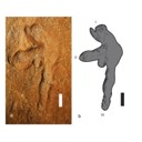
Pterosaurs (Pterosauria) from the Cerro del Pueblo Formation (Late Campanian) of Coahuila, Mexico
Published online: 21/11/2025
Keywords:
Azhdarchoidea; Coahuila; Mexico; Pterodactyloidea; Pterosauria
https://doi.org/10.18563/pv.48.2.e1
Abstract
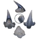
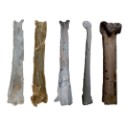
The Late Campanian Cerro del Pueblo Formation, located in southeastern Coahuila, Mexico, has produced a diverse array of vertebrate fossils. However, pterosaur remains from this unit are notably scarce. In this study, we describe new pterosaur material from the formation. The specimens include a fragmentary vertebra identified as belonging to an indeterminate, but derived pterodactyloid, along with the distal condyle of a left metacarpal, referable to an azhdarchoid pterosaur, and a left manus print. While these specimens provide additional evidence of pterosaur presence in the region during the Late Cretaceous, their fragmentary nature limits precise taxonomic and ichnotaxonomic identification. Nevertheless, they highlight the potential for future discoveries that could refine our understanding of the diversity and distribution of pterosaurs in Mexico.
PV article infos
Published in 48-2 (2025)
|
PDF
|
|
Revision of the historical collections of Pliocene-Pleistocene large mammals from Le Riège and Saint-Palais localities, near Pézenas (Southern France)
Published online: 23/06/2025
Keywords:
Hérault; Mammalia; Montpellier; Neogene; Quaternary
https://doi.org/10.18563/pv.48.1.e2
Abstract
Numerous “Quaternary” large-mammal fossils have been collected since the 1830s along the Le Riège stream, near Pézenas (Southern France). More than 120 specimens are stored in the collections of the Université de Montpellier (UM) under the name “Le Riège”. A major operation aiming at relocating the palaeontological collections of the University has made it possible to group together all the specimens of interest and launch their systematic revision for the first time. The fossils belong to the Reboul (1839; 51 samples) and de Christol (1865; 18 samples) Collections and 17 samples compose the Crochet & Ivorra Collection (1998). The remaining 38 samples have no mention about the exact time and location of their finding. We provide a critical inventory with literal transcription of inscriptions on specimens and historical labels. This revision confirms the presence of two distinct faunal assemblages under the name of “Le Riège”: Saint-Palais (Early Pliocene, MN14–15) and Le Riège sensu stricto (late Early Pleistocene, most likely MNQ19). The former assemblage, with coastal affinities, is composed of the ruminants Alephis sp. and Procapreolus cf. pyrenaicus, the rhinocerotid Pliorhinus megarhinus, the gomphotheriid Anancus arvernensis and marine mammals, all emblematic taxa for the Early Pliocene of Montpellier and Perpignan. The latter assemblage documents a late Early Pleistocene fluvio-volcanic sequence, yielding the bovid Bison (Eobison) spp., the cervid Eucladoceros cf. giulii, the hippopotamid Hippopotamus antiquus, the rhinocerotid Stephanorhinus etruscus, the equid Equus cf. altidens, and the elephantid Mammuthus cf. meridionalis, plus a few specimens of uncertain taxonomic affinities. This revision underscores the interest of revisiting historical collections and further provides a starting point for future research.
PV article infos
Published in 48-1 (2025)
|
PDF
S.I. Data
|
|
Enigmatic rodents from Lavergne, a late middle Eocene (MP 16) fissure-filling of the Quercy Phosphorites (Southwest France)
Published online: 08/04/2024
Keywords:
diversity; late Bartonian; Rodentia; taxonomy; Theridomyidae
https://doi.org/10.18563/pv.47.2.e1
Abstract
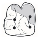
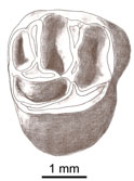
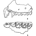
Two somewhat “odd” taxa of theridomyid rodents, one formerly known (Bernardia marandati Vianey-Liaud, 1991) and the other new (Idicia vidalenci gen. et sp. nov.) are discussed from a taxonomical and taphonomical perspectives. These two rodents were found at Lavergne, a late middle Eocene (MP16) “phosphatière” from the Quercy (Southwest France). The genus Bernardia, being preoccupied by a scale insect (Bernardia Ashmead, 1881), is here renamed Burgia. We benefit from this nomenclatural change to describe additional new dental specimens of this patriotheridomyine species, including a previously undescribed locus (P4). The other theridomyid from Lavergne, Idicia vidalenci gen. et sp. nov., so far documented by a mandible preserving two teeth (m2-m3) is a new taxon of peculiar occlusal morphology, and whose subfamilial affinities remain unknown. These two peculiar theridomyids recorded at Lavergne are found nowhere else, whether in coeval localities in Quercy or elsewhere in Western Europe. We discuss the possible causes of their unique presence at Lavergne.
PV article infos
Published in 47-2 (2024)
|
PDF
|
|
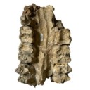
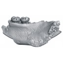
A partial skeleton of Metaxytherium medium from the middle Miocene of La Morfassière quarry (Indre-et-Loire, France)
Published online: 16/01/2025
Keywords:
Faluns; France; Metaxytherium; Miocene; Sirenia
https://doi.org/10.18563/pv.48.1.e1
Abstract
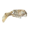
Sirenians are among the common marine fossil remains found in the Faluns deposits of western France. We describe new material of a Dugongidae sirenian from the middle Miocene Savignean facies of La Morfassière quarry (Indre-et-Loire, northwestern France) that includes a well preserved and almost complete skull associated with its mandible, several vertebrae and ribs. The cranial remains exhibit features that allow to attribute them to Metaxytherium medium, a species recorded from the middle and early late Miocene of European and Mediterranean coasts. The discovery of an associated skull and mandible of this species is unusual in this area and deserves to be reported, mostly because its preservation contributes to a better knowledge of the variable structure of its skull and teeth anatomy. For the first time the body size and weight of M. medium can be assessed through regression equations based on skull measurements. The particularly good condition of the material can be explained by the calm and deep marine environment in which it was deposited.
PV article infos
Published in 48-1 (2025)
|
PDF
|

|
Book of Abstracts of the XXII Annual Meeting of the European Association of Vertebrate Palaeontologists, 30 June–5 July 2025, Kraków, Poland
Published online: 20/06/2025
Keywords:
Abstracts; EAVP
https://doi.org/10.18563/pv.eavp2025
Abstract
Book of Abstracts of the XXII Annual Meeting of the European Association of Vertebrate Palaeontologists, 30 June–5 July 2025, Kraków, Poland.
PV article infos
Published in 48-1 (2025)
|
PDF
|
|
Lissamphibians from Dams (Quercy, SW France): Taxonomic identification and evolution across the Eocene-Oligocene transition
Published online: 25/06/2025
Keywords:
Eocene-Oligocene; Grande Coupure; Lissamphibia; Quercy Phosphorites
https://doi.org/10.18563/pv.48.1.e3
Abstract
The locality of Dams (Quercy, southwestern France) has yielded two fossil assemblages, one from the late Eocene and another from the early Oligocene, making it one of the few localities with infillings across the Eocene-Oligocene transition. At least 24 taxa (13 mammals, 11 snakes) have been identified in this locality. Study of the lissamphibian remains from Dams yields an Eocene and an Oligocene assemblage, with a total of eight taxa. The Eocene assemblage includes two unnamed salamandrine species, one unnamed pelobatid species and one pyxicephalid species (Thaumastosaurus). The Oligocene assemblage includes two unnamed pleurodeline species, one salamandrine species (Salamandra sansaniensis) and an unnamed pelobatid species. Among the eight taxa from Dams, one Eocene salamandrine and one Oligocene pleurodeline are identified for the first time in the Quercy. A review of the lissamphibians from the Quercy area identifies eleven taxa for the Late Eocene (MP19) and eight taxa for Early Oligocene (MP22), with a major turnover at the Eocene-Oligocene transition. This turnover occurs in a time of major climatic changes, with a significant decrease in temperature and precipitation and concurrent increase in seasonality in Europe, likely affecting specialized taxa.
PV article infos
Published in 48-1 (2025)
|
PDF
|
|
First report of Cylindracanthus (Osteichthyes) from the Eocene of India
Published online: 25/03/2024
Keywords:
Cylindracanthus; Eocene; histology; rostrum; Umarsar mine.
https://doi.org/10.18563/pv.47.1.e2
Abstract
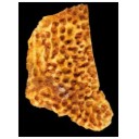
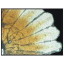
Fossils of the endangered sturgeons and peddlefishes are widely distributed. We here report for the first time the presence of one of the extinct osteichthyes genus Cylindracanthus (Liedy 1856a) from the Early Eocene lignite-bearing successions of the Kutch Basin, India. The present well preserved rostrum is characterised by numerous wedge-shaped components encircling the central canal that runs along its length, paired at the base and each wedge contributing to the formation of a ridge. The rostrum lacks teeth. The present find extends the palaeobiogeographical distribution of Cylindracanthus considerably and supports its Eocene age as dental remnants preserved in Cylindracanthus sp. shows a decrease in remanent dentition and tooth bases from the Cretaceous to the Eocene. Cylindracanthus is an useful palaeoenvironmental indicator as it has been found associated typically with deposits of nearshore marine environments.
PV article infos
Published in 47-1 (2024)
|
PDF
|
|
Preliminary report on the fishes (Chondrichthyes & Teleostei) from the lower Oligocene (Rupelian) Red Bluff Clay at site AMo-9, Monroe County, Alabama, USA
Published online: 26/06/2024
Keywords:
Batomorphii; Elasmobranchii; Galeomorphi; Gulf Coastal Plain; Vicksburg Group
https://doi.org/10.18563/pv.47.2.e2
Abstract
Herein we describe a small but relatively diverse assemblage of fossil fishes derived from the lower Oligocene (Rupelian) Red Bluff Clay at site AMo-9 in Monroe County, Alabama, USA. Identified amongst the remains are 15 unequivocal taxa representing 11 families within five orders, and one additional taxon represents an unknown order and family. Taxa identified include Eostegostoma sp., Otodus (Carcharocles) sp., Mitsukurinidae/Carchariidae indet., Macrorhizodus praecursor, Galeorhinus sp., Negaprion gilmorei, Physogaleus sp., “Sphyrna” sp., Galeocerdo sp., cf. “Aetobatus” sp., Sphyraena sp., Xiphiorhynchus kimblalocki, Xiphiorhynchus sp., Cylindracanthus ornatus, and C. rectus. Several additional fossils could not be identified beyond Lamniformes, Carcharhiniformes, and Teleostei, but they likely belong to one of the identified taxa within this paleofauna. All of the fishes previously reported from the Red Bluff Clay within the entirety of the Gulf Coastal Plain of the USA are otolith-based, and each of the 15 unequivocal taxa reported herein are important new records for this lithostratigraphic unit. In particular, the Eostegostoma sp. and Xiphiorhynchus spp. specimens represent the first occurrences of these taxa in Alabama. The specimens of C. ornatus, Eostegostoma sp., and X. kimblalocki are stratigraphic and temporal range extensions from the middle and late Eocene into the Rupelian Stage of the Oligocene. Other described taxa may represent transitional forms between those described from the late Eocene and late Oligocene within the region. This study provides a tantalizing preliminary view into faunal transitions that occurred amongst marine fishes across the Eocene/Oligocene boundary within the Gulf Coastal Plain of the USA.
PV article infos
Published in 47-2 (2024)
|
PDF
|

|
Morphological description and identification of an extraordinary new elephant cranium from the early Pliocene of Ileret, Kenya
Published online: 21/10/2021
Keywords:
Elephantidae; Loxodonta adaurora; cranium; early Pliocene; Ileret; Kenya
https://doi.org/10.18563/pv.44.2.e3
Abstract
Abstract: Paleontological exploration in the Turkana Basin near Ileret, Kenya yielded the most complete adult elephant cranium (KNM-ER 63642) known from the late Miocene to mid-Pliocene. KNM-ER 63642 derives from the lower Lonyumun Mb. of the Koobi Fora Fm. and dates to the early Pliocene, >4.3 Ma. The cranium is immense in size and preserves most of its structures including left and right M2-3, permitting its comprehensive comparative study and secure taxonomic assignment to Loxodonta adaurora. Features distinctive of the species and exhibited by KNM-ER 63642 include very elongate, divergent tusk alveoli, a short, biconvex cranial roof, anterosuperior angulation of the occipital planum, non-inflated occipital planum and absence of supralateral parietal "bossing," broad, flat premaxillary nasal processes, broad, laterally downturned nasal aperture superior to the level of the orbits, and M3s with wide, subhypsodont plates that are parallel-faced and separated by U-shaped transverse valleys. The M3s also exhibit characteristic L. adaurora traits of greatest width at their bases, rounded cross-sectional shape, thick enamel, abundant cementum, and strong anterior and posterior accessory conules. Of extant taxa, KNM-ER 63642 most closely resembles crania of African elephants. Its inclusion in the Loxodonta clade is tenuous, however, because shared features are either symplesiomorphic or are difficult to test for synapomorphy due to the poor fossil record of crania of late Miocene-early Pliocene elephants. Overall, the cranial morphology of KNM-ER 63642 is unexpectedly advanced for an elephant of its antiquity. Its anteroposterior compression and height are concordant with efficient proal masticatory action, indicating that by the early Pliocene L. adaurora evolved craniodental adaptations in phase with feeding preference for C4 grasses. The advantage of synchrony of morphology and behavior is reflected by the dominance of the species in the greater Turkana Basin during that interval.
PV article infos
Published in 44-2 (2021)
|
PDF
|

|
Révision des Chiroptères Lutériens de Messel (Hesse, Allemagne).
Published online: 04/04/1970
Keywords:
Chiroptera; Lutetian; Messel
https://doi.org/10.18563/pv.3.4.83-182
Abstract
The revision of the Lutetian chiropterans from Messel, first described by Revilliod in 1917, is based on the anatomy of the teeth and the skeleton. A figuration or refiguration of thematerial utilized accompanies the new description, which goes beyond that of the original monograph.
The study shows a certain variability of the dental structure within the genera Palaeochiropteryx Revilliod and Archaeonycteris Revilliod, as well as a general resemblance of the two forms. The morphology of the teeth permits, however, the verification of the validity of the different species: Palaeochiropteryx tupaiodon Revilliod, P. spiegeli Revilliod, Archaeonycterís trigonodon Revilliod, and Archaeonycteris revilliodi, n. sp.
Some differences of the skeletal and dental anatomy tend to indicate a stage of evolution less advanced for the genus Archaeonycteris.
The comparison of the chiropterans of Messel with the principal groups of living chiropterans, as well as with different Eocene fossíls (notably Cecílionycteris Heller and Icaronycteris Jepsen) leads to a more precise idea of the anatomy of primitive chiropterans. This comparison also permits the proposition that the oid forms so far described by integrated in a superfamily, the Palaeochiropterygoidea and allows a general phylogenetic hypothesis to be advanced for the order Chiroptera.
PV article infos
Published in Vol. 03, Fasc. 4 (1970)
|
PDF
|
|
A late Eocene palaeoamasiine embrithopod (Mammalia, Afrotheria) from the Adriatic realm (Island of Rab, Croatia)
Published online: 14/12/2023
Keywords:
Balkanatolia; Grande Coupure; Great Adria; Paleobiogeography; Systematics
https://doi.org/10.18563/pv.47.1.e1
Abstract
A cheek tooth recently unearthed in the Lopar Sandstone unit, of late Eocene age, in the northern part of Rab Island, Croatia, is one of the very few Eocene mammalian remains found in the Adriatic area. Thorough comparison of this tooth with those of Old-World Palaeogene mammalian orders suggests that it is a M3 belonging to an embrithopod afrothere. The specimen is referred to as Palaeoamasia sp. This genus was formerly known only in Eocene deposits of Anatolia but with close relatives in Romania among Palaeoamasiinae. The geographical distribution of this subfamily perfectly matches the recently-named Balkanatolian landmass, which experienced in-situ evolution of endemic mammals prior to the Grande Coupure event that occurred around the Eocene–Oligocene transition. This last event is characterised by massive Asian immigration in Western Europe and the supposed extinction of many endemic Central and Western European mammals, including Palaeoamasiinae.
PV article infos
Published in 47-1 (2024)
|
PDF
|

|
Les sélaciens du Miocène de la région de Montpellier
Published online: 15/12/1970
Keywords:
Ichtyofauna; Miocene; Montpellier
https://doi.org/10.18563/pv.3.ext.1-139
Abstract
The utilization of screen-washing and attack by dilute acetic acid has permitted the collecting, in the Miocene of the department of Hérault (France), of a very rich ichthyofauna. This fauna is presently comprised of about 60 studied species, of which 11 are new, and represents, in the present state of knowledge, the most varied Miocene selachian fauna described in the world.
The abondance of material has allowed an overall revision to be made; it has thus been possible to complete the description and the figuration of species that were poorly known until now, and to synonymize species that were established on simple morphotypes. Paleo-ecologic study of the ichthyofauna has permitted conclusions to be drawn relative to climate and bathymetry; it was thus possible to show that the Miocene fauna of Hérault was a fauna of a subtropical sea, essentially neritic with rare pelagic contributions.
Knowing the stratigraphic position of the localities, it has been possible to distinguish three faunal assemblages based on associations of species. Some hypotheses on the evolution of certain lineages have been expressed.
The comparison of this fauna with that of other regions permitted the relationships of two diflerent faunal provinces to be specified: the first belongs to the northern domain, characterized by a fauna still subtropical but with numerous temperate water elements; the leoond belongs to the Mesogean domain characterized by warm water forms. It has also lhovm that contemporary faunas could be very different according to the bathymetric zone in which they lived, which furnishes valuable information for the paleogeographic reconstruction of sedimentary basins.
PV article infos
Published in Vol. 3, Ext (1970)
|
PDF
|
|
Rodent paleocommunities from the Oligocene of Ulantatal (Inner Mongolia, China)
Published online: 10/06/2014
Keywords:
late Paleogene; Mammalia; Mongolian Plateau; Rodentia; Systematics
https://doi.org/10.18563/pv.38.1.e3
Abstract
The Oligocene deposits of the Ulantatal area in Inner Mongolia (China) contain among the richest mammalian faunas from Asia. To date, only some parts of the rodent faunas have been described. Here, we propose to review the rodent faunal lists for each site, including the description of a few new rodent specimens. We describe three additional rodent species: the Cylindrodontidae Anomoemys lohiculus, the Eomyidae Asianeomys sp., and the Dipodidae Litodonomys huangheensis. This study allows us to constrain the stratigraphic range of Anomoemys lohiculus, which ranged from the late Early Oligocene to the early Late Oligocene in this area. Asianeomys sp. and Litodonomys huangheensis are dated from the latest Oligocene. These Oligocene deposits consist now of more than 70 species of mammals if we include the fauna from Kekeamu. This latter corresponds to the basal part of the Ulantatal Formation and could be dated biochronologically from the earliest Oligocene. When compared to the faunas from the Valley of Lakes in Central Mongolia, the Ulantatal faunas present a great majority of rodents, and this difference can be partly explained by sampling and description biases regarding macro-mammals. This study also shows that variations existed between Inner and Central Mongolia, especially regarding the composition of the rodent paleocommunities. However, the assessment of their evolutionary history in this part of Asia with respect to the important climate and environment changes, require further precisions and more material than current data allow.
PV article infos
Published in Vol.38-1 (2014)
|
PDF
|
|
Reconstruction of the cervical skeleton posture of the recently-extinct litoptern mammal Macrauchenia patachonica Owen, 1838
Published online: 02/05/2023
Keywords:
biomechanics; cervical posture; functional anatomy; Litopterna; Macrauchenia
https://doi.org/10.18563/pv.46.1.e1
Abstract
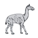
Macrauchenia patachonica was among the largest litopterns. It had a long neck with elongated cervical vertebrae, unique among endemic South American ungulates. We calculated the pattern of stress in the joints between the vertebral centra along the neck of the recently-extinct litoptern mammal M. patachonica for various hypothetical neck postures to determine which one is optimal. We also determined the zygapophyseal alignment positions for the neck, assuming a wide range of values for the thickness of the intervertebral discs. We concluded that a vertical posture is the one that best meets the requirements of nearly constant stress. This upright posture was probably a frequently adopted posture by M. patachonica while feeding or standing. It is also possible that occasionally it could adopt a gerenuk-like posture. In almost any other position, the standard deviations of stress values (SD) divided by mean stress (MS) have values between 0.4 and 0.5. Since it was a mixed feeder, M. patachonica probably used different postures to reach resources at different heights. However, an almost horizontal posture was required for the optimal articulation of the neck vertebrae. It probably represents the posture during fast locomotion, as suggested in a previous biomechanical study of locomotion.
PV article infos
Published in 46-1 (2023)
|
PDF
|

|
Muridae (Rodentia) du Pliocène supérieur d'Espagne et du midi de la France.
Published online: 20/09/1969
Keywords:
Anthracomys meini; Castillomys crusafonti; Pliocene; Rodents; Valerymys ellenbergeri
https://doi.org/10.18563/pv.3.1.1-25
Abstract
The murid fauna of the terminal Pliocene of southwest Europe is rich in at least eight genera and ten species. With the species belonging to the genera Apodemus, Rhagapodemus, and Stephanomys not being studied here, the study of the other murids resulted for one thing in the description of three new genera and three new species: Castillomys crusafonti n. g., n. sp., Occitanomys brailloni n. g., n. sp., Anthracomys meini n. sp., Valerymys ellenbergerí (THALER) n. g., and for another thing in the recognition of a form hitherto unknown in this region, Micromys praeminutus KRETZOI. Systematic study has shown that certain species of the terminal Pliocene fauna had their ancestors in the Turolian fauna presently known in Spain. The evolutionary lineages thereby recognized have been studied more in detail and a list of the evolutionary tendencies of the dendal characters has been given. A chart of the probable phyletic relationships between the different murids of the Pliocene faunas of southwest Europe (With the genus Rhagapodemus and Apodemus dominans being excluded) is given in conclusion of this work.
PV article infos
Published in Vol. 03, Fasc. 1 (1969)
|
PDF
|

|
Field trip guides of the 20th Annual Conference of the European Association of Vertebrate Palaeontologists, 26th June – 1st July 2023, Sabadell (Barcelona), Spain
Published online: 16/06/2023
Keywords:
https://doi.org/10.18563/pv.eavp2023fieldtrip
Abstract
PV article infos
Published in special issue 1-2023 (2023)
|
PDF
|

|
A New caseid Synapsid from the Permian (Guadalupian) of the Lodève basin (Occitanie, France)
Published online: 18/07/2022
Keywords:
; Caseidae; France; Guadalupian; semi-aquatic lifestyle
https://doi.org/10.18563/pv.45.2.e2
Abstract
Lalieudorhynchus gandi gen. nov. and sp. nov. is a new caseid synapsid from the Permian of the Lodève Basin, Occitanie, France. This new taxon is represented by a partial but well-preserved postcranial skeleton, and is characterized by the following apomorphies: a transverse section of the sacral and anterior caudal neural spines with a very thin keel-like process anteriorly, a slender dorsal tip of the dorsal and caudal spines, a narrow distal end of the first sacral rib, a fossa on triceps process of metacoracoid, and a very large distal tarsal 1 of same width than the astragalus, with nearly all sides being shallowly concave.
The skeleton corresponds to a sub-adult individual that was excavated from the La Lieude Formation dated as Roadian-Capitanian (Guadalupian). A sedimentological and taphonomical analysis of the type locality, together with preliminary osteohistological observations, suggest that this new French caseid was rather aquatic, as already hypothesised for other large forms.
A phylogenetic analysis of caseids is performed to test the position of this new taxon and to better understand the evolution of the clade: interestingly, Lalieudorhynchus gandi gen. nov. et sp. nov. is closer to the NorthAmerican “Cotylorhynchus” hancocki than to the other French caseids Ruthenosaurus and Euromycter from the Artinskian of the geographically closer Rodez Basin. These two last caseids document the Artinskian radiation of the clade, which remained diverse until Olson’s extinction. Caseids survived, as Lalieudorhynchus is one of the youngest representatives of the clade, and may have used novel ecological strategies to access their vegetarian food sources.
PV article infos
Published in 45-2 (2022)
|
PDF
|
|
Les Chiroptères du Miocène inférieur de Bouzigues. 1- Etude systématique.
Published online: 17/04/1968
Keywords:
bats
https://doi.org/10.18563/pv.1.3.65-133
Abstract
In recent years, the techniques of chemical preparing have permitted a rich paleontologic material to be obtained from the phosporitic sediment of Bouzigues (Hérault, France). The fauna of this locality is comprised of quite varied microvertebrates, amphibians, reptiles, birds, mammals. Twenty five species of the latter, belonging to seven orders, are today known from the site. Among them, the numerous rodents have allowed L. Thaler to chronologically situate this fauna in the Zone of Laugnac (<< late Aquitanian ›> of some authors).
The chiropterans are, with the rodents, the best represented of the locality's mammals. Three families comprise the bat fauna, with nearly complete dominance by one of them (Hippoxideridae) over the two others (Megadermatidae and Vespertílionidae)
Six forms are described, of which three are new species and one a new sub-genus.
Megaderma braillomi n. sp., an animal of rather large size, shows like the Miocene megaderms several evolved dental characters, translating the adaptation of these animals to a partially carnivorous regime. The Bouzigues species seems, however, to represent a particular lineage.
Hipposideros (Brachipposideros n. subgen.) dechaseauxi n. sp. and Hípposideros (Brachipposideros) cf. collongenris Depéret, small sized forms, belong to a group rather well represented in the late Oligocene and early Miocene of Europe, and not distinguished until now within the genus Hipposideros.
Hipposideros (Pseudorhinolophus) bouziguensis n. sp., is the most abundant mammal in the locality and, occuring at the Oligocene-Miocene limit, the last representative known of the subgenus Pseudorhínolophus, common in Europe from the middle Eocene.
However, beyond Neogene and Quaternary times, certain among the numerous living species of Hipposideros are close to Pseudorhinolophus and others to Brachipposíderos. 'This fact would in the future justify a global revision of the genus, on the basis of comparative anatomy of the squeleton and of the teeth.
The bat fauna of Bouzigues is completed by two small Vespertilionidae, rare forms, Myoris sp. I and sp. II.
PV article infos
Published in Vol. 01, Fasc. 3 (1968)
|
PDF
|
|
The geologically youngest remains of an ornithocheirid pterosaur from the late Cenomanian (Late Cretaceous) of northeastern Mexico with implications on the paleogeography and extinction of Late Cretaceous ornithocheirids
Published online: 21/07/2020
Keywords:
Coahuila; Late Cenomanian; north-eastern Mexico; Ornithocheiridae; Pterosauria
https://doi.org/10.18563/pv.43.1.e4
Abstract
Ornithocheirid pterosaurs were the largest of the toothed pterodactyloids and had a worldwide distribution, although their fossil record is fragmentary, with the exception of the north-eastern Brazilian Crato and Santana Formations (Aptian, ?Albian, Early Cretaceous). With Istiodactylidae, they were also the only toothed pterosaurs that survived into the Cenomanian (Late Cretaceous), becoming extinct at the end of this period. Here we report on an ornithocheirid metacapus from the Late Cenomanian laminated limestone of north-eastern Mexico discovered about 120 km north-west of Ciudad Acuña, northern Coahuila at the south banks of Rio Bravo. The specimen comprises a fragmentary distal syncarpal, a crushed but complete metacarpal IV, two fragmentary preaxial metacarpals and a possible fragmentary terminal left wing finger phalanx. It represents the geologically youngest known ornithocheirid worldwide. We suggest that ornithocheirid pterosaurs may have become extinct because of massive sea level fluctuations during the mid to late Cretaceous that may have obliterated their breeding sites on coastal plains and low lying islands.
PV article infos
Published in Vol 43-1 (2020)
|
PDF
|
