|
Abstract book of the 18th Conference of the European Association of Vertebrate Palaeontologists (EAVP), 5-9 July 2021, Benevento, Italy
Published online: 12/07/2021
Keywords:
2021; Abstracts; Benevento; EAVP
https://doi.org/10.18563/pv.eavp2021
Abstract
Welcome to the 18th conference of the EAVP, the first online meeting of our association. The pandemic emergency made it impossible to organize the in-person meeting in Benevento as we all had hoped. However, we couldn’t miss another EAVP meeting. Therefore, this year we are meeting online, trying to make the experience the closest to the in-person meeting possible, in order to offer the delegates the opportunity to share knowledge, build new networks and reinforce the old ones. We have received 137 communications, with more than 150 delegates from 24 countries. All the abstracts have passed a peer review process and are part of this special volume of Palaeovertebrata, the official journal of the EAVP. This year we are also offering a variety of workshops, roundtables and symposia on different topics. These include the annual “Pride EAVP: An LGBTQ+ Roundtable” and “Women in Palaeontology Roundtable Discussion”, together with the workshops on “Gendered Perspective in Palaeontological Research: from Definition to Action”, “International Palaeontology Education: Virtual Teaching and Real-World Learning”, “Stepping out of Academia: Why, When and How?”, “Introduction to Hypothesis Testing in Statistics”, “The Early-Middle Pleistocene Transition: Marked Mammal Turnover and Ecosystem Dynamic” (included in the early event for the XXI INQUA Congress in Rome 2023, “A Mediterranean Perspective on Quaternary Sciences”). To conclude, we are hosting two symposia on “Palaeoart: Diversity on and behind the Canvas” and “3D fossils, Robotic and Experimental Palaeontology”. We wish you all a happy and productive meeting. And see you in Benevento next year!
PV article infos
Published in Special Volume 1-2021 (2021)
|
PDF
|

|
Les Dipodidae (Mammalia, Rodentia) d'Europe occidentale au Paléogène et au Néogène inférieur: origine et évolution.
Published online: 01/10/1980
Keywords:
Dipodidae; Late Oligocene; Quercy Phosphorites
https://doi.org/10.18563/pv.9.ext.302-342
Abstract
The study of three new populations of Plesiosminthuspromyarion from the "phosphorites du Quercy" and of material from "Auvergne" brings new data on european oligocene Dipodidae. They appear in Western Europe at the beginning of late Oligocene. Evolutionary trends of the group are drawn and particularly the emergence of morphotypes announcing P. schaubi, from the Coderet level, is revealed among the most recent populations of P. promyarion. Differences are attempted to be drawn between the three species : P. promyarion, P. myarion and P. schaubi by restudying the type-population of P. myarion from the aquitanian deposits of Chavroches (Allier) in comparison with two other populations from the same age and the same region. Relationships between early european, american and asiatic Dipodidae are discussed.
PV article infos
Published in Vol. 9, Ext (1980)
|
PDF
|
|
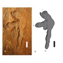
Pterosaurs (Pterosauria) from the Cerro del Pueblo Formation (Late Campanian) of Coahuila, Mexico
Published online: 21/11/2025
Keywords:
Azhdarchoidea; Coahuila; Mexico; Pterodactyloidea; Pterosauria
https://doi.org/10.18563/pv.48.2.e1
Abstract
The Late Campanian Cerro del Pueblo Formation, located in southeastern Coahuila, Mexico, has produced a diverse array of vertebrate fossils. However, pterosaur remains from this unit are notably scarce. In this study, we describe new pterosaur material from the formation. The specimens include a fragmentary vertebra identified as belonging to an indeterminate, but derived pterodactyloid, along with the distal condyle of a left metacarpal, referable to an azhdarchoid pterosaur, and a left manus print. While these specimens provide additional evidence of pterosaur presence in the region during the Late Cretaceous, their fragmentary nature limits precise taxonomic and ichnotaxonomic identification. Nevertheless, they highlight the potential for future discoveries that could refine our understanding of the diversity and distribution of pterosaurs in Mexico.
PV article infos
Published in 48-2 (2025)
|
PDF
|

|
Compléments sur les Chiroptères de l'Eocène moyen d'Europe. Les genres Palaeochiropteryx et Cecilionycteris.
Published online: 01/10/1980
Keywords:
Chiroptera; Geiseltal; Messel; Middle Eocene
https://doi.org/10.18563/pv.9.ext.91-126
Abstract
New dental and skeletal material referable to Palaeochiropteryx tupaiodon from the Middle Eocene locality of
Messel (G.F.R.) is studied, which provides additions to the previously gained knowledge of this european genus. Dental specimens from Geiseltal (G.D.R.), also of Middle Eocene age, allow us to analyze Cecilionycteria prisca. Some of these are separated to establish a new genus, Matthesia, and two new species, M. germanica and M. ? insolita.
PV article infos
Published in Vol. 9, Ext (1980)
|
PDF
|

|
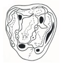
Nouvelle quantification de l'Hypsodontie chez les Theridomyidae : l'exemple de Theridomys ludensis nov. sp.
Published online: 30/12/1985
Keywords:
Dental morphology; evolution; Hypsodonty; Oligocene; Theridomyidae
https://doi.org/10.18563/pv.15.3.159-172
Abstract
A new example of parallelism in the dental pattern ofthe Theridomyidae is illustrated by the description ofa new species: Theridomys Iudensis from the standard level of Antoingt (middle Oligocene). Considering the occurence ofthis parallelism phenomenon. the use of numerous qualitative and quantitative criteria is essential to characterize the different stages ofthe different evolutive lineages. Thus, a new simple parameter is proposed (CHY = H+l/0,5 L) to estimate hypsodonty of the medium hypsodont Rodentia.
PV article infos
Published in Vol. 15, Fasc. 3 (1985)
|
PDF
|

|
Révision des Chiroptères Lutériens de Messel (Hesse, Allemagne).
Published online: 04/04/1970
Keywords:
Chiroptera; Lutetian; Messel
https://doi.org/10.18563/pv.3.4.83-182
Abstract
The revision of the Lutetian chiropterans from Messel, first described by Revilliod in 1917, is based on the anatomy of the teeth and the skeleton. A figuration or refiguration of thematerial utilized accompanies the new description, which goes beyond that of the original monograph.
The study shows a certain variability of the dental structure within the genera Palaeochiropteryx Revilliod and Archaeonycteris Revilliod, as well as a general resemblance of the two forms. The morphology of the teeth permits, however, the verification of the validity of the different species: Palaeochiropteryx tupaiodon Revilliod, P. spiegeli Revilliod, Archaeonycterís trigonodon Revilliod, and Archaeonycteris revilliodi, n. sp.
Some differences of the skeletal and dental anatomy tend to indicate a stage of evolution less advanced for the genus Archaeonycteris.
The comparison of the chiropterans of Messel with the principal groups of living chiropterans, as well as with different Eocene fossíls (notably Cecílionycteris Heller and Icaronycteris Jepsen) leads to a more precise idea of the anatomy of primitive chiropterans. This comparison also permits the proposition that the oid forms so far described by integrated in a superfamily, the Palaeochiropterygoidea and allows a general phylogenetic hypothesis to be advanced for the order Chiroptera.
PV article infos
Published in Vol. 03, Fasc. 4 (1970)
|
PDF
|
|
The beginning of the adaptive radiation of Theridomorpha (Rodentia) in Western Europe: morphological and phylogenetic analyses of early and middle Eocene taxa; implications for systematics
Published online: 20/09/2021
Keywords:
characters analyses; Dental morphology; Eocene; Rodentia; variability
https://doi.org/10.18563/pv.44.2.e2
Abstract
This paper provides a revision of the early and middle Eocene European rodents previously referred to as Ischyromyoidea, including taxa considered to be at the origin of the Theridomorpha. The use of an accurate dental terminology and a better understanding of the size and shape of their infra-orbital foramen (i.o.f.) led us to a substantial revision of this group, which allowed to better characterize them and to appreciate their variability. On these bases, phylogenetic analyses (cladistic and standard Bayesian
approaches) of early Ypresian to late Priabonian European rodent species were undertaken in order to highlight the root of the early Theridomorpha and its content. In this paper, the phylogeny was established based on 343 characters (338 dental) through 45 early Paleogene taxa using both cladistic and bayesian analyses. The ingroup included on one hand a few North American genera (Reithroparamys, Microparamys, and Acritoparamys) and European ones (Eogliravus, Ailuravus, Corbarimys, Meldimys, Euromys, Plesiarctomys, and Pseudoparamys) considered until now as being related with the North American superfamily Ischyromyoidea. On the other hand, it included genera close to the root of the Theridomorpha (Sparnacomys, Pantrogna, and Hartenbergeromys) and early Theridomyoidea (Masillamys, Protadelomys, and some Pseudosciuridae). The phylogenetic results obtained via the two
distinct reconstruction approaches are consistent in virtually all relationships. The proposed systematics here derives from these phylogenetic results. This phylogenetic context led us to change the suprafamilial, familial, subfamilial or generic attribution of several species. Characters of Theridomorpha, like the obliquely developed postprotocristid allied with the occurrence of a metalophulid I, have been found in genera previously considered as Ischyromyidae (Pseudoparamys, Euromys, Sparnacomys, Meldimys, Pantrogna, and Hartenbergeromys) as well as the large i.o.f., when preserved (Pseudoparamys, Hartenbergeromys, and Masillamys). Based on these morphological observations and new phylogenetic considerations, the content of the Theridomorpha clade is here enlarged, thereby extending back the first theridomorph radiations to the early Eocene. Aside, a new taxon (Reinomys rhomboides gen and sp. nov.) is described from Avenay. In addition, a new genus, Auroremys, is created for the species subita (Comte et al., 2012) from Chery-Chartreuve.
PV article infos
Published in 44-2 (2021)
|
PDF
S.I. Data
|

|
Decouverte d'un nouveau Diacodexis (Artiocactyla, Mammalia) dans l'Eocène inférieur de Silveirinha, Portugal.
Published online: 15/09/1989
Keywords:
Artiodactyla; Eocene; Migration; Portugal; Silveirinha
https://doi.org/10.18563/pv.19.1.29-44
Abstract
A new artiodactyl, Diacodexis antunesi n.sp., is described from the early Eocene of Silveirinha, Portugal. Comparisons are made with Diacodexis gazini GODINOT, 1978, D. varleti SUDRE et al., 1983, D. cf. varleti from Paris Basin sites, D. sp. from Dormaal and from localities in Spain and England, D. secans from North America and D. pakistanensis from Asia; affinities and evolutive tendencies are discussed. The presence of Diacodexis in the locality of Silveirinha confirms the very early Eocene age of the latter. As Diacodexis antunesi appears ta be more primitive than D. gazini from Rians (early Eocene of France), it lends corroboration to the interpretation (essentially based previously on condylarths) of the Silveirinha assemblage as the oldest Eocene fauna known in Europe and supports the hypothesis that early artiodactyls migrated from Europe to North America.
PV article infos
Published in Vol. 19, Fasc. 1 (1989)
|
PDF
|

|
La poche à phosphate de Ste-Néboule (Lot) et sa faune de vertebres du Ludien supérieur. 9- Primates et Artiodactyles
Published online: 25/09/1978
Keywords:
Eocene; Quercy Phosphorites
https://doi.org/10.18563/pv.8.2-4.269-290
Abstract
La faune d'artiodactyles de Ste-Néboule, qui comprend neuf espèces, présente de nombreux
points communs avec les faunes habituellement rattachées au Ludien supérieur, telles celles de La Débruge ou de Montmartre. A l'inverse de ces localités, on ne connaît pourtant, à Ste-Néboule, ni Oxacronae, ni Anoplotheriinae. Parmi les espèces du gisement se trouve une nouvelle espèce du genre Mouillacitherium (M. schlosseri n. sp.), cette forme devant être interprétée comme le dernier représentant du rameau. Nous faisons connaître par ailleurs la presque totalité de la denture du Dacrytherium saturninii, espèce qui était à ce jour très imparfaitement documentée. Ste-Néboule est, d'autre part, la seule localité où l'on peut signaler l'association probable de deux lignées du genre Amphimeryx. Les primates sont peu diversifiés à Ste-Néboule, puisque le groupe est limité au seul genre Adapis. Les genres Microchoerus et Pseudoloris, que l'on sait pourtant être représentés dans des gisements dâge voisin (Microchoerus ornatus à San Cugat de Gavadons et Mormont-Entreroches, Pseudoloris reguanti à San Cugat de Gavadons, Pseudoloris cf. reguanti à Neustadt ; cf. Louis et Sudre 1975) sont absents dans les faunes du Quercy de cette période.
PV article infos
Published in Vol. 08, Fasc. 2-4 (1978)
|
PDF
|

|
Artiodactyla aus den Eozänen Braunkohlen des Geiseltales bei Halle (DDR)
Published online: 04/12/1989
Keywords:
Artiodactyles; Eocene; Europe; Paleoecology; Stratigraphy; taxonomy
https://doi.org/10.18563/pv.19.3.131-160
Abstract
The present study of Artiodactyla from the Middle Eocene of the Geiseltal lignite beds concems systematics, biostratigraphy, and palaeoecology on the basis of 174 specimens: isolated remains to more complete skeletons. Instead of the formerly known five species of two families are now recognized 14 species of the Diacodexeidae, Dichobunidae, Cebochoeridae, and Haplobunodontidae. New species are Aumelasia maniai, Anthracobunodon neumarkensis, Masillabune franzeni. Four species of the Geiseltalfauna are definitely known from elswere, and five species are closely related to those from other European localities. Evidently the faunal situation of artiodactyls during the Middle Eocene of Europe was largely uniform. The distribution of artiodactyls within the sequence of the Geiseltal strata corroborates the biostratigraphical concept of the land mammal age Geiseltalian (Franzen & Haubold l986b) as well as the mammalian reference levels MP 11-13 (Franzen 1987). Reconstructions of the skulls and skeletons allow conclusions on the functional morphology and palaeoecology of the artiodactyls of the European Middle Eocene
PV article infos
Published in Vol. 19, Fasc. 3 (1989)
|
PDF
|

|
A new rodent from Quaternary deposits of the Canary Islands and its relationships with Neogène and recent murids of Europe and Africa.
Published online: 15/12/1988
Keywords:
Canary Islands; Holocene; Island evolution; Muridae; PHYLOGENY; Rodents; Spain
https://doi.org/10.18563/pv.18.4.241-262
Abstract
A peculiar new rodent, Malpaisomys insularis nov. gen., nov. sp., is described from subfossil deposits of the eastern Canary Islands. The species shows some highly specialized skull features although its molars exhibit a mixture of primitive and derived characters among which a partial stephanodonty is most notable. A comparison of the new rodent with several Miocene to Holocene Muridae shows that Malpaisomys possibly shares a common ancestor with Acomys and Uranomys.
PV article infos
Published in Vol. 18, Fasc. 4 (1988)
|
PDF
|

|
Les mammifères Montiens de Hainin (Paléocène moyen de Belgique) Part III : Marsupiaux
Published online: 30/09/1983
Keywords:
Belgium; Marsupials; Paleobiogeography; Paleocene
https://doi.org/10.18563/pv.13.3.51-64
Abstract
The oldest european marsupials are described from some specimens (isolated upper molars) recently found from the Hainin sediment (Middle Paleocene of Belgium). These fossils document a new species of the Peradectes genus. They give evidence of a much older occurrence of the marsupials in Europe than it was assumed. They allow us to postulate a didelphid dispersal from South America towards the western-holarctic area operating in two phases : the first one of the Peradectes genus at the end of the Cretaceous; the second one of the Didelphíni tribe at the end of the Paleocene. A central american crossing is likely for the first one, whereas a transafrican way is tentatively argued for the second one.
PV article infos
Published in Vol. 13, Fasc. 3 (1983)
|
PDF
|

|
Study of the Turolian hipparions of the lower Axios valley (Macedonia, Greece). 4. Localities of Dytiko.
Published online: 15/12/1988
Keywords:
Equidae; Greece; Hipparion; Lower Axios Valley; Macedonia; Mammalia; Turolian
https://doi.org/10.18563/pv.18.4.187-239
Abstract
The hipparions from the Dytiko localities of the lower Axios valley (Macedonia, Greece) are studied. The material comes from three localities Dytiko-l, 2, 3 (DTK, DIT, DKO), which are situated near the village of Dytiko, about 60 km northwest to Thessaloniki. Three species have been determined, the medium-sized H. mediterraneum, the small-sized H. matthewi and the very small-sized H. periafricanum. The determined Hipparion species, their morphological characters and their comparison with the other Axios valley material indicate a Late Turolian age for the Dytiko localities.
PV article infos
Published in Vol. 18, Fasc. 4 (1988)
|
PDF
|

|
Les gisements de Robiac (Eocène supérieur) et leurs faunes de Mammifères.
Published online: 05/04/1969
Keywords:
Fauna; Late Eocene; Mammalia; Robiac
https://doi.org/10.18563/pv.2.3.95-156
Abstract
Designated the type-locality of a late Eocene paleomammal zone, Robiac has recently been the object of important excavations. The first results of the new collecting, as well as a revision of the material in old collections, are given in this work.
Two stratigraphic section, cutting through the two sites presently distinguished (Robiac-Nord and Robiac-Sud) reveal the lithologic variation at the base of the Fons Limestone and the localization of three fossiliferous beds at Robiac-Sud.
The molluscan fauna and the flora (charophytes), which have already been described, as well as that of the lower vertebrates, have been listed.
A list of 46 mammalian species (only 16 species were known previous to 1964) has been established. The micro-mammals, nearly all new in this fauna (marsupials, insectivores, bats, rodents, primates, and some smallsized artiodactyls), Were obtained only after screen-washing of the matrix; about 4 tons of sediment were thus treated.
The artiodactyls have been the most extensively analyzed; 6 genera, of which one is new, have been recognized. The latter is described as Robiacina minum n.g., n.sp., and represents a very small artiodactyl of the family Anoplotheriidae. The taxonomie status of certain species formerly described has been clarified by the designation of lectotypes (Cebochoerus robiucensis, Catodonerium robiacense, Xiphodon castrense).
The paleontologic corrélations at present possible between the late Eocene faunas have allowed the relative positions of te different French localities of this age to be established; the Guépelle locality, it seems, could define in the future a new paleomammal zone.
PV article infos
Published in Vol. 02, Fasc. 3 (1969)
|
PDF
|
|
Autopsie d’une radiation adaptative : Phylogénie des Theridomorpha, rongeurs endémiques du Paléogène d’Europe - histoire, dynamique évolutive et intérêt biochronologique
Published online: 15/12/2016
Keywords:
Diversification; Extinction; Paléoenvironnements; Rodentia; Theridomyoidea
https://doi.org/10.18563/pv.40.3.e1
Abstract
Résumé :
L’étude des rongeurs Theridomorpha permet de suivre le déroulement d’une radiation adaptative pendant toute sa durée (Eocène moyen-Oligocène terminal), sur un territoire restreint à l’extrémité ouest de l’Europe Occidentale. Dans ce papier, la phylogénie de ce groupe est établie à partir d’une analyse cladistique reposant sur l’examen de 315 caractères (310 dentaires). Le groupe d’intérêt comprend 110 des 132 espèces (24 genres) de Theridomyoidea et deux genres encore inclus jusqu’ici dans les Reithroparamyinae qui rejoignent les Theridomorpha. Les groupes externes comprennent des Glires basaux, Cocomys, Tanquammys et 16 Ischyromyiformes. Un cadre phylogénétique robuste est produit, qui permet de clarifier la systématique des Theridomorpha. La position des Remyoidea (nov. sup.fam.) au sein des Ischyromyiformes, extérieure aux Theridomorpha, est confortée. Les Protadelomys et Tardenomys sont à la base des Theridomyoidea, avant la séparation en deux clades correspondant aux familles Pseudosciuridae et Theridomyidae. Les sous-familles sont consolidées : Pseudosciurinae et Sciuroidinae pour les Pseudosciuridae ; Issiodoromyinae, Oltinomyinae, Columbomyinae, Theridomyinae, auxquelles s’ajoute au moins une nouvelle sous-famille (Patriotheridomyinae), pour les Theridomyidae. La topologie des chrono-espèces (sensu Simpson), traitées antérieurement comme lignées évolutives, apparaît dans la plupart des cas sous forme de clades successifs dans lesquels les espèces sont le plus souvent arrangées de manière pectinée, émergeant dans l’ordre stratigraphique. L’analyse des caractères aux principaux nœuds permet de consolider les caractères diagnostiques des taxons et les tendances évolutives, ainsi que de discuter des divers parallélismes et convergences dans l’évolution des structures et patrons dentaires (e.g., émail des incisives unisérié chez les Issiodoromyinae et les Patriotheridomyinae, ou pseudo-multisérié chez les Blainvillimys les plus hypsodontes, les Protechimys et Archaeomys ; patrons dentaires téniodontes ; allongement des dents déciduales chez les Patriotheridomyinae, Issiodoromyinae et Theridomyidae ; sélénodontie ou lophodontie). Les dynamiques évolutives traduites par les changements morphologiques sont mises en relation avec les variations environnementales. Enfin, les implications biochronologiques de l’évolution des Theridomyoidea sont discutées.
Abstract:
The adaptive radiation of the rodents Theridomorpha occurred during a limited time window (middle Eocene to late Oligocene), on an area restricted to Western Europe. In this paper, the phylogeny of this group is established via a cladistic assessment of 315 morphological characters (310 dental). The group of interest encompasses 110 upon 132 species (24 genera) of Theridomyoidea, and two genera formerly included within the Reithroparamyinae, and which are included here within the Theridomorpha. The outgroups include basal Glires, Cocomys, Tanquammys and 16 Ischyromyiformes. A robust phylogenetic frame is produced, which allows clarifying the systematics of the Theridomorpha. Within the Ischyromyiformes, the Remyoidea (nov. supfam.) are set apart from the Theridomorpha. Protadelomys and Tardenomys represent the earliest offshoots of the Theridomyoidea, before the dichotomy between Pseudosciuridae and Theridomyidae. It supports the former subfamilies Pseudosciurinae and Sciuroidinae within the Pseudosciuridae; and for the Theridomyidae: the Issiodoromyinae, Oltinomyinae, Columbomyinae, Theridomyinae, with at least one new subfamily (Patriotheridomyinae). The topologies of the chronospecies (sensu Simpson), formerly considered as evolutionary lineages, appear in most cases as successive clades, in which the species are generally pectinately arranged and emerging in the stratigraphic order. The analysis of characters supporting the main nodes allow consolidating the diagnosic characters of the taxa and their evolutionary trends, as well as discussing the various cases of parallelism and convergence in the evolution of structures and dental patterns (e.g., uniserial incisor enamel for Issiodoromyinae and Patriotheridomyinae, or pseudo-multiserial for the most hypsodont Blainvillimys, Protechimys and Archaeomys; taeniodont dental patterns; lengthening of deciduous premolars for Patriotheridomyinae, Issiodoromyinae and Theridomyidae; selenodonty or lophodonty).
Evolutionary dynamics are analysed with respect to environmental changes. Finally, biochronological implications of the evolution of Theridomyoidea are discussed.
PV article infos
Published in Vol 40-3 (2016)
|
PDF
S.I. Data
|

|
Arvicolinae (Rodentia) du Pliocène terminal et du quaternaire ancien de France et d'Espagne.
Published online: 30/10/1971
Keywords:
Arvicolinae; France; Pleistocene; Pliocene; Spain
https://doi.org/10.18563/pv.4.5.137-214
Abstract
Two steps can be distinguished in the history of the first invasion of western and south western Europe by the arvicolines. The first step corresponds to the installation of these rodents with the immigration of Promimonys inxuliferus Kowalski, then of Mimomys stehlini Kormos and of Mimomys gracilis (Kretzoi). The second is characterized by the establishment of a geographic differentiation in the arvicoline fauna between the south of France and Spain, from where are described new species of Mimomys (Mimamys cappettai, Mimomys septimanus, Mimonys medasensis), and the rest of France, where are found only elements already known from central Europe or England (Mimomys polonicus Kowalski, Mimomys pliocaenicus F. Major, Mimomys reidi Hinton, or forms very close to the latter). This geographic differentiation, which is very certainly the consequence of the division of Europe into distinct climatic provinces, one of them being the southern province comprising at least Spain and southern France, could result from a cladogenetic evolution of Mimomys stehlini and Mimomys gracilis after their immigration. The present work is also a contribution to the search for correlations between the diverse micromammal localities of the latest Pliocene (or early Villafranchian) and of the early Quaternary of Europe.
PV article infos
Published in Vol. 04, Fasc. 5 (1971)
|
PDF
|

|
La plus ancienne faune de mammifères du Quercy : Le Bretou
Published online: 01/12/1974
Keywords:
Le Bretou; Quercy Phosphorites
https://doi.org/10.18563/pv.6.3-4.177-196
Abstract
Redécouvertes en 1968 dans le bois du Bretou, les deux poches à phosphorites qui ont livré cette faune sont parmi les plus méridionales du plateau quercynois. De faible profondeur, huit mètres, les cavités sont de petite taille et font partie d'un complexe de sept fissures réparties dans les bois du Bretou. Les autres trous n'ont livré aucun fossile, soit parce qu'ils avaient été intégralement vidés de leur sédiment, soit que des venues d'eau en interdisent l'accès.
Dans les deux poches fossilifères, proches de quelques mètres, le sédiment que l'on rencontre en placage sur les parois présente un aspect particulier. Il s'agit en effet d'une brèche très indurée, où la calcite domine, et contenant assez peu de sédiments sidérolithiques. Les ossements contenus dans cette brèche sont assez nombreux, très souvent brisés. Les dents isolées y sont relativement rares. Exploitées séparément, les deux poches ont livré des faunes identiques, aucune différence chronologique ne pouvant être décelée entre elles. Les formes rencontrées dans une seule des cavités sont rares mais la poche la plus riche est celle dénommée Le Bretou 2.
L'intérêt de la faune récoltée réside dans son ancienneté. On sait en effet que H.G. Stehlin avait signalé dans les collections du Quercy des restes attribuables à Lophiodon lautricense, mais la provenance exacte de ces fossiles reste inconnue. Au Bretou si nous n'avons pas rencontré à ce jour le Lophiodon, il n'en demeure pas moins que l'âge de la faune récoltée doit être proche de celui de la faune de Robiac.
PV article infos
Published in Vol. 06, Fasc. 3-4 (1975)
|
PDF
|

|
Sur les empreintes de pas des gros mammifères de l'Eocène supérieur de Garrigues-ste-Eulalie (Gard)
Published online: 01/10/1980
Keywords:
Eocene; Euzet; Footprints; Ichnofauna
https://doi.org/10.18563/pv.9.ext.37-78
Abstract
Is hereby described an impressive lchnoiauna belonging to the Lower to Middle Ludian of the Gard (S. France). The slab, already cleaned over a length of 18 m, is located near the top of the Potamides aporoschema lacustrine limestone (Lower Ludian, Euzet zone). It is therefore older than the Célas sandstone deposit, and still more than the Melanoides albigensis and M. acutus marly limestone corresponding to the Upper Levels of the Ludian stage. Although biostratigraphically older than the La Débruge and Montmartre zone, the biotope shows already a sampling of very tall Artiodactyles, Perissodactyles and Carnivorous. One of the most « majestic ›› Artiodactyles, Anopolotheriipus lavocati, nov., points out a huge size type. To mention also among the Ichnotypes described, 10, the big Perissodactyle Palaeotheriipus similimedius, nov., and the big Carnivorous Hyaenodontipus praedator, nov.
PV article infos
Published in Vol. 9, Ext (1980)
|
PDF
|

|
Muridae (Rodentia) du Pliocène supérieur d'Espagne et du midi de la France.
Published online: 20/09/1969
Keywords:
Anthracomys meini; Castillomys crusafonti; Pliocene; Rodents; Valerymys ellenbergeri
https://doi.org/10.18563/pv.3.1.1-25
Abstract
The murid fauna of the terminal Pliocene of southwest Europe is rich in at least eight genera and ten species. With the species belonging to the genera Apodemus, Rhagapodemus, and Stephanomys not being studied here, the study of the other murids resulted for one thing in the description of three new genera and three new species: Castillomys crusafonti n. g., n. sp., Occitanomys brailloni n. g., n. sp., Anthracomys meini n. sp., Valerymys ellenbergerí (THALER) n. g., and for another thing in the recognition of a form hitherto unknown in this region, Micromys praeminutus KRETZOI. Systematic study has shown that certain species of the terminal Pliocene fauna had their ancestors in the Turolian fauna presently known in Spain. The evolutionary lineages thereby recognized have been studied more in detail and a list of the evolutionary tendencies of the dendal characters has been given. A chart of the probable phyletic relationships between the different murids of the Pliocene faunas of southwest Europe (With the genus Rhagapodemus and Apodemus dominans being excluded) is given in conclusion of this work.
PV article infos
Published in Vol. 03, Fasc. 1 (1969)
|
PDF
|

|
Les nouvelles faunes de rongeurs proches de la limite mio-pliocène en Roussillon. Implications biostratigraphiques et biogéographiques
Published online: 29/04/1991
Keywords:
Arvicolidae; Cricetidae; Gliridae; Miocene; Muridae; Pliocene; Rodents; Southern France
https://doi.org/10.18563/pv.20.4.147-174
Abstract
Three new fossiliferous localities, two of karstic origin, Castelnou 3 and Font Estramar, respectively Late Upper Miocene and Lower Pliocene, and one of lacustrine origin, Thuir, Lower Pliocene, add data about the transition between Miocene and Pliocene faunas of rodents in southern France. An unexpected association of taxa was present in the late Upper Miocene, including between others, Myocricetodon, Hispanomys, Ruscinomys, Cricetus barrierei, Promimomys and a new species of Stephanomys, S. dubari nov. sp. Myocricetodon is still known in the Lower Pliocene. It is shown that the large field-mice known since the Late Upper Miocene belong to two different lineages, on one side, A. jeanteti, on the other side, A. gudrunae followed by A. gorafensis. Biochronological and biogeographical implications are discussed.
PV article infos
Published in Vol. 20, Fasc. 4 (1991)
|
PDF
|
