|
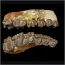
Les Périssodactyles (Mammalia) du gisement Bartonien supérieur de Robiac (Éocène moyen du Gard, Sud de la France)
Published online: 04/05/2015
Keywords:
Chasmotherium; new species; Palaeotheriidae; paleoenvironments
https://doi.org/10.18563/pv.39.1.e3
Abstract
We present here a new updated counting of the perissodactyls of Robiac, the type locality of the MP 16 level of the biochronological scale of paleogene mammals and that of the Robiacian stage of Eocene Land Mammals Ages in Western Europe.
The outcrop of Robiac consists actually of two 500m apart loci, Robiac-Nord and Robiac-Sud, considered of the same age according to the current discriminating power, and is dated from -38,7 MA after the last faunal, magnetostratigraphic and climatic calibrations.
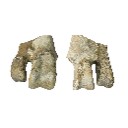
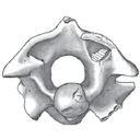
It has yielded a very abundant and rich of 21 taxa perissodactyl fauna, topped by the giant Lophiodon lautricense, last representative of the family Lophiodontidae, of which it is the last proved deposit. The Palaeotheriidae are much diversified with 5 genera and 9 species of "Pachynolophinae", 3 genera and 10 species of Palaeotheriinae. Nine taxa have been defined from Robiac: Chasmotherium depereti n. sp., Palaeotherium castrense robiacense Franzen, 1968, the genus Leptolophus Remy, 1965 with the species L. stehlini Remy, 1965 and L. magnus Remy, 1998, Anchilophus (Paranchilophus) jeanteti Remy, 2012, Metanchilophus chaubeti Remy, 2012, Lophiotherium robiacense Depéret, 1917 and Pachynolophus gaytei n. sp.
The faunal Robiac cenogram with the associated flora testify to a hot, wet and forestal environment, likely corresponding to a short warming climatic phase; the broken up fossil bones should have been carried away and then gathered in swamp areas along the banks of a meandering river.
The swarm of mammals of Robiac, the richest of contemporaneous deposits, has been followed by a drastic drop in perissodactyl diversity at the MP 17A level; a crisis which could have originated in a renewal of the global Eocene cooling. Fons 4, the type-locality of this level, is largely scarcer in perissodactyls and its cenogram testifies to a less diversified fauna, with on the whole smaller species, that likely means a cooler and drier climatic environment; a new perissodactyl diversification occurred but later.
PV article infos
Published in Vol.39-1 (2015)
|
PDF
|

|
Mammals and stratigraphy : the Paleocene of Europe
Published online: 01/12/1982
Keywords:
Europe; Mammalia; Mammalian biochronology; Paléogène; Stratigraphy
https://doi.org/10.18563/pv.12.ext
Abstract
The mammalian faunas of the Paleogene of Europe and their localities are reviewed with comments on problems of European stratigraphy (epoch, stage and substage limits) and on the possibilities of faunal migrations. Radiometric dating is discussed. A stratigraphic scale for the Paleogene is presented, as well as a refined system of sequential faunal levels.
PV article infos
Published in Vol. 12, Ext (1982)
|
PDF
|

|
Functional aspects of the evolution of rodent molars
Published online: 01/10/1980
Keywords:
Chewing; Muridae; Rodents; Wear facets
https://doi.org/10.18563/pv.9.ext.249-262
Abstract
The wear facets of primitive rodents can be homologized with those of primitive primates and ungulates. As in primates, the jaw movement was ectental, with an increased anterior component in the lingual phase (phase ll). The buccal phase (phase I) in rodents approaches the horizontal and it tends to be reduced in importance in comparison with the lingual phase. ln more advanced rodents the efficiency of grinding is increased by the development of additional cutting edges of enamel (e.g. enlargement of hypocone, development of mesoloph and lingual sinus). The buccal phase movement becomes lined up with the lingual phase movement to form a single oblique chewing stroke,resulting in planation of the crown. As the stroke becomes more longitudinal (propalinal) the enamel edges become more transverse. In Muridae propalinal chewing evolved before the loss of cusps, facets were reorientated and additional cusps developed.
PV article infos
Published in Vol. 9, Ext (1980)
|
PDF
|
|
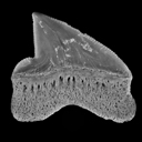
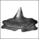
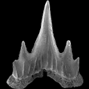
New Squalicorax species (Neoselachii: Lamniformes) from the Lower Maastrichtian of Ganntour phosphate deposit, Morocco
Published online: 05/12/2014
Keywords:
Anacoracidae; Chondrichthyes; Maastrichtian; Morocco; New taxa
https://doi.org/10.18563/pv.38.2.e3
Abstract
Two new Squalicorax species, S. benguerirensis nov. sp. and S. microserratus nov. sp. are described from the Lower Maastrichtian of the Benguérir phosphate open mine, Ganntour deposit, Morocco. The species S. benguerirensis nov. sp. was classically assigned to S. yangaensis since Arambourg (1952) and has been also recognized in coeval deposits from eastern USA to Mid-East. The species S. microserratus nov. sp. correspond to the lateral teeth of S. kaupi as reported by Arambourg (1952) and which is now referred in fact to S. bassanii. The comparison of these two new species with other Anacoracids, known in Moroccan or elsewhere, allows highlighting the great taxonomic and ecological diversities of this family during the Cretaceous.
PV article infos
Published in Vol.38-2 (2014)
|
PDF
|

|
New records of terrestrial Mammals from the upper Eocene Qasr el Sagha Formation, Fayum Depression, Egypt
Published online: 16/12/1996
Keywords:
Egypt; Eocene; Fossil mammals; Qasr el Sagha Formation
Abstract
New records of terrestrial mammals from the Qasr el Sagha Formation, Fayum Depression, Egypt are reported, and the stratigraphic occurrences of these fossils noted. These include additional specimens of Moeritheríum, Barytherium, and anthracotheres, as well as the oldest record of a hyracoid in the Fayum.These Eocene mammals occur almost exclusively in the alluvial deposits of the Dir Abu Lifa Member of the Qasr el Sagha Formation and show close affinities to the faunas from the lower sequence of the Jebel Qatrani Formation. There is no evidence of a more marked faunal discontinuity between the Qasr el Sagha and Jebel Qatrani Formations than there is across any of the three major breaks in sedimentation that exist within the Jebel Qatrani Formation. The faunal similarities between fossils of the lower sequence of the Jebel Qatrani Formation and of the upper part of the Qasr el Sagha Formation is consistent with recent paleomagnetic dating that suggests that these rocks differ in age by only one to two million years.
PV article infos
Published in Vol. 25, Fasc. 2-4 (1996)
|
PDF
|
|
A new Desmodillus (Gerbillinae, Rodentia) species from the early Pliocene site of Langebaanweg (South-western Cape, South Africa)
Published online: 20/01/2017
Keywords:
Lower Pliocene; Muridae; Rodentia; RSA
https://doi.org/10.18563/pv.41.1.e1
Abstract
Situated in the Cape region of the Republic of South Africa (RSA), the paleontological site of Langebaanweg is dated to 5.1 Myr and is famous for having yielded an abundant vertebrate assemblage, including numerous rodent species from the Mio-Pliocene transition. Based on molar morphology and skull anatomy, the single Gerbillinae taxon identified at Langebaanweg and described in this paper is allocated to Desmodillus, which is a modern monotypic South African endemic genus. It is significant in being the oldest representative of the genus in Africa. We describe here a new species of this genus which is larger than the modern D. auricularis, but nevertheless retains some of its main characteristics, namely the shape of the maxilla and mandible, the presence of poorly fused alternating cusps, and no longitudinal crest. This taxon differs from modern South African Gerbilliscus representatives in some mandibular and maxillary characters, in the m1 prelobe cusp, and in having less fused cusps. Two fossil Gerbillinae discovered in the Upper Miocene of Africa and Asia, Abudhabia and Protatera, have been compared with the new species. We discuss their relationships with modern and Plio-pleistocene Gerbillinae and conclude that Abudhabia could be the sister taxon of Desmodillus and that around 6-5 Myr a vicariance event allowed Gerbillinae to diversify into modern Desmodillus in South Africa, and Gerbilliscus in East Africa. The murine/gerbilline ratio, which is a good indicator of rainfall, supports other proxies which suggest that at 5.1 Myr the climate in the Langebaanweg region was more humid than today.
PV article infos
Published in Vol 41-1 (2018)
|
PDF
S.I. Data
|

|
Palaecarcharodon orientalis (Sinzow) (Neoselachii : Cretoxyrhinidae), from the Paleocene of maryland, USA.
Published online: 15/09/1989
Keywords:
Maryland; Palaeocarcharodon; Paleocene; Selachian; Systematics; U.S.A.
https://doi.org/10.18563/pv.19.1.1-6
Abstract
Recent collecting of fossil vertebrate remains from the lowermost member of the Aquia Formation (Paleocene), has enabled me to report here for the very fIrst time, the earliest occurrence for the teeth of Palaeocarcharodon in the fossil record of the New World.
This report represents only one species of neoselachian from this locality, the remaining fauna of which will subsequently be described.
PV article infos
Published in Vol. 19, Fasc. 1 (1989)
|
PDF
|
|
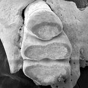
A new species of Propalaeotherium (Palaeotheriidae, Perissodactyla, Mammalia) from the Middle Eocene locality of Aumelas (Hérault, France).
Published online: 24/05/2016
Keywords:
Eocene; new species; Palaeotheriidae; Propalaeotherium
https://doi.org/10.18563/pv.40.2.e1
Abstract
A new Propalaeotherium species, clearly distinct from the genus Eurohippus, is described. It is characterized by having a similar size as P. voigti from the German Geiseltal localities (MP 11 to MP 13 reference-level), but differs in several features suggesting a slighty more derived morphology. It presents indeed less brachyodont crowns with less prominent and less elevated cingula, slightly larger relative surface of premolars, and a more marked metaconid splitting on cheek teeth. This new species is unknown from other European localities except the nearby Saint-Martin de Londres locality which has been considered older than the MP 13 level.
PV article infos
Published in Vol.40-2 (2016)
|
PDF
S.I. Data
|

|
New late Paleocene rodents (Mammalia) from Big Multi Quarry, Washakie Basin,Wyoming.
Published online: 16/12/1996
Keywords:
Clarkforkian; North America; Paleocene; Rodentia
Abstract
The earliest North American rodents occur in basal Clarkforkian beds of the Fort Union Formation at Big Multi Quarry near Bitter Creek, northern Washakie Basin, Sweetwater County, Wyoming, and in closely correlative Fort Union beds formerly accessible in the Eagle Coal Mine near Bear Creek, northern Clark's Fork Basin, Carbon County, Montana. Two new species of early Clarkforkian rodents, Paramys adamus and Alagomys russelli, are described from Big Multi Quarry. Paramys adamus is represented by virtually complete upper and lower dentitions, which demonstrate that this species is one of the most primitive North American paramyids yet discovered. These specimens form the basis for a reevaluation of the content and stratigraphic range of P. atavus, which is known with certainty only from Bear Creek. Alagomys russelli is the first North American record for the enigmatic rodent family Alagomyidae, otherwise known from ?late Paleocene-early Eocene localities in Mongolia and China. Phylogenetic analysis of dental and gnathic traits suggests that Alagomyidae form the sister group of all other undoubted rodents. At least two rodent clades, alagomyids and basal paramyids, seem to have invaded North America from Asia at the beginning of Clarkforkian time, but only the paramyids persisted to undergo a significant evolutionary radiation in North America.
PV article infos
Published in Vol. 25, Fasc. 2-4 (1996)
|
PDF
|

|
Mammifères de l'Ilerdien Moyen (Eocène inférieur) des Corbières et du Minervois (Bas-Languedoc, France). Systématique, Biostratigraphie, Corrélations.
Published online: 25/01/1991
Keywords:
Biostratigraphy; Corbières; correlations; Early Eocene; Ilerdian; Mammalia; Minervois; Paleobiogeography; Southern France
https://doi.org/10.18563/pv.20.2-3.55-144
Abstract
Mammal-bearing localities have been discovered in the marine and lacustrine series of the middle Ilerdian (Lowermost Eocene) from Southem France (Minervois and Corbières). In the localities of Fordones, Monze, Fournès, and La Gasque, thirty mammal species have been identified. Among others, they include ischyromyid rodents (Microparamys and Pseudoparamys), paromomyid and adapid primates (Arcius and Donrussellia), new insectivores, condylarths, and a dyspternine pantolestid. These faunas provide new informations on the early Eocene Mesogean faunas of Rians and Palette. The assemblages of primates and rodents from Fordones support good correlations with Palette which was recently placed near the standard-level of Dormaal (MP 7). In fact, Palette and Fordones could be even older than Dormaal. Consequently, there seems to be a relatively important temporal gap between the late Paleocene of Cernay and the Sparnacian of Dormaal. This gap could be partly filled with the Mesogean faunas of Palette, Fordones, and Silveirinha. On the basis of these new mammal faunas the marine middle Ilerdian is proved to be older than the Cuisian stage of the Paris Basin. With regards to the position of the Fordones fauna at the top of the NP 10 calcareous nannoplankton biozone, the westem European paleomammalogists Paleocene/Eocene boundary could be situated between the NP 9 and NP 10 biozones.
PV article infos
Published in Vol. 20, Fasc. 2-3 (1991)
|
PDF
|
|
Batoids (Rajiformes, Torpediniformes, Myliobatiformes) from the Sülstorf Beds (Chattian, Late Oligocene) of Mecklenburg, northeastern Germany: a revision and description of three new species
Published online: 24/06/2015
Keywords:
Batoids; Chattian; Elasmobranchii; North Sea Basin; Oligocene
https://doi.org/10.18563/pv.39.2.e2
Abstract
Bulk-sampling of fossil-rich tempestites from the Chattian Sülstorf Beds of
Mecklenburg, north-eastern Germany, yielded a rich selachian fauna in which batoids
predominate by the abundance of teeth but are subordinate by the number of taxa. Thirteen
taxa are identified, among which rajiform batoids are the most diverse (six species). One
genus and three species are newly described: Raja thiedei sp. nov., Oligoraja pristina gen. et
sp. nov., and Torpedo chattica sp. nov. Two species are reallocated: Atlantoraja cecilae
(Steurbaut & Herman, 1978) new comb., and Dipturus casieri (Steurbaut & Herman, 1978)
new comb. Ontogenetic heterodonty is documented for the first time in the dental pattern of
Myliobatis sp. Stratigraphical ranges of batoid taxa in the period from Rupelian to Langhian
are presented and partly discussed in context with the palaeoclimatic evolution and
palaeogeographic situation of the North Sea Basin.
PV article infos
Published in Vol.39-2 (2015)
|
PDF
|

|
Historical and new perspectives on the parataxonomyof fossil eggs.
Published online: 15/12/2003
Keywords:
amniotic eggshells; Parataxonomy
Abstract
A critical review on the literature about the parataxonomy of amniote eggshells reasserts the great interest of this systematics tool for the progress of dinosaur eggshell paleontology. However, shedding light on its limits, we propose to give up the use of the basic types - morphotypes key system.
PV article infos
Published in Vol. 32, Fasc. 2-4 (2003)
|
PDF
|

|
La morphologie dentaire des Thalattosuchia (Crocodylia, Mesosuchia).
Published online: 15/12/1997
Keywords:
Dental morphology; Dental types; feeding habits.; Jurassic; Metriorhynchidae; Systematics; Teleosauridae; Thalattosuchia
Abstract
The tooth morphology of the Thalattosuchia (marine crocodilians from the Jurassic and the Early Cretaceous) is analysed. The Callovian from Poitou and the Kimmeridgian from Quercy have yielded many remains of Metriorhynchus, Steneosaurus and Machimosaurus. These remains allow us to study the variations of tooth morphology during ontogenic growth, tooth replacement and the location of the teeth. We have defined different tooth types for these genera. In Metriorhynchus, the two tooth types defined do not coincide with the two groups recognized in the Callovian (broad-skulled and narrow-skulled metriorhynchids) but reflect the prey preferences of these forms. In Steneosaurus and Machimosaurus the five tooth types deñned are in agreement with the main taxa known from the Bathonian to the Early Cretaceous. This study allows to precise the function and the prey preference of the Thalattosuchia during the Jurassic and the Early Cretaceous.
PV article infos
Published in Vol. 26, Fasc. 1-4 (1997)
|
PDF
|
|
Two new scyliorhinid shark species (Elasmobranchii, Carcharhiniformes, Scyliorhinidae), from the Sülstorf Beds (Chattian, Late Oligocene) of the southeastern North Sea Basin, northern Germany.
Published online: 30/04/2014
Keywords:
Chattian; Elasmobranchii; North Sea Basin; Scyliorhinidae; Scyliorhinus
https://doi.org/10.18563/pv.38.1.e1
Abstract
Based on isolated teeth two new scyliorhinid shark species, Scyliorhinus biformis nov. sp. and Scyliorhinus suelstorfensis nov. sp., are described from the Sülstorf Beds, early-middle Chattian, of Mecklenburg, northeastern Germany. They form part of a speciose assemblage of necto-benthic sharks and batoids which populated the warm-temperate to subtropical upper shelf sea of the south-eastern North Sea Basin.
PV article infos
Published in Vol.38-1 (2014)
|
PDF
|

|
Le genre Plagiolophus (Palaeotheriidae, Perissodactyla, Mammalia): révision systématique, morphologie et histologie dentaires, anatomie crânienne, essai d'interprétation fonctionnelle
Published online: 15/12/2004
Keywords:
New taxa; Paléogène; perissodactyls; skull anatomy; tooth histology
Abstract
The genus Plagiolophus is documented, almost solely in Western Europe, from the middle Eocene up to the mid Oligocene (MP 12 to MP 25), i.e. more than for 15 MY. Seventeen species are now recorded whose two of them are new, P. ringeadei nov. sp. and P. mamertensis nov. sp. Some anatomical variations and the deflection of certain evolutionary trends justify the distinction of three subgenera, Paloplotherium, Fraasiolophus nov. and Plagiolophus s.s. The genus displays a wide range in size and weight (between 10 and 150 kg). The detailed description of the skull of several species is here given for the first time.
Despite important evolutionary drifts during this long time span, the dentition shows a great structural homogeneity, which renders difficult the determination of fragmentary specimens or isolated teeth. It is characterized by a great heterodonty; premolars are little molarized and present a certain regression through time with paradoxically some progress in the molarization. The hypsodonty increases: the first Plagiolophus are hardly less brachyodont than Propalaeotherium, and the last ones are nearly as hypsodont as Merychippus from the early Miocene. The upper molars change from a wide crown pattern, with an open occlusal surface, lightly oblique transverse lophs and rounded internal cusps, to a narrower pattern, with a frontally constricted occlusal surface and internal lophs aligned parallel to the ectoloph. The M3/3 become always longer.
The dental enamel displays horizontal Schreger-bands with imprecise limits occupying only the middle part of the enamel layer. The dentine is remarkable by its high rate of pericanalicular dentine. The crown cementum, lacking in earlier forms, increases to the point where it fills the occlusal valleys of the
teeth.
The masticatory musculature shows a increasing prominence of the temporal, with probably an important role devoted to the pterygoid muscles in lateral movements related to a two-phase type of chewing.
The evolution of the dentition, of the masticatory musculature and of the repartition of masticatory forces indicate that the Plagiolophus have known different diets through their long evolutionary history; at first browsers they became mixed feeders and finally grazers. Their relatively long neck allowed these animals to reach different vegetal layers. The strength of the nuchal crests also suggests that they were able to have strong backwards movements of the head to pull up their food.
This evolution of diet seems related to the slow degradation of environmental conditions attested during this period in western Europe, with the generalization of more open landscapes, increasing aridity and more marked seasons.
Besides, a remodeling of the face is ontogenetically and along time observed, in relation with the evolution of the masticatory apparatus and especially with that of the mandibular lever arm. The postcanine diastemata become longer in the course of evolution; the free extremities of the nasals are always relatively long which contradicts the hypothesis according to which Paloplotherium may have had a trunk. At last the lineage Fraasiolophus can be distinguished by the presence of a deep malar fossa, probably related to a strong development of the maxillo-labialis superior muscle.
The orbit is always large and tends to increase in size, which indicates a good development of the vision and its increasing role in the life relations. A peculiar type of epitympanic sinus could have been used as a resonance chamber insuring a certain amplification of sounds before their transmission to the eardrum. The endocranial cast reveals a relatively large brain with an advanced degree of gyrencephaly. Beside the role eventually played in food research and social relations, these neurophysiological abilities, also related to an advance in cursorial fitness, could have contributed to the survival of these animals facing the predation pressure of the first fissipede carnivores and the competition with new immigrant herbivores after the "Grande Coupure".
On the basis of some shared apomorphies with the Pachynolophinae, which prevent from considering the latter as Equidae (molarization of the premolars, reduction of the premaxilla dorsal apophysis, peculiar epitympanic sinus, splitting of the jugular process), the hypothesis of an autochthonous origin of Plagiolophus issued from a form near Propalaeotherium, is once again proposed and discussed. Finally, intra-generic relationships are taken into consideration.
PV article infos
Published in Vol. 33, Fasc. 1-4 (2004)
|
PDF
|
|
First report of Cylindracanthus (Osteichthyes) from the Eocene of India
Published online: 25/03/2024
Keywords:
Cylindracanthus; Eocene; histology; rostrum; Umarsar mine.
https://doi.org/10.18563/pv.47.1.e2
Abstract
Fossils of the endangered sturgeons and peddlefishes are widely distributed. We here report for the first time the presence of one of the extinct osteichthyes genus Cylindracanthus (Liedy 1856a) from the Early Eocene lignite-bearing successions of the Kutch Basin, India. The present well preserved rostrum is characterised by numerous wedge-shaped components encircling the central canal that runs along its length, paired at the base and each wedge contributing to the formation of a ridge. The rostrum lacks teeth. The present find extends the palaeobiogeographical distribution of Cylindracanthus considerably and supports its Eocene age as dental remnants preserved in Cylindracanthus sp. shows a decrease in remanent dentition and tooth bases from the Cretaceous to the Eocene. Cylindracanthus is an useful palaeoenvironmental indicator as it has been found associated typically with deposits of nearshore marine environments.
PV article infos
Published in 47-1 (2024)
|
PDF
|

|
The Quaternary avifauna of Crete, Greece.
Published online: 01/09/1988
Keywords:
Avifauna; Crete; Quaternary; Systematics
https://doi.org/10.18563/pv.18.1.1-94
Abstract
Pleistocene bird fossils have been studied from nine localities on Crete. Part of this material was described earlier by the author (Weesie, 1982) and will not be treated here in extenso, the results will be incorporated. More than one third of the over 10,000 fossil bird bones available could be identified ; they were found to represent at least 65 bird species. The following species of the Pleistocene Cretan avifauna are new to the fauna of Crete : Branta ruficollis, Haliaeetus albicilla, Gyps melitensis, Aquila chrysaetos simurgh n. ssp., Ketupa zeylomensis, Aegolius funereus, Dendrocopos leucotos, Zoothera dauma, Turdus iliacus and Pyrrhula pyrrhula. The Pleistocene Cretan avifauna differs less from comparable mainland avifaunas than (fossil) avifaunas from oceanic islands do. Still, the Pleistocene Cretan avifauna has two qualities that are characteristic of island avifaunas : the almost complete absence of a group of birds (the Galliformes) and the presence of two endemic (sub)species : the giant eagle Aquila chrysaetos simurgh n. ssp. and the long-legged owl Athene cretensis (Weesie, 1982). The new subspecies is described in the present study.
These endemic birds of prey were found in association with their supposedly principal prey species (now extinct as well) : endemic mice for the owl and endemic deer for the eagle. Endemic mammals have been found in association with endemic birds of prey on many islands, not only in the Mediterranean. There is evidence that the size of endemic birds of prey becomes optimally adapted to their feeding on certain endemic mammals, especially rodents. Another characteristic of the Pleistocene Cretan avifauna is the great number of species of birds of prey. This appears to be a common characteristic of fossil avifaunas from caves on Mediterranean islands as well as from caves on the European mainland. However, we think that ecological conditions on Pleistocene Crete (especially the abundant presence of mice) helped to account for the high representation of birds of prey. Furthemore, the fossil avifauna enables us to draw some conclusions about the climate and vegetation on Pleistocene Crete : it is concluded that the climate was cooler than today and that Crete was largely covered with forests. Finally, the reasons for the extinction or disappearance from Crete of some bird species of the Pleistocene Cretan avifauna are discussed.
PV article infos
Published in Vol. 18, Fasc. 1 (1988)
|
PDF
|
|
A reassessment of the giant birds Liornis floweri Ameghino, 1895 and Callornis giganteus Ameghino, 1895, from the Santacrucian (late Early Miocene) of Argentina.
Published online: 13/12/2016
Keywords:
Argentina; Aves; Callornis; Liornis; Miocene
https://doi.org/10.18563/pv.40.2.e3
Abstract
The status of the giant bird taxa Liornis floweri and Callornis giganteus from the Santa Cruz Formation (late Early Miocene) of Patagonia, first described by Ameghino (1895) is reassessed on the basis of a re-examination of the type material at the Natural History Museum, London. Liornis floweri, which lacks a Pons supratendineus on the tibiotarsus and has an unbifurcated Canalis interosseus distalis on the tarsometatarsus, is clearly a brontornithid and is considered as a junior synonym of Brontornis burmeisteri. Ameghino’s replacement of Callornis by Eucallornis is unjustified. Callornis giganteus is a chimera based on a phorusrhacid tarsometatarsus (probably belonging to Phorusrhacos longissimus) and a brontornithid tibiotarsus. The latter can be considered as the lectotype of Callornis giganteus, which may represent a small morph of Brontornis burmeisteri or a distinct taxon. It is referred to here as Brontornithidae indet. The tarsometatarsus described by Dolgopol de Saez (1927a,b) as Liornis minor and considered by her as a gracile brontornithid apparently has a bifurcated Canalis interosseus distalis and should therefore be placed among the Phorusrhacidae.
PV article infos
Published in Vol.40-2 (2016)
|
PDF
|
|
Fossil snakes from the Palaeocene of São José de Itaboraí, Brazil Part III. Ungaliophiinae, Booids incertae sedis, and Caenophidia. Summary, update and discussion of the snake fauna from the locality
Published online: 16/12/2008
Keywords:
booid-grade incertae sedis; Brazil; Caenophidia; New taxa; Palaeocene; Russellophiidae; Snakes; tropidophiids; Ungaliophiinae
https://doi.org/10.18563/pv.36.1-4.37-73
Abstract
Aside from Madtsoiidae, anilioids, and Boidae that were studied previously, the middle Palaeocene of ltaborai (BraziI) has produced Ungaliophiinae ("tropidophiids"), booid-grade snakes incertae sedis, and a possible Russellophiidae (Caenophidia) that are described in the present article. This article is the third and final report on the snakes from the locality. The Ungaliophiinae (Paraungaliophis pricei gen. et sp. nov.) are rare whereas the booid-grade snakes incertae sedis (ltaboraiophis depressus gen. et sp. nov., Paulacoutophis perplexus gen. et sp. nov.) are more frequent. A single vertebra is referred to the Russellophiidae (Caenophidia) with reservation. An update of the whole fauna of snakes from ltaborai is provided. Hechtophis austrinus that was tentatively referred to the erycine Boidae is now regarded as a Boidae incertae sedis. Most snakes from Itaborai are known only from the locality. Astonishingly, only the ailioids Coniophis cf. C. precedens gives possible evidence of interchanges between South and North America. The fauna of snakes from Itaborai, as well as the other Palaeocene faunas of snakes from South America are distinct from those of the Cretaceous and the Eocene of South America; they appear to be more different from the Cretaceous faunas than from those of the Eocene. The fauna from Itaborai is the richest and most diverse assemblage of snakes from the Palaeocene worldwide; it shares only a few taxa with other Palaeocene localities.
PV article infos
Published in Vol. 36, Fasc. 1-4 (2008)
|
PDF
|

|
Rythme et modalités de l'évolution chez les rongeurs à la fin de l'Oligocène-leurs relations avec les changements de l'environnement.
Published online: 15/12/2000
Keywords:
Environment; evolution; Oligocene; Rodents; Systematics
Abstract
The analysis of oxygene isotope variations as well as paleobotanical data suggest that the Oligocene/Miocene boundary corresponds to a transitional period marked by floristical and climatic variations. During this period, the pyreneo-alpine tectonics has contribued to modify the geography and western Europe landscapes. Faunal changes (appearances, extinctions, migrations) are observed in different mammalian groups, notably in the rodents. A study of the evolutionary trends and patterns in paleogene rodents is involved for the period ranging from level MP 28 of the Late Oligocene to the Early Miocene, including the Oligo-Miocene boundary.
The Rodents fauna from the sites of Venelles (Bouches-du-Rhône District, France) and Thezels (Lot, France), previously mentionned in litterature, have been studied. The first description of the Eomyidae of La Milloque (MP 29) has been completed. These faunas are compared to those from various localities dating from the considered period. In La Milloque, a new representative of the Eomys species is described next to a form close to Rhodanomys hugueneyae ENGESSER, 1987. It is the Eomys milloquensis nov. sp., the likely descendant of Eomys quercyi COMTE & VIANEY-LIAUD, 1987. Two new species are also described in Thezels: Eucricetodon thezelensis nov. sp., resulting from a likely and local evolution of Eucricetodon praecursor (SCHAUB, 1925) from La Milloque, which, in the same geographic area, could be at the origin of Eucricetodon hesperius ENGESSER, 1985 from Paulhiac. Plesiosminthus admyarion nov. sp., quite distinct from Plesiosminthus schaubi VIRET, 1926, which announces Plesiosminthus myarion SCHAUB 1930. Venelles 'Plesiosminthus schaubi population is considered as a sub-species, named Plesiosminthus schaubi meridionalis nov. subsp. New phylogenetic patterns are proposed. Among the Eomyidae, a quantification of various features of the M1-2/ crown (hypsodonty, degree of abrasion, occlusal angle, state of development of the I and V anticlines), and a comparison with the occlusal diagram of the other teeth among various other populations allows a more efficient separation of Eomys and Rhodanomys genera. In Western Europe, and within this period, it finally does not seem possible to gradually connect the genus Eomys to the genus Rhodanomys. The evolution of the Eomys quercyi - milloquensis lineage seems to underline a similar evolution to that which may have led from the Eomys to the Rhodanomys form. The latter which appears totally accomplished at level MP 29 of the Oligocene is considered as an immigrant. If we compare the most representative species of the Venelles, Thezels, and Coderet sites, (i.e. Rhodanomys, Eucricetodon, Adelomyarion, Peridyromys, Plesíosminthus), it becomes impossible to confirm their biochronological separation. The noticeable differences between the populations may be interpreted as geographical variations. An explanation to these variations, and to fauna's evolution during the Late Oligocene and Early Miocene can be found in the environmental modifications, supported by isotopic, paleobotanical and sedimentologic analysis. A tentative reconstruction of the environments is attempted by the cenogram method. The analysis of the fluctuations of fauna's diversity shows variations which may be correlated to a drop in temperature at MP 29, during the Late Oligocene, followed by an increase in temperature along with an aridity phenomenom, during the basal Miocene (MN O).The confrontation of various methods give the opportunity of reconstituting and comparing the evolution of the environment of three sequences of sites chosen from different regions. Ecological affinities of various rodents' species are being examined. It is possible to consider that the integration of all the conclusions resulting from this study should lead to an explanation to the evolution of rodents for the period around the Oligocene-Miocene boundary. The site of Coderet- level 3- would be posterior to the latter, at the beginnig of the Miocene, and would mark the level MN 0 of the Aquitanian.
PV article infos
Published in Vol. 29, Fasc. 2-4 (2000)
|
PDF
|
