|
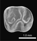
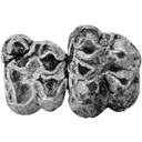
A new and primitive species of Protophiomys (Rodentia, Hystricognathi) from the late middle Eocene of Djebel el Kébar, Central Tunisia
Published online: 02/06/2014
Keywords:
Adaptive radiation; Bartonian; Dental morphology; North Africa; Paleobiogeography
https://doi.org/10.18563/pv.38.1.e2
Abstract
Based on fossil discoveries and phylogenetic studies, an Eocene Asian origin for hystricognathous rodents and anthropoid primates has gained strong support in recent years. The two groups then invaded both Africa and South America, which promoted their evolutionary success. However, the fossil record has so far failed to constrain the nature and precise timing of these pivotal dispersal events. In Africa, given the apparent absence of hystricognaths and anthropoids in early to early middle Eocene localities, it is suggested that these mammal groups dispersed from Asia to Africa sometime during the middle Eocene. In this paper, we report the discovery of several isolated teeth of a rodent from a new vertebrate locality situated in central Tunisia (Djebel el Kébar, KEB-1), dating from the late middle Eocene (Bartonian, ~39.5 Myr). These fossils document a diminutive new species of Protophiomys (P. tunisiensis nov. sp.), a basal genus of hystricognathous rodents which is well known from several North African mammalian-bearing localities of the end of the Eocene. The teeth of P. tunisiensis display a suite of anatomical details comparable with those observed in the other species of the genus, but with a lesser degree of development. Such an apparent primitive evolutionary stage is corroborated by the greater antiquity of this Tunisian species. P. tunisiensis nov. sp. is so far the most ancient representative of hystricognaths in Africa. However, it can be expected that hystricognaths were already present on that landmass given the new data on early caviomorphs recently reported from South America (at ~41 Myr). The arrival of hystricognaths in Africa from South Asia certainly predates the depositional period of the Kébar sediments, but perhaps not by much time.
PV article infos
Published in Vol.38-1 (2014)
|
PDF
|

|
Rongeurs nouveaux de l'Oligocène Moyen d'Espagne.
Published online: 15/09/1969
Keywords:
Cricetidae; Oligocene; Pseudocricetodon; Rodents; Theridomys
https://doi.org/10.18563/pv.2.5.191-207
Abstract
Description of four new rodents from a recently discovered locality at Montalban. Theridomys crusafonti nov. sp. is considered as the ancestry of T. Iembronicus. Theridomys varian: nov. sp. includes «Theridomys» morphotypes and «Blainvilllimys» morphotypes; it could be ancestral to B. blainvillei. Pseudoltinomys nanus nov. sp. represents a new lineage paralleling in evolution that of P. gaillardi (which is equally found at Montalban). Pseudocricetodon montalbanensis nov. gen., nov. sp. designates a lineage of very small Cricetidae accompanying Eucricetodon. With these well defined new species and six others present in the locality, Montalban appears as the best faunal reference point within the biochronologic zone of La Sauvetat.
As an annex, discussion of two rodent specimens from the classic localíty of Tárrega, close in age to that of Montalban.
PV article infos
Published in Vol. 02, Fasc. 5 (1969)
|
PDF
|
|
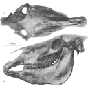
Old world hemiones and new world slender species (Mammalia, Equidae)
Published online: 16/12/2008
Keywords:
Amerhippus; biometry; Equus; Holocene; New World; Old World; Osteology; Pleistocene; Pliocene
https://doi.org/10.18563/pv.36.1-4.159-233
Abstract
Morphological and biometrical description of skulls, teeth, and limb bones of extant and fossil Old World herniones (including E. hydruntinus) and of New World 'stilt-Iegged' and other slender species from Blancan to Holocene. An Appendix presents ways in which the approximate size of some missing bones or dimensions may be deduced from available ones.
The discussed and/or illustrated fossils were found in Bolivia (Tarija), Canada (Yukon), China (Choukoutien, Gulongshan, Jiling, Loufangzi), Ecuador (Oil Fields), Ethiopia (Melka Kunturé), France (Lunel-Viel), Germany (Süssenborn), Greece (Agios Georgios, Petralona), Hungary (Dorog), Italy (Romanelli), Mexico (Cedazo, San Josecito), Mongolia (Sjara-osso-gol), Spain (Venta Micena), ex-Soviet Union (Akhalkalaki, Binagady, Chokurcha, Chukochya, Kabazi, Kolyma, Krestovka, Kurtak, Staroselie, Tologoj), USA (Alaska, Arkalon, Cedar Meadow, Channing, Conkling, Dry Mountains, Hay Springs, Leisey Shell Pit A, Lissie Formation, Natural Trap, Pool Branch, Powers Ranch, Rock Creek, San Diego, Santo Domingo, Seymour Formation, Shelter, Slaton, Trinity River). Numerous raw or statistically elaborated data are given in Tables.
There is no evidence for the existence of Old World hemiones in the New World nor of 'stilt-Iegged' equids in the Old World. The first 'stilt-Iegged' equid was found at Santo Domingo, New Mexico, and is believed to be Late Blancan. It was probably at the origin of E. calobatus (Arkalon, Rock Creek) and of the smaller E. semiplicatus (Channing, Rock Creek). Slender, but not 'stilt-Iegged', equids found at Natural Trap, Wyoming, ca. 12 ky ago belong to Amerhippus. AlI these species share with Oid World Sussemiones (and some hemiones) peculiar patterns on the lower cheek teeth.
The slender Equus sp. B of Leisey Pit A, Florida, ca. 1.2 Ma, as weIl as Amerhippus francisci and E. tau (probably a senior synonym of E. quinni) share conventional lower cheek teeth patterns. The skulls of A. francisci and E. tau, however, are quite different.
Paleontological data suggest a common origin of Amerhippus, Sussemiones, and 'stilt-Iegged' equids during the late Blancan. Old World hemiones seem to have differentiated later.
PV article infos
Published in Vol. 36, Fasc. 1-4 (2008)
|
PDF
|
|
Latest Early-early Middle Eocene deposits of Algeria (Glib Zegdou, HGL50), yield the richest and most diverse fauna of amphibians and squamate reptiles from the Palaeogene of Africa
Published online: 08/02/2021
Keywords:
Africa; Algeria; amphibians; Eocene; squamates
https://doi.org/10.18563/pv.44.1.e1
Abstract
HGL50 is a latest Early-early Middle Eocene vertebrate-bearing locality located in Western Algeria. It has produced the richest and most diverse fauna of amphibians and squamate reptiles reported from the Palaeogene of Africa. Moreover, it is one of the rare faunas including amphibians and squamates known from the period of isolation of Africa. The assemblage comprises 17 to 20 taxa (one gymnophionan, one probable caudate, three to six anurans, seven ‘lizards’, and five snakes). Two new taxa were recovered: the anuran Rocekophryne ornata gen. et sp. nov. and the snake Afrotortrix draaensis gen. et sp. nov. The locality has also yielded the first confirmed anilioid snake, the first Palaeogene gymnophionan, and probably the first caudate from the Palaeogene (and possibly from the Tertiary) of Africa. The presence of a caudate at that time in Africa would be of particular interest; unfortunately, the available material does not permit a definitive identification. The fauna comprises Gondwanan and more specifically West Gondwanan vicariants, probably autochthonous groups and a Eurasian immigrant (assuming that the identification of the caudate is accurate). The fauna from HGL50 is clearly distinguished from the few other Eocene assemblages of Africa. However, if this results largely from differences in geological ages, geographic positions of the localities and mainly differences in environments took a part in the composition of the faunas.
PV article infos
Published in 44-1 (2021)
|
PDF
|
|
Designating a lectotype for Mesacanthus pusillus (Gnathostomata: Acanthodii)
Published online: 03/03/2021
Keywords:
acanthodians; Chordata; Devonian; Midland Valley; Orcadian Basin
https://doi.org/10.18563/pv.44.1.e2
Abstract
The early gnathostome genus Mesacanthus is well represented in both Lower Old Red Sandstone and Middle Old Red Sandstone assemblages of northern and central Scotland. This ‘acanthodian’ taxon is currently thought to comprise two valid species: M. mitchelli and M. pusillus. Although the whereabouts of the holotype of M. mitchelli (NHMUK PV P560) is known, the syntype material for M. pusillus has long been thought lost. Here we identify at least one specimen that formed part of the original syntype material for M. pusillus, albeit in a slightly different condition than when it was originally figured. This specimen is ROM 25872, which is here designated as the lectotype. A second specimen – ELGNM 1978.191.1 – could represent another of the syntype specimens, but poor preservation quality makes it impossible to be certain.
PV article infos
Published in 44-1 (2021)
|
PDF
S.I. Data
|
|
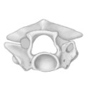
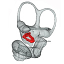
Fallen in a dead ear: intralabyrinthine preservation of stapes in fossil artiodactyls
Published online: 09/03/2016
Keywords:
allometry; bony labyrinth; inner ear; middle ear ossicles
https://doi.org/10.18563/pv.40.1.e3
Abstract
The stapes is the last of the middle ear ossicle chain and the smallest bone of the mammalian skeleton. Because it rests on the membrane of the fenestra vestibuli during life, the stapes may often fall within the bony labyrinth cavity when soft structures decay after death. In this work, we highlight the unexpected role that the bony labyrinth plays in the preservation of the stapes. Systematic investigation of the bony labyrinth of 50 petrosal bones of extinct and extant artiodactyls led to the discovery of eight cases of “intralabyrinthine” stapes. Three dimensional reconstructions of these stapes allow documenting stapes morphology of four extinct artiodactyl taxa: Microstonyx erymanthius (Suidae), Elomeryx borbonicus (Hippopotamoidea), ?Helohyus plicodon (Helohyidae), and an undetermined Cainotheriidae; and four extant ones Choeropsis and Hippopotamus (Hippopotamidae), and Tayassu and Phacochoerus (Suoidea). ?Helohyus plicodon from the Middle Eocene documents the oldest stapes known for the order Artiodactyla. Morphological study and metric analyses of our sample of artiodactylan stapes show that stapes are likely to carry relevant phylogenetic characters/signal within artiodactyls, and a potential Euungulata signature.
PV article infos
Published in Vol.40-1 (2016)
|
PDF
S.I. Data
|
|
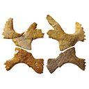
New material of “Eurysternidae” (Thalassochelydia, Pan-Cryptodira) from the Kimmeridgian of the Swiss Jura Mountains
Published online: 25/06/2020
Keywords:
Eurysternidae; Late Jurassic; morphology; Switzerland; Testudines; Thalassochelydia
https://doi.org/10.18563/pv.43.1.e2
Abstract
The region of Porrentruy (Swiss Jura Mountains) is known for its rich and diverse assemblage of Late Jurassic coastal marine turtles (Thalassochelydia). Dominated by the “Plesiochelyidae”, this assemblage also includes representatives of the two other thalassochelydian groups, the “Thalassemydidae” and “Eurysternidae.” In this study, we present new shell-based material from Porrentruy referable to eurysternids. One specimen represents a juvenile individual or a relatively small taxon, and is notably characterized by a well fenestrated plastron exhibiting a wider than long central plastral fontanelle. Two other specimens are much larger and possibly represent the largest eurysternids known to date. The fourth specimen is characterized by a unique plastral morphology otherwise only known in very small juveniles. This is the first time this unique plastral morphology is known to persist in an adult or subadult. The new material described herein represents at least three distinct taxa, all of them probably new. However, we refrain from naming new species based on this incomplete material in order to avoid adding confusion to an already complex taxonomical situation. This study provides new insights into the great diversity of eurysternids during the Late Jurassic.
PV article infos
Published in Vol 43-1 (2020)
|
PDF
|

|
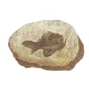
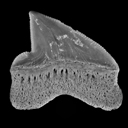
Physogaleus hemmooriensis (Carcharhinidae, Elasmobranchii), a new shark species from the early to middle Miocene of the north sea basin.
Published online: 15/10/2006
Keywords:
Carcharhinidae; Early Miocene; Elasmobranchii; Hemmoorian; new species; North Sea Basin; Physogaleus
https://doi.org/10.18563/pv.34.e14
Abstract
A new carcharhinid shark species, Physogaleus hemmooriensis sp. nov., is described from the Lower Hemmoorian (Behrendorfian, late Burdigalian, early Miocene) of Werder, Lower Saxony, Germany. P. hemmooriensis also occurs in the Edegem and Antwerpen Sands Members of the Berchem Formation, Belgium, and in the Miste Bed, Aalten Member of the Breda Formation, The Netherlands, which have an early to middle Miocene age. In the Western Atlantic region, the taxon is present in the early Miocene Calvert Formation of Delaware, U.S.A, which is largely contemporaneous with the Hemmoorian.
PV article infos
Published in Vol. 34, Fasc. 1-2 (2006)
|
PDF
|
|
A new study of the anthracotheres (Mammalia, Artiodactyla) from pondaung formation, Myanmar: systematics implications
Published online: 16/12/2008
Keywords:
Anthracohyus; Anthracokeryx; Anthracotherium; Pondaung Formation; sexual dimorphism; Siamotherium; South East Asia; taxonomy
https://doi.org/10.18563/pv.36.1-4.89-157
Abstract
Anthracotheres from the Pondaung Formation, Myanmar, are considered as one of the most primitive artiodactyl groups and they represent the oldest known record in the world. Thus, the understanding of this group has numerous implications for evolutionary biology and biochronological correlations. However, the systematlcs of these mammals has been interpreted in different ways, and the main debate focuses on the number of taxa represented in the Pondaung Formation. The revised taxonomy proposed here is mainly based on the relative development of the upper molar W-shaped ectoloph, system of crests and stylar cusps, and on body size. On the basis of these characters, they are classified into four genera including six different species. Two well-known genera, Anthracotherium and Anthracokeryx, are validated and more precisely diagnosed. Anthracokeryx possesses a better developed W-shaped ectoloph, system of crests and stylar cusps than Anthracotherium, which displays notable differences with the more derived representatives of this genus. Both of these Pondaung genera show evidence for sexual dimorphism. However, the incompleteness of fossil material fueled a debate concerning the status of two additional Pondaung anthracotheres, Siamotherium and Anthracohyus. The latter genus is of uncertain affinities, but it has been considered as a hippopotamid ancestor. Despite new material attributed to these two forms, additional discoveries are still required to establish their taxonomic status. The hypothesis that Southeast Asia was the centre of origin of Anthracotheriidae is supported by the retention of numerous primitive dental characters in these taxa and by the antiquity of the Pondaung Formation, to which an age of 37 My is now generally accepted.
PV article infos
Published in Vol. 36, Fasc. 1-4 (2008)
|
PDF
|

|
Les poissons crétacés et tertiaires du bassin des Iullemmeden (République du Niger)
Published online: 15/09/1972
Keywords:
Actinopterygians; Cenozoic; Cretaceous; Dipnoans; Selachians
https://doi.org/10.18563/pv.5.5.179-251
Abstract
The present work is devoted to the study of the Cretaceous and Tertiary fishes (teeth of Selachians, Actinopterygians and Dipnoans) collected during a recent expedition in Niger. The Maestrichtian localities have yielded a new genus and a new subspecies of Selachian: Igdabatis sigmodon nov. gen., nov. sp. and Lamna biauriculata nigeriana nov. subsp. The locality of Sessao, which has been attributed to the Thanetian by means of the study of the fish, has furnished by screen-washing an interesting fauna wherein six new species are described: Raja Iouisi, Dasyatis sessaoensis, D. sudrei, D. russelli, Hypolophites thaleri and Ceratodus casieri. Comparison of these faunas with contemporary faunas of Africa has brought out a certain endemism in the Iullemmeden Basin during the late Cretaceous and the early Tertiary.
PV article infos
Published in Vol. 05, Fasc. 5 (1972)
|
PDF
|

|
Rythme et modalités de l'évolution chez les rongeurs à la fin de l'Oligocène-leurs relations avec les changements de l'environnement.
Published online: 15/12/2000
Keywords:
Environment; evolution; Oligocene; Rodents; Systematics
Abstract
The analysis of oxygene isotope variations as well as paleobotanical data suggest that the Oligocene/Miocene boundary corresponds to a transitional period marked by floristical and climatic variations. During this period, the pyreneo-alpine tectonics has contribued to modify the geography and western Europe landscapes. Faunal changes (appearances, extinctions, migrations) are observed in different mammalian groups, notably in the rodents. A study of the evolutionary trends and patterns in paleogene rodents is involved for the period ranging from level MP 28 of the Late Oligocene to the Early Miocene, including the Oligo-Miocene boundary.
The Rodents fauna from the sites of Venelles (Bouches-du-Rhône District, France) and Thezels (Lot, France), previously mentionned in litterature, have been studied. The first description of the Eomyidae of La Milloque (MP 29) has been completed. These faunas are compared to those from various localities dating from the considered period. In La Milloque, a new representative of the Eomys species is described next to a form close to Rhodanomys hugueneyae ENGESSER, 1987. It is the Eomys milloquensis nov. sp., the likely descendant of Eomys quercyi COMTE & VIANEY-LIAUD, 1987. Two new species are also described in Thezels: Eucricetodon thezelensis nov. sp., resulting from a likely and local evolution of Eucricetodon praecursor (SCHAUB, 1925) from La Milloque, which, in the same geographic area, could be at the origin of Eucricetodon hesperius ENGESSER, 1985 from Paulhiac. Plesiosminthus admyarion nov. sp., quite distinct from Plesiosminthus schaubi VIRET, 1926, which announces Plesiosminthus myarion SCHAUB 1930. Venelles 'Plesiosminthus schaubi population is considered as a sub-species, named Plesiosminthus schaubi meridionalis nov. subsp. New phylogenetic patterns are proposed. Among the Eomyidae, a quantification of various features of the M1-2/ crown (hypsodonty, degree of abrasion, occlusal angle, state of development of the I and V anticlines), and a comparison with the occlusal diagram of the other teeth among various other populations allows a more efficient separation of Eomys and Rhodanomys genera. In Western Europe, and within this period, it finally does not seem possible to gradually connect the genus Eomys to the genus Rhodanomys. The evolution of the Eomys quercyi - milloquensis lineage seems to underline a similar evolution to that which may have led from the Eomys to the Rhodanomys form. The latter which appears totally accomplished at level MP 29 of the Oligocene is considered as an immigrant. If we compare the most representative species of the Venelles, Thezels, and Coderet sites, (i.e. Rhodanomys, Eucricetodon, Adelomyarion, Peridyromys, Plesíosminthus), it becomes impossible to confirm their biochronological separation. The noticeable differences between the populations may be interpreted as geographical variations. An explanation to these variations, and to fauna's evolution during the Late Oligocene and Early Miocene can be found in the environmental modifications, supported by isotopic, paleobotanical and sedimentologic analysis. A tentative reconstruction of the environments is attempted by the cenogram method. The analysis of the fluctuations of fauna's diversity shows variations which may be correlated to a drop in temperature at MP 29, during the Late Oligocene, followed by an increase in temperature along with an aridity phenomenom, during the basal Miocene (MN O).The confrontation of various methods give the opportunity of reconstituting and comparing the evolution of the environment of three sequences of sites chosen from different regions. Ecological affinities of various rodents' species are being examined. It is possible to consider that the integration of all the conclusions resulting from this study should lead to an explanation to the evolution of rodents for the period around the Oligocene-Miocene boundary. The site of Coderet- level 3- would be posterior to the latter, at the beginnig of the Miocene, and would mark the level MN 0 of the Aquitanian.
PV article infos
Published in Vol. 29, Fasc. 2-4 (2000)
|
PDF
|

|
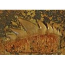
Contribution à la classification des Pistes de Vertébrés du Trias : les types du Stormberg d'Afrique du Sud (2 ème Partie: le Stormberg supérieur - 1. Le biome de la zone B/1 ou niveau de Moyeni: ses biocénoses).
Published online: 01/12/1974
Keywords:
biocenosis; Footprints; South Africa; Stormberg; Trias
https://doi.org/10.18563/pv.6.ext
Abstract
Les Pistes de Vertébrés du Stormberg Supérieur ("Trias terminal à Rhétien"), ou Quthingien
Si les zones du Stormberg inférieur se sont révélées contenir de nombreuses traces, surtout dans les faciès dits "Molteno moyen et supérieur", représentant apparemment la base du Keuper, il est frappant de voir pratiquement l'ensemble de cette grosse faune "Molteno" disparaître avec la fin de cette période, que nous avons appelée le "Maphutsengien".
Dès les premières zones du Stormberg supérieur, que nous nommons le"Quthingien" la zoocénose et la phytocénose, en même temps que les données d'ensemble manifestées par l'environnement, sont modifiées. Nous ne verrons plus guère de dépôts marécageux à flore riche et variée, parfois même luxuriante. Les fougères elles-mêmes ont disparu. Elles sont remplacées par de maigres plantes, aux feuilles très souvent filiformes qui paraissent témoigner d'un climat continental. Le sol est devenu de plus en plus rouge, avec des variations latérales beaucoup plus accusées. Les fleuves amenant des galets des monts du "Grand Sud" ont tari. La faune va en subir les conséquences. Certaines des espèces se révèleront sautillantes ou coureuses, pour un grand nombre plus légères et pour la quasi-totalité d'apparence carnivore ou entomophages, les phytophages devant se contenter d'un régime ingrat,difficile ou à tout le moins irrégulier,les dépôts le montrent.
C'est dans ces conditions que s'inaugure notre Etage nouveau,quelque peu discordant sur les zones A/5, A/6 ou A/7 du Stormberg inférieur (Maphutsengien). Le Stormberg supérieur (ou Quthingien) commence avec le paléopaysage remarquable dit de Moyeni, que nous allons maintenant étudier, typologiquement, avec ses homologues du même âge. Quelques 38 types d'animaux tous nouveaux vont défiler à nos yeux lors de la zone de base de cet Etage, ou zone B/1.
L'on nous avait proposé d'intituler ce Ile Tome de la série : "La grande Dalle de Moyeni et ses homologues. Paléo-spectacles, scènes et paysages animaux au Lesotho à l'approche du Trias finissant". Nous avons préféré garder le sous-titre plus haut, peut-être plus prosaïque.
Un llle Tome est en préparation : "Les développements ultérieurs et terminaux de la faune du Gondwana".
PV article infos
Published in Vol. 6, Ext (1974)
|
PDF
|
|
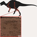
Comparative bone histology of rhabdodontid dinosaurs
Published online: 17/11/2014
Keywords:
bone histology-based ontogeny; Mochlodon; Rhabdodon; skeletal maturation; Zalmoxes
https://doi.org/10.18563/pv.38.2.e1
Abstract
A comparative bone histological study of the three known genera of the endemic European ornithopod dinosaur family, Rhabdodontidae, is presented here in an ontogenetic context. Investigated specimens were assigned to different ontogenetic stages based exclusively on the histological indicators of osteologic maturation during diametrical bone growth; an entirely size-independent method as opposed to most previous studies. Qualitative comparison of bone histology of corresponding ontogenetic stages and elements among the three valid rhabdodontid genera, Mochlodon, Zalmoxes, and Rhabdodon, revealed some consistent patterns. Genus specific histological differences within Rhabdodontidae are most expressed between Rhabdodon and the Mochlodon-Zalmoxes clade. These indicate a prolonged phase of fast growth and a less constrained cyclicity in the growth dynamics of Rhabdodon, as opposed to the slower and more regulated growth strategy reflected in the bones of Mochlodon and Zalmoxes. These genus specific differences are consistent with the phylogenetic interrelation of the genera and are most probably related to the pronounced differences in body size. However, when compared to other ornithopods, most detected histological features in rhabdodontids do not seem to reliably reflect either phylogenetic relations or body size. A notable common feature of all rhabdodontid genera irrespective of body size is the ontogenetically early onset of cyclical growth and secondary remodelling; a pattern that more resembles the condition found in derived ornithopods than that described in more basal taxa which are closer relatives of rhabdodontids. The recognition of taxon-specific histological patterns as well as patterns indicative of ecological and thereby functional traits clearly requires more accurate, preferably quantitative evaluations.
PV article infos
Published in Vol.38-2 (2014)
|
PDF
|

|
Observations sur l'anatomie crânienne du genre Palaeotherium (Perissodactyla, Mammalia): mise en évidence d'un nouveau sous-genre, Franzenitherium
Published online: 01/12/1992
Keywords:
Palaeotherium; Paléogène; Perissodactyla; skull anatomy; Systematics
https://doi.org/10.18563/pv.21.3-4.203-224
Abstract
The skull remains referred to the genus Palaeotherium are recorded and described. Biometrical tests are made to elucidate intrageneric allometric relationships and to allow comparisons with various other perissodactyls. Apart from the well known shortness of post canine diastems and deepness of the narial opening, the genus is characterized by a great lengthening of the splanchnocranium, owing to a spreading of the post-orbital facial region, by a reduced area of the eye-socket and by the prevalence of the temporal muscle with regard to the masseter; this original shape of the masticatory apparatus needs to be related to the morphology of the jugal teeth and particularly to their asymmetrical semi-hypsodonty.
These animals, whose running ability was evidently poor, appear to have been adapted to rather closed environments, feeding on relatively soft vegetable matter; olfactory sense was likely to play a leading part in interindividual and environmental relationships. Such evolutionary trends might explain the disappearance of most of them, as clirnatic conditions deteriorated at the end of the Eocene, before the "Grande Coupure" which affected mammalian faunas at that time.
Although the present paper is not directly concemed with phylogenetics, it invalidates the supposed ancestor-descendant relationship between P. castrense and P. magnum, and it suggests a possible emergence of the P. medium lineage from a P. siderolithicum stock. Moreover, the structure of the post-orbital facial area allows the erection of a new sub-genus, Franzenitherium, for the species lautricense and duvali.
PV article infos
Published in Vol. 21, Fasc. 3-4 (1992)
|
PDF
|

|
Reflections on some Russian eotheriodonts (Reptilia, Synapsida, Therapsida)
Published online: 28/02/1972
Keywords:
Reptilia; Russia; Synapsida; Therapsida
https://doi.org/10.18563/pv.5.3.79-109
Abstract
As a result of the enrichment of eotheriodont material by one of us (P.K.T.), these specimens (essentially Biarmosuchur and Eotitanosuchur) are reexamined and refigured. A reevaluation of their particularities supports the distinction of two families, for which new diagnoses are proposed. This leads us to discuss the affinities of these families, with respect to the sphenacodonts on one hand, and to the South African primitive theriodonts on the other (gorgonopsids and ictidorhinids). This study contains inherent paleogeographic consequences which are considered in conclusion.
PV article infos
Published in Vol. 05, Fasc. 3 (1972)
|
PDF
|

|
Contribution à l'étude des Cricétidés oligocènes d'Europe occidentale
Published online: 20/01/1972
Keywords:
Cricetidae; Europe; Oligocene
https://doi.org/10.18563/pv.5.1.1-44
Abstract
Of the ten cricetid species from the Oligocene of Western Europe, attributed until now to the genus Eucricetodon, only four prove to be utilizable - E. atavus, E. huberi, E. praecursor, E. collatum - to which it is possible to add two forms newly described: E. huerzeleri and E. quercyi. The evolullon of the genus Pseudocricetodon is also the subject of new observations. The study of the dental morphology allows us to distinguish in these two genera three lineages beginning in the middle Oligocene:
- Lineage P. montalbenensis-P. thaleri (from Montalban to Goderet), of small size, without
increase in size.
- LineageE. atavus-E. infralactorensis (from Hoogbulsel to Estrepouy), of middle size, with a regular increase in size.
- Lineage E. huerzeleri-E. haslachense (level of Montalban to that of Estrepouy), of large size, with an increase in size.
Two other forms are equally represented in these loealitles: Heterocricetodon aff. helbengi and Melissiodon quercyi. It has been possible to attribute a precise age (zone of "Cournon") to the last species, which has been defined by Schaub (l925) from material in the Old Quercy collections. The genera Pseudocricetodon, Eucricetodon, ? "Cricetodon", Leydimys, Eumys, differentiated at the beginning of the middle Oligocene in Europe, Asia and North America, seem to derive from a common ancestral group. The place of origin of this group could be situated in Asia.
PV article infos
Published in Vol. 05, Fasc. 1 (1972)
|
PDF
|
|
New Squalicorax species (Neoselachii: Lamniformes) from the Lower Maastrichtian of Ganntour phosphate deposit, Morocco
Published online: 05/12/2014
Keywords:
Anacoracidae; Chondrichthyes; Maastrichtian; Morocco; New taxa
https://doi.org/10.18563/pv.38.2.e3
Abstract
Two new Squalicorax species, S. benguerirensis nov. sp. and S. microserratus nov. sp. are described from the Lower Maastrichtian of the Benguérir phosphate open mine, Ganntour deposit, Morocco. The species S. benguerirensis nov. sp. was classically assigned to S. yangaensis since Arambourg (1952) and has been also recognized in coeval deposits from eastern USA to Mid-East. The species S. microserratus nov. sp. correspond to the lateral teeth of S. kaupi as reported by Arambourg (1952) and which is now referred in fact to S. bassanii. The comparison of these two new species with other Anacoracids, known in Moroccan or elsewhere, allows highlighting the great taxonomic and ecological diversities of this family during the Cretaceous.
PV article infos
Published in Vol.38-2 (2014)
|
PDF
|
|
Discovery of the most ancient Notidanodon tooth (Neoselachii: Hexanchiformes) in the Late Jurassic of New Zealand. New considerations on the systematics and range of the genus
Published online: 21/09/2018
Keywords:
Chondrichthyes; Hexanchiformes; new genus; New Zealand; Tithonian
https://doi.org/10.18563/pv.42.1.e1
Abstract
This paper describes the first hexanchid tooth from the Tithonian (Late Jurassic) of New Zealand. For the moment, this tooth represents the earliest representative of the fossil genus Notidanodon in the world and one of the most ancient neoselachians in the Southern Hemisphere. Despite the perfect state of preservation of the unique tooth, the species is left in open nomenclature, pending the discovery of additional specimens. Few nominal species have been assigned to the genus Notidanodon. Four from Cretaceous deposits: N. antarcti Grande & Chatterjee, 1987, Notidanodon dentatus (Woodward, 1886), Notidanodon lanceolatus (Woodward, 1886), Notidanodon pectinatus (Agassiz, 1843) and only two from Paleocene: Notidanodon brotzeni Siverson, 1995, and Notidanodon loozi (Vincent, 1876). Considering the important morphological variations observed between some of these species, it seems obvious that the genus Notidanodon is not monophyletic and will need a revision in the future.
PV article infos
Published in Vol 42-1 (2019)
|
PDF
|
|
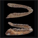
A mandible of the hyracoid mammal Titanohyrax andrewsi in the collections of the Muséum National d'Histoire Naturelle, Paris (France) with a reassessment of the species
Published online: 18/04/2016
Keywords:
Afro-Arabia; Fayum; Oligocene; Titanohyracidae
https://doi.org/10.18563/pv.40.1.e4
Abstract
An unpublished mandible of the large hyracoid Titanohyrax andrewsi from the early Oligocene Jebel Qatrani Formation, Fayum Depression, Egypt is described. This specimen has a twofold importance. Firstly, it opens an unexpected window on early paleontological research in the Fayum because it was discovered as early as 1904 by the French paleontologist René Fourtau during an expedition to the Fayum organized by the Muséum National d’Histoire Naturelle, Paris (MNHN). This expedition has remarkably never been mentioned in the literature. Secondly, the mandible documents the best-preserved specimen of T. andrewsi, permitting a revision of one of the very rare Paleogene hyracoids. Interestingly, the new mandible was discovered two years before the first report of the species by Charles W. Andrews. The hypodigm of T. andrewsi is reviewed and the dentition as a whole is compared in detail, notably with other Titanohyrax species from the Fayum. The validity of the large Titanohyrax “schlosseri” species is discussed, but a pronounced sexual size dimorphism for T. andrewsi is favoured.
PV article infos
Published in Vol.40-1 (2016)
|
PDF
|

|
New remains of the very small cuckoo, Chambicuculus pusillus (Aves, Cuculiformes, Cuculidae) from the late Early/early Middle Eocene of Djebel Chambi, Tunisia
Published online: 15/02/2016
Keywords:
Cuckoos; Eocene; Fossil bird
https://doi.org/10.18563/pv.40.1.e2
Abstract
Abstract: A very tiny cuckoo, Chambicuculus pusillus, was recently described after a few fragments of tarsometatarsi from the late Early/early Middle Eocene of Djebel Chambi, Tunisia. New remains, notably a coracoid, confirm the attribution of this genus to the recent family Cuculidae. This coracoid shows a very elongate and strap-like processus procoracoideus. This morphological feature is otherwise only known in the family Cuculidae. The characteristics of the coracoid and tarsometatarsi show that Chambicuculus is morphologically more advanced over the other stem cuculids described in Europe and North America. Chambicuculus is the oldest Cuculidae known so far.
PV article infos
Published in Vol.40-1 (2016)
|
PDF
|
