|
The digital endocast of Necrolemur antiquus
Published online: 24/07/2020
Keywords:
brain evolution; Eocene; Omomyiforms; Primates
https://doi.org/10.18563/pv.43.2.e1
Abstract
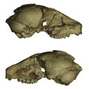
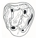
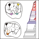
The study of endocasts, or casts of the endocranial space, have played an important role in shaping understanding of mammalian, and particularly primate, brain evolution. Recently, the reconstructions of three-dimensional virtual endocasts from high-resolution computed tomography images have allowed for the visualization and quantification of endocasts in several Paleocene and Eocene primate species. Here we present the virtual endocast of MaPhQ 289 (informally known as the Montauban 9 skull), a specimen of Necrolemur antiquus Filhol 1873, a middle to late Eocene European primate of the family Microchoeridae. The virtual endocast of MaPhQ 289 reveals a lissencephalic surface morphology with expanded temporal poles and minimal overlap of the cerebellum or olfactory bulb by the cerebrum, which closely resembles the morphology of the endocast of its contemporary relative, Microchoerus erinaceus (Primates, Microchoeridae). MaPhQ 289 yields an endocranial volume (ECV) of 2.36 cm3, about 60% smaller than the volume of the most commonly cited ECV of N. antiquus. Thus, the size of the brain of N. antiquus relative to its body size is likely to be smaller than has been reported in previous literature, highlighting the importance of corroborating older ECV estimates with new evidence using 3-D imaging techniques.
PV article infos
Published in 43-2 (2020)
|
PDF
|
|
Small sauropod tracks in the Hettangian of Southern France – A case of ichnite fossilization in an intertidal zone
Published online: 28/06/2022
Keywords:
Intertidal zone; Lower Jurassic; Sauropods; Southern France; Tracks
https://doi.org/10.18563/pv.45.2.e1
Abstract
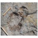
This paper presents the description and the interpretation of recently discovered traces on a Lower Hettangian dolomitic outcrop in the Bédarieux area, Southern France. One trace set immediately attracted the attention by its resemblance to a small sauropod pes-manus couple but no trackway was visible. As the other traces have a variety of shapes with no obvious significance, it took a thorough examination of the 3D and sedimentological data to come to the conclusion that most traces likely were sauropod tracks made under diverse conditions. Sedimentological and ichnological data indicate that the tracks have been made in the intertidal zone of a carbonated tidal flat shortly before an emersion period. It appears that that the variety of trace shapes is due to a variety of water depths: the sauropods were punting when the water level was high. The lack of trackways seems due to the combination of an underprint situation, buoyancy effects and the small size of the track-bearing slab. Several hypotheses can be considered for explaining the very small size of the tracks, such as insular dwarfism or the immaturity of the trackmakers.
PV article infos
Published in 45-2 (2022)
|
PDF
|

|
Book of Abstracts of the 20th Annual Conference of the European Association of Vertebrate Palaeontologists, 26th June – 1st July 2023, Sabadell (Barcelona), Spain
Published online: 15/06/2023
Keywords:
Abstracts; EAVP
https://doi.org/10.18563/pv.eavp2023
Abstract
PV article infos
Published in special issue 1-2023 (2023)
|
PDF
|
|
Les Palaeotheridae (Perissodactyla) de la faune de Mammifères de Fons 1 (Eocène supérieur).
Published online: 15/06/1967
Keywords:
Anchilophus; Eocene; Pachynolophus; Palaeotheriidae; Perissodactyla
https://doi.org/10.18563/pv.1.1.1-46
Abstract
The locality of Fons 1, one of the fossiliferous outcrops in the late Eocene limestones of Fons-outre-Gardon (Gard), has yielded varied remains of mammals. The specimens were prepared by dilute acetic acid attack on the rock and by impregnation with an acrylic resin.
This fauna, rich with about 20 species, can be included in the biochronologic zone of Euzet. The Palaeotheridae (sensu Simpson 1945), represented by 6 species, occupy a predominant place; Plagiolophus annectens is particularly abundant, comprising 55% of all the specimens found.
The abundance of this species has permitted a detailed comparative study with the corresponding form from Euzet. The quantitative tests brought out a slight but highly sígnificant difference between the average dimensions of the teeth in the two populations. Moreover and in spite of the feeble rate of evolution in the Plagialophus dentition, diverse archaic particularities can be seen which permit the conclusion that the form from Fons 1 is a little the older. This conclusion is corroborated by observations on the other palaeotherids, particularly Lophiotherium aff. cervulum, whose dental evolutionary stage is intermediate between those of the corresponding forms known from Robiac and Euzet.
A new species of Pachynolophus is described, P. garimondi n.sp., the last representative known of the genus. lts teeth are distinguished by their size, a great heterodonty, and the weakness
of their cingula.
PV article infos
Published in Vol. 01, Fasc. 1 (1967)
|
PDF
|
|
Les Paramyidae (Rodentia) de l'Eocène inférieur du bassin de Paris.
Published online: 15/07/1968
Keywords:
Ailuraviinae; Eocene; Paramyinae; Rodents
https://doi.org/10.18563/pv.1.4.135-193
Abstract
The exploitation of new early Eocene localities in the Paris Basin has resulted in the collecting of numerous mammalian remains, among which are about 300 isolated teeth representing the rodents. They belong, for the most part, to the paramyid group. Only the latest level of the early Eocene has yielded rodents belonging to the pseudosciurid group. The paramyids, the object of this study, are represented by at least 5 genera and 10 species; they are distributed among 4 clearly dilferentiated subfamilies : Paramyinae Simpson 1945, Pseudoparamyinae Michaux 1964, Ailuraviínae n. subf., Microparamyinae Wood1962.
It results from this study that the principal types of rodents in the middle and late Eocene of Europe are clearly related to the forms described here: Plesiarctomys Bravard 1850 is related to Pseudoparamys Michaux 1964, Ailuravus Rütimeyer1891 to Meldimys n. gen., Gliravus hammeli Thaler 1966 to Microparamys nanus (Theilard1927), and some Masillamys Tobien 1954 to Microparamys russelli Michaux 1964 and to M. sp. 1. Gliravus and Masillamys are the oldest representatives of the Gliridae and the Pseudosciurídae, respectively, families which will progressively replace the Paramyidae in the course of the middle and late Eocene.
Two stages can be recognized in the history of te European Paramyidae fauna during the course of the early Eocene: the older is characterized by the Mutigny fauna, the younger by the Cuis fauna.
The Mutigny fauna appears already rather diversified and does not completely correspond, point by point, to the homologous fauna of North America. A geographic differentiation seems to have been manifested rapidly, unless the fauna established in Europe was already a little different from that which established itself in North America.
PV article infos
Published in Vol. 01, Fasc. 4 (1968)
|
PDF
|
|
The beginning of the adaptive radiation of Theridomorpha (Rodentia) in Western Europe: morphological and phylogenetic analyses of early and middle Eocene taxa; implications for systematics
Published online: 20/09/2021
Keywords:
characters analyses; Dental morphology; Eocene; Rodentia; variability
https://doi.org/10.18563/pv.44.2.e2
Abstract
This paper provides a revision of the early and middle Eocene European rodents previously referred to as Ischyromyoidea, including taxa considered to be at the origin of the Theridomorpha. The use of an accurate dental terminology and a better understanding of the size and shape of their infra-orbital foramen (i.o.f.) led us to a substantial revision of this group, which allowed to better characterize them and to appreciate their variability. On these bases, phylogenetic analyses (cladistic and standard Bayesian
approaches) of early Ypresian to late Priabonian European rodent species were undertaken in order to highlight the root of the early Theridomorpha and its content. In this paper, the phylogeny was established based on 343 characters (338 dental) through 45 early Paleogene taxa using both cladistic and bayesian analyses. The ingroup included on one hand a few North American genera (Reithroparamys, Microparamys, and Acritoparamys) and European ones (Eogliravus, Ailuravus, Corbarimys, Meldimys, Euromys, Plesiarctomys, and Pseudoparamys) considered until now as being related with the North American superfamily Ischyromyoidea. On the other hand, it included genera close to the root of the Theridomorpha (Sparnacomys, Pantrogna, and Hartenbergeromys) and early Theridomyoidea (Masillamys, Protadelomys, and some Pseudosciuridae). The phylogenetic results obtained via the two
distinct reconstruction approaches are consistent in virtually all relationships. The proposed systematics here derives from these phylogenetic results. This phylogenetic context led us to change the suprafamilial, familial, subfamilial or generic attribution of several species. Characters of Theridomorpha, like the obliquely developed postprotocristid allied with the occurrence of a metalophulid I, have been found in genera previously considered as Ischyromyidae (Pseudoparamys, Euromys, Sparnacomys, Meldimys, Pantrogna, and Hartenbergeromys) as well as the large i.o.f., when preserved (Pseudoparamys, Hartenbergeromys, and Masillamys). Based on these morphological observations and new phylogenetic considerations, the content of the Theridomorpha clade is here enlarged, thereby extending back the first theridomorph radiations to the early Eocene. Aside, a new taxon (Reinomys rhomboides gen and sp. nov.) is described from Avenay. In addition, a new genus, Auroremys, is created for the species subita (Comte et al., 2012) from Chery-Chartreuve.
PV article infos
Published in 44-2 (2021)
|
PDF
S.I. Data
|

|
Les rongeurs du site Pliocène à Hominidés de Hadar (Ethiope)
Published online: 15/02/1982
Keywords:
Ethiopia; hominids; Muridae; Pliocene
https://doi.org/10.18563/pv.12.1.1-56
Abstract
The intensive exploration of the Pliocene Hadar Formation, rich in hominid remains, led us to the discovery of several micromammals levels. ln some of them, rodents are very abundant. The stratigraphic repartition of these levels do not cover the whole fossiliferous series of the formation but takes place only in the sedimentary members from Sidi Hakoma and Denen-Dora (rancing from 3.1 - 3.2 MY to 2.8 - 2.9 MY, according to the recent geochronological data). During this gap of time, the species do not show morphological changes, what allowed us to gather, in the same taxa, forms of slighty different ages.
Two striking facts, giving a lot of indications, characterize these small rodents'faunas. First, we notice the domination of the Muridae, as well on a qualitative way (number of species) as on a quantitative one (number of individuals). Then, it appears that, until now, two genera of these murids were known only in the south-western asiatic regions. So, we can suppose continuous biotops between Africa and Indian Subcontinent before 3 MY. In this hypothesis, the hominids had already the possibility to leave their african « cradle ››. Finally, almost all studied genera are still represented at the present time. This fact, previously observed in Laetolil, Omo, Olduvai contributes to remove hope of establishing a biochronological scale based on rodents, in tropical zone. Nethertheless, that allows to try a reconstruction of the palaeoenvironnement, by using the principle of actualism.
PV article infos
Published in Vol. 12, Fasc. 1 (1982)
|
PDF
|

|
Mammals of the Eocene locality Toru Ajgyr (Kyrgyzstan)
Published online: 15/12/2006
Keywords:
Eocene; Kyrgyzstan; Mammalia; Olsenia; Palaeoecology; Stratigraphy; taxonomy
https://doi.org/10.18563/pv.34.e12
Abstract
Morphological descriptions are given of Eocene mammals from the locality Toru Ajgyr (NEKyrgyzstan) that were excavated in 1997 and 1998 in a cooperation between the Martin-Luther-University Halle (Germany), the Zoological Institute in St. Petersburg (Russia) and the Seismological Institute in Bishkek (Kyrgyzstan). The species found belong mostly to perissodactyls, as Lophialetes sp., Teleolophus sp. and brontotheres. The primitive ungulate family Olseniidae is represented by a complete foot skeleton of cf. Olsenia sp. In addition, postcranial materials of Gobiatherium mirificum (Dinocerata) and of artiodactyls have been collected and are described herein. Based on mammals, the locality is part of the Asian Land Mammal Age Arshantan and is stratigraphically equivalent with the Bridgerian Land Mammal Age in North America and with the lower and middle Geiseltalian of the European Middle Eocene.
PV article infos
Published in Vol. 34, Fasc. 3-4 (2006)
|
PDF
|
|
A new species of bat (Chiroptera: Vespertilionidae) from the early Oligocene global cooling period, Brule Formation, North Dakota, USA
Published online: 09/12/2019
Keywords:
Eocene-Oligocene global cooling; Mammalia; Oligocene; Plecotini; Quinetia
https://doi.org/10.18563/pv.42.2.e2
Abstract
We report the first confirmed fossil bats from North Dakota, including a new species referable to the Vespertilionidae represented by a maxilla with P4-M3 from the Brule Formation, Fitterer Ranch local fauna, early Oligocene, Whitneyan North American Land Mammal Age. Unassociated postcranial fragments of the humerus and femur also represent a vespertilionoid, but appear to reflect a different, unidentified species. The new taxon, Quinetia frigidaria sp. nov., is referred to the genus Quinetia, previously known only from approximately contemporaneous deposits in Europe. The new species is larger than Quinetia misonnei from the early Oligocene of Belgium. It is similar in some morphological characters to Chadronycteris rabenae (Chiroptera incertae sedis) of the late Eocene (Chadronian) of northwestern Nebraska and to Stehlinia species (?Palaeochiropterygidae) from the Eocene and Oligocene of Europe, but differs from each in morphological details of the dentition and maxilla. An unassociated talonid of a lower molar from Fitterer Ranch shows myotodont morphology, unlike the nyctalodont lower molars in Q. misonnei, and thus represents a second chiropteran taxon in the fauna. Quinetia frigidaria is a member of a Paleogene radiation of bats near the low point of the Eocene-early Oligocene decline in global temperatures, increased seasonal aridity, and loss of tropical floras from mid-latitude North America.
PV article infos
Published in Vol 42-2 (2019)
|
PDF
|
|
The skull of Tetraceratops insignis (Synapsida, Sphenacodontia)
Published online: 09/01/2020
Keywords:
cranium; pelycosaur; Permian; therapsid origins
https://doi.org/10.18563/pv.43.1.e1
Abstract
Tetraceratops insignis is known from a single, crushed skull from the Lower Permian of Texas. Its unique proportions and osteological details gained central meaning in the question of the origins of Therapsida since this early synapsid has been determined as the oldest and less derived therapsid. Apart from Tetraceratops, the ‘mammal-like’ Therapsida and their sister, the pelycosaur-grade Sphenacodontidae, are separated by one of the longest ghost lineages in tetrapod fossil record. However, the minor, though well justified critique faced insistent publication regarding the therapsid hypothesis. A carefull re-evaluation of the holotypic skull reveals that therapsid traits cannot be supported, including a rejection of the formerly supposed adductor shelf in the temporal fenestra. Increased understanding of ‘pelycosaur’ character variation underlines a haptodontine-grade or, less likely, sphenacodontid position for Tetraceratops.
PV article infos
Published in Vol 43-1 (2020)
|
PDF
|
|
Additions to the elasmobranch assemblage from the Bandah Formation (middle Eocene, Bartonian), Jaisalmer District, Rajasthan, India, and the palaeobiogeographic implications of the fauna
Published online: 23/06/2021
Keywords:
Chondrichthyes; Elasmobranchii; Indian Ocean; Palaeogene; South Asia
https://doi.org/10.18563/pv.44.2.e1
Abstract
Isolated elasmobranch teeth (sharks and rays) from the middle Eocene (Bartonian) Bandah Formation in the Jaisalmer District of Rajasthan, India are described. The remains improve our knowledge of the environment represented by this lithostratigraphic unit and the ecology preserved therein. Seventeen unequivocal taxa were identified, including Nebrius sp., Striatolamia aff. S. macrota, Brachycarcharias atlasi, B. lerichei, cf. Jaekelotodus sp., Carcharhinus mancinae, Rhizoprionodon sp., Physogaleus sp., Galeocerdo clarkensis, G. eaglesomei, Odontorhytis aff. O. pappenheimi, “Rhinobatos” sp., “Dasyatis” sp., Coupatezia sp., “Aetomylaeus” sp., “Rhinoptera” sp., and Ouledia aff. O. lacuna. Of these, “Aetomylaeus” sp., B. atlasi, C. mancinae, G. clarkensis, G. eaglesomei, cf. Jaekelotodus sp., Nebrius sp., Odontorhytis aff. O. pappenheimi, Ouledia aff. O. lacuna, and “Rhinoptera” sp. are reported from the middle Eocene of India for the first time. The Bandah Formation elasmobranch palaeofauna has close affinities to the Palaeocene-Eocene Tethyan/Paratethyan faunas of Africa, Madagascar, Asia, and Europe, and some taxa indicate a western hemisphere influence from North America. The Bandah Formation palaeofauna indicates that deposition occurred in a moderately shallow marine environment. The Bartonian age is primarily based on foraminifera but is corroborated by the presence of elasmobranch taxa that also occur in contemporaneous deposits elsewhere. The marine regression started during the early Palaeogene, and our study indicates that the sea completely withdrew from the Jaisalmer Basin after the deposition of the Bandah Formation. This event may have been synchronous with the middle Eocene uplift of the Himalayan-Tibetan Plateau.
PV article infos
Published in 44-2 (2021)
|
PDF
|
|
Autopsie d’une radiation adaptative : Phylogénie des Theridomorpha, rongeurs endémiques du Paléogène d’Europe - histoire, dynamique évolutive et intérêt biochronologique
Published online: 15/12/2016
Keywords:
Diversification; Extinction; Paléoenvironnements; Rodentia; Theridomyoidea
https://doi.org/10.18563/pv.40.3.e1
Abstract
Résumé :
L’étude des rongeurs Theridomorpha permet de suivre le déroulement d’une radiation adaptative pendant toute sa durée (Eocène moyen-Oligocène terminal), sur un territoire restreint à l’extrémité ouest de l’Europe Occidentale. Dans ce papier, la phylogénie de ce groupe est établie à partir d’une analyse cladistique reposant sur l’examen de 315 caractères (310 dentaires). Le groupe d’intérêt comprend 110 des 132 espèces (24 genres) de Theridomyoidea et deux genres encore inclus jusqu’ici dans les Reithroparamyinae qui rejoignent les Theridomorpha. Les groupes externes comprennent des Glires basaux, Cocomys, Tanquammys et 16 Ischyromyiformes. Un cadre phylogénétique robuste est produit, qui permet de clarifier la systématique des Theridomorpha. La position des Remyoidea (nov. sup.fam.) au sein des Ischyromyiformes, extérieure aux Theridomorpha, est confortée. Les Protadelomys et Tardenomys sont à la base des Theridomyoidea, avant la séparation en deux clades correspondant aux familles Pseudosciuridae et Theridomyidae. Les sous-familles sont consolidées : Pseudosciurinae et Sciuroidinae pour les Pseudosciuridae ; Issiodoromyinae, Oltinomyinae, Columbomyinae, Theridomyinae, auxquelles s’ajoute au moins une nouvelle sous-famille (Patriotheridomyinae), pour les Theridomyidae. La topologie des chrono-espèces (sensu Simpson), traitées antérieurement comme lignées évolutives, apparaît dans la plupart des cas sous forme de clades successifs dans lesquels les espèces sont le plus souvent arrangées de manière pectinée, émergeant dans l’ordre stratigraphique. L’analyse des caractères aux principaux nœuds permet de consolider les caractères diagnostiques des taxons et les tendances évolutives, ainsi que de discuter des divers parallélismes et convergences dans l’évolution des structures et patrons dentaires (e.g., émail des incisives unisérié chez les Issiodoromyinae et les Patriotheridomyinae, ou pseudo-multisérié chez les Blainvillimys les plus hypsodontes, les Protechimys et Archaeomys ; patrons dentaires téniodontes ; allongement des dents déciduales chez les Patriotheridomyinae, Issiodoromyinae et Theridomyidae ; sélénodontie ou lophodontie). Les dynamiques évolutives traduites par les changements morphologiques sont mises en relation avec les variations environnementales. Enfin, les implications biochronologiques de l’évolution des Theridomyoidea sont discutées.
Abstract:
The adaptive radiation of the rodents Theridomorpha occurred during a limited time window (middle Eocene to late Oligocene), on an area restricted to Western Europe. In this paper, the phylogeny of this group is established via a cladistic assessment of 315 morphological characters (310 dental). The group of interest encompasses 110 upon 132 species (24 genera) of Theridomyoidea, and two genera formerly included within the Reithroparamyinae, and which are included here within the Theridomorpha. The outgroups include basal Glires, Cocomys, Tanquammys and 16 Ischyromyiformes. A robust phylogenetic frame is produced, which allows clarifying the systematics of the Theridomorpha. Within the Ischyromyiformes, the Remyoidea (nov. supfam.) are set apart from the Theridomorpha. Protadelomys and Tardenomys represent the earliest offshoots of the Theridomyoidea, before the dichotomy between Pseudosciuridae and Theridomyidae. It supports the former subfamilies Pseudosciurinae and Sciuroidinae within the Pseudosciuridae; and for the Theridomyidae: the Issiodoromyinae, Oltinomyinae, Columbomyinae, Theridomyinae, with at least one new subfamily (Patriotheridomyinae). The topologies of the chronospecies (sensu Simpson), formerly considered as evolutionary lineages, appear in most cases as successive clades, in which the species are generally pectinately arranged and emerging in the stratigraphic order. The analysis of characters supporting the main nodes allow consolidating the diagnosic characters of the taxa and their evolutionary trends, as well as discussing the various cases of parallelism and convergence in the evolution of structures and dental patterns (e.g., uniserial incisor enamel for Issiodoromyinae and Patriotheridomyinae, or pseudo-multiserial for the most hypsodont Blainvillimys, Protechimys and Archaeomys; taeniodont dental patterns; lengthening of deciduous premolars for Patriotheridomyinae, Issiodoromyinae and Theridomyidae; selenodonty or lophodonty).
Evolutionary dynamics are analysed with respect to environmental changes. Finally, biochronological implications of the evolution of Theridomyoidea are discussed.
PV article infos
Published in Vol 40-3 (2016)
|
PDF
S.I. Data
|

|
Le genre Leptolophus (Perissodactyla, Mammalia): morphologie et histologie dentaires, anatomie cranienne, implications fonctionnelles.
Published online: 15/09/1998
Keywords:
dental histology; Eocene; functional anatomy; Palaeotheriidae; skull anatomy; Southern France; Systematics
Abstract
A strong lophodonty, an extreme heterodonty, some hypsodonty and regular overlayings of coronal cement are prominent features of the genus Leptolophus (Palaeotheriinae = Palaeotheriidae s.s.). The histological pattern of the teeth unusually joins type II enamel prisms, characteristic of advanced ungulates, together with archaic features, such as an almost complete lack of Hunter-Schreger zonation and a weak expanse of peritubular dentine. The skull is narrow and slender, with an elongated ante-orbital facial region, a moderately notched nasal aperture, a rather elongated post-canine diastem, parallel zygornatic arches and a fairly dorsally located squamoso-mandibular joint.The functional analysis brings to light "ectolophodont" masticatory cycles with two phases, in which maximum power was applied, contrary to equíds, on hindmost teeth; likewise, skull accomodations to increasing height of the teeth are quite different. This study leads to the assumption that Leptolophus may have been light mammals, living in rather open surroundings, browsing on herbaceous plants or leaves cropped close to the ground. Moreover, it appears that it could have been some inadequacy of dental structures to the dietary, which leaded to quick wear of the teeth and to many enamel notches, but had been somewhat balanced by the early increase of hypsodonty, not induced in such a case by a biotop deterioration (as it will happen at the end of the Eocene). This ínadaptation might account for the short duration of the genus Leptolophus, whose the 3 species, L. stehlini, L. nouletí and L. magnus n. sp. are indeed confined in the level MP 16. Its geographical spreading (as far as known, South of western Europe) and the morphological pattern of its dentition suggest that this genus would have been related to early upper Eocene endemic spanish forms.
PV article infos
Published in Vol. 27, Fasc. 1-2 (1998)
|
PDF
|

|
Norselaspis glacialis n.g., n.sp, et les relations phylogénétiques entre les kiaeraspidiens (Osteostraci) du dévonien inférieur du Spitsberg.
Published online: 15/06/1981
Keywords:
Devonian; kiaeraspids; Osteostraci; Spitsbergen
https://doi.org/10.18563/pv.11.2-3.19-131
Abstract
The anatomy of Norselaspis glacialis n.g., n.sp., a primitive kiaeraspidian from the Lower Devonian of Spitsbergen, is described on the basis of spécimens studied by grinding sections or prepared with dilute formic acid. This study yielded some new anatomical details, including the presence of a canal prolonging posteromedially the canal alloted to the facial nerve by Stensiö. This posterior prolongation of the « facial canal ›› into the posterolateral part of the labyrinth cavity is consistent with the hypothesis put forward by Allis, Lindström, Jefferies and Whiting, that this canal housed the glossopharyngeus nerve. Furthermore, in N. glacialis, the foramen usually referred to as the foramen for the œsophagus opens posteriorly into a cavity in the postbranchial wall, referred to here as the intramural cavity, and which is interpreted as having housed the heart. Consequently, the œsophagus probably accompanied the dorsal aorta through the aortic canal. Finally, the foramen generally interpreted as having transmitted the ventral afferent arterial trunk is here considered as having housed the hepatic vein, which emptied into the venous sinus of the heart. The ventral afferent arterial trunk may thus have passed through the former «œsophageal ›› foramen.
The problem of the position of the dorsal nerves in the Osteostraci is discussed, and it is suggested that the three foremost nerve canals opening into the oralobranchial cavity housed the maxillary ramus of the trigeminus, the facial nerve and the glossopharyngeus nerve respectively. The mandibular ramus of the trigeminus must have accompanied one of the two foremost nerves, but for the moment it is impossible to decide which.
The problem of the nature of the interbranchial crests of the Osteostraci is briefly discussed. Comparison with the branchial apparatus of the Petromyzontida does not support the hypothesis that the interbranchial crests are part of the branchial arches, incorporated into the endoskeletal shield. A different hypothesis is proposed, that the branchial skeleton of the Osteostraci was situated entirely inside the oralobranchial cavity, and was attached to the endoskeletal shield only by the ventromedial processes. The grooves classically allotted to the efferent branchial arteries would thus have housed extrabranchial arteries, branching off from the dorsal aorta, and irrigating the ventral branchial musculature.
A phylogeny and a classification of the kiaeraspidians are proposed. The evolution of this monophyletic group is characterized by, e.g., reduction of cornual processes, shortening of the abdominal division of the shield, subdivision of the lateral fields, and enlargement of the supraoral fossae.
The phylogenetic position of the kiaeraspidians within the Osteostraci remains uncertain. Their sister-group may be either the benneviaspidiens or the thyestidians, or Thyestes alone (in which case they would have to be included within the thyestidians).
PV article infos
Published in Vol. 11, Fasc. 2-3 (1981)
|
PDF
|
|
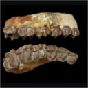
Les Périssodactyles (Mammalia) du gisement Bartonien supérieur de Robiac (Éocène moyen du Gard, Sud de la France)
Published online: 04/05/2015
Keywords:
Chasmotherium; new species; Palaeotheriidae; paleoenvironments
https://doi.org/10.18563/pv.39.1.e3
Abstract
We present here a new updated counting of the perissodactyls of Robiac, the type locality of the MP 16 level of the biochronological scale of paleogene mammals and that of the Robiacian stage of Eocene Land Mammals Ages in Western Europe.
The outcrop of Robiac consists actually of two 500m apart loci, Robiac-Nord and Robiac-Sud, considered of the same age according to the current discriminating power, and is dated from -38,7 MA after the last faunal, magnetostratigraphic and climatic calibrations.
It has yielded a very abundant and rich of 21 taxa perissodactyl fauna, topped by the giant Lophiodon lautricense, last representative of the family Lophiodontidae, of which it is the last proved deposit. The Palaeotheriidae are much diversified with 5 genera and 9 species of "Pachynolophinae", 3 genera and 10 species of Palaeotheriinae. Nine taxa have been defined from Robiac: Chasmotherium depereti n. sp., Palaeotherium castrense robiacense Franzen, 1968, the genus Leptolophus Remy, 1965 with the species L. stehlini Remy, 1965 and L. magnus Remy, 1998, Anchilophus (Paranchilophus) jeanteti Remy, 2012, Metanchilophus chaubeti Remy, 2012, Lophiotherium robiacense Depéret, 1917 and Pachynolophus gaytei n. sp.
The faunal Robiac cenogram with the associated flora testify to a hot, wet and forestal environment, likely corresponding to a short warming climatic phase; the broken up fossil bones should have been carried away and then gathered in swamp areas along the banks of a meandering river.
The swarm of mammals of Robiac, the richest of contemporaneous deposits, has been followed by a drastic drop in perissodactyl diversity at the MP 17A level; a crisis which could have originated in a renewal of the global Eocene cooling. Fons 4, the type-locality of this level, is largely scarcer in perissodactyls and its cenogram testifies to a less diversified fauna, with on the whole smaller species, that likely means a cooler and drier climatic environment; a new perissodactyl diversification occurred but later.
PV article infos
Published in Vol.39-1 (2015)
|
PDF
|
|
Eocene Teleostean Otoliths, Including a New Taxon, from the Clinchfield Formation (Bartonian) in Georgia, USA, with Biostratigraphic, Biogeographic,
and Paleoecologic Implications
Published online: 03/01/2022
Keywords:
climate; Congridae; Ophidiidae; Sciaenidae; tectonics
https://doi.org/10.18563/pv.45.1.e1
Abstract
Investigations of the Clinchfield Formation (middle Eocene, upper Bartonian) exposed at the Hardie Mine (Wilkinson County, Georgia, USA), produced 4,768 actinopterygian otoliths representing 14 taxa and increased the number of bony fishes threefold from the site. The somewhat limited richness was characterized by bonefishes, mud eels, conger eels, sea catfishes, cusk-eels, snooks, grunts, drums and croakers, and porgies. The assemblage had a relatively even distribution with Ophidiidae, Congridae, and Sciaenidae most common. Included in the otolith taxa was a new sciaenid genus and species, Eosciaena ebersolei, with unknown relationships to other Sciaenidae. The Clinchfield otoliths were compared to other middle and late Eocene in age otolith assemblages in Alabama, Mississippi, and Louisiana utilizing percentage similarity measurements. Analysis indicated that the Clinchfield otoliths were not greatly similar or greatly unlike the Moodys Branch and Yazoo Clay otolith assemblages. However, the Clinchfield showed little relationship to the slightly older Lisbon Formation in adjacent Alabama and is postulated to be related to global climatic and plate tectonic events. Biostratigraphically, the Clinchfield otolith taxa are essentially the same as the other formations except for the Lisbon, which has at least ten unique species. Abundances of Clinchfield otolith taxa indicate a possible sub-bioprovince in the eastern Gulf Coastal Plain. The Clinchfield otoliths indicate a tropical to perhaps subtropical, soft substrate, mainly normal marine to slightly reduced salinities, inner shelf (0–20 m) paleoenvironment with indications of proximal continental coastlines. This investigation represents an initial step in addressing the immensely understudied Paleogene otolith assemblages in Georgia.
PV article infos
Published in 45-1 (2022)
|
PDF
|
|
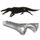
First record of the family Protocetidae in the Lutetian of Senegal (West Africa)
Published online: 05/12/2014
Keywords:
innominate; Lutetian; Protocetid; Senegal
https://doi.org/10.18563/pv.38.2.e2
Abstract
The earliest cetaceans are found in the early Eocene of Indo-Pakistan. By the late middle to late Eocene, the group colonized most oceans of the planet. This late Eocene worldwide distribution clearly indicates that their dispersal took place during the middle Eocene (Lutetian). We report here the first discovery of a protocetid fossil from middle Eocene deposits of Senegal (West Africa). The Lutetian cetacean specimen from Senegal is a partial left innominate. Its overall form and proportions, particularly the well-formed lunate surface with a deep and narrow acetabular notch, and the complete absence of pachyostosis and osteosclerosis, mark it as a probable middle Eocene protocetid cetacean. Its size corresponds to the newly described Togocetus traversei from the Lutetian deposits of Togo. However, no innominate is known for the Togolese protocetid, which precludes any direct comparison between the two West African sites. The Senegalese innominate documents a new early occurrence of this marine group in West Africa and supports an early dispersal of these aquatic mammals by the middle Eocene.
PV article infos
Published in Vol.38-2 (2014)
|
PDF
|

|
Contribution à l'étude des genres Gliravus et Microparamys (Rodentia) de l'Eocène d'Europe.
Published online: 15/03/1971
Keywords:
Eocene; Gliravus; Microparamys; Rodentia
https://doi.org/10.18563/pv.4.4.97-135
Abstract
Based on material found in about 15 localities the relationships of the genera Microparamys and Glirarus have been studied. One new genus, two subgenera and three species [Microparamys (Sparnacomys) chandoni n. subgen. and n. sp., Microparamys (Pantrogna) russelli n. subgen., Eoglirarus wildi n. gen. and n. sp., Gliravus meridionalis n. sp.] as well as the publication
of numerous new facts concerning species previously reported, support the phyletic scheme proposed. The latter shows that the origin of the Gliravinae is to be sought among the very small and still rather poorly known Microparamys species of the early Eocene. Gliravus: robiacensis can be considered as the common ancestor to different lineages not only of Glirarus but also of modern genera (Peridyromys, Glirudinus and Microdyromyx), at the origin of which Peridyromys micio, although difficult to interpret, occupies a similar place.
The stratigraphic conclusions permit more detail in the chronologie succession of the localities studied. The paleoecologic and biogeographic aspects lead one to the problem of the oligocene "Grande Coupure" through the study of this group.
PV article infos
Published in Vol. 04, Fasc. 4 (1971)
|
PDF
|

|
Les Dipodidae (Mammalia, Rodentia) d'Europe occidentale au Paléogène et au Néogène inférieur: origine et évolution.
Published online: 01/10/1980
Keywords:
Dipodidae; Late Oligocene; Quercy Phosphorites
https://doi.org/10.18563/pv.9.ext.302-342
Abstract
The study of three new populations of Plesiosminthuspromyarion from the "phosphorites du Quercy" and of material from "Auvergne" brings new data on european oligocene Dipodidae. They appear in Western Europe at the beginning of late Oligocene. Evolutionary trends of the group are drawn and particularly the emergence of morphotypes announcing P. schaubi, from the Coderet level, is revealed among the most recent populations of P. promyarion. Differences are attempted to be drawn between the three species : P. promyarion, P. myarion and P. schaubi by restudying the type-population of P. myarion from the aquitanian deposits of Chavroches (Allier) in comparison with two other populations from the same age and the same region. Relationships between early european, american and asiatic Dipodidae are discussed.
PV article infos
Published in Vol. 9, Ext (1980)
|
PDF
|

|
A survey of Cretaceous tribosphenic mammals from middle Asia (Uzbekistan, Kazakhstan and Tajikistan), of their geological setting, age and faunal environment
Published online: 20/05/1994
Keywords:
Cretaceous; Environment; Middle Asia; Sharks; Tribosphenic mammals
https://doi.org/10.18563/pv.23.1-4.51-92
Abstract
This paper is an English concentrate of various Russian publications by the senior author presenting the mammaIian taxa from the Cretaceous (Albian through Santonian) of the region termed Middle Asia by Soviet geographers. The diagnoses are the unmodified, literal translation of the original version, but are followed with short complementary remarks; most of the species are illuslrated anew with SEM photographs, others are by normal photography. The fossiliferous formations are cited and arguments for their dating are given. Finally, the main vertebrate groups accompanying mammaIs are listed and the environment and climate at the time of deposition are suggested. In conclusion, an hypothesis on the origin and high diversity of tribosphenic mammals on the Cretaceous coastal plains of southwest Asia is proposed. In appendix the taxon Khuduklestes bohlini novo gen. novo sp. is formally defined.
PV article infos
Published in Vol. 23, Fasc. 1-4 (1994)
|
PDF
|
