
|
Les nouvelles faunes de rongeurs proches de la limite mio-pliocène en Roussillon. Implications biostratigraphiques et biogéographiques
Published online: 29/04/1991
Keywords:
Arvicolidae; Cricetidae; Gliridae; Miocene; Muridae; Pliocene; Rodents; Southern France
https://doi.org/10.18563/pv.20.4.147-174
Abstract
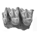
Three new fossiliferous localities, two of karstic origin, Castelnou 3 and Font Estramar, respectively Late Upper Miocene and Lower Pliocene, and one of lacustrine origin, Thuir, Lower Pliocene, add data about the transition between Miocene and Pliocene faunas of rodents in southern France. An unexpected association of taxa was present in the late Upper Miocene, including between others, Myocricetodon, Hispanomys, Ruscinomys, Cricetus barrierei, Promimomys and a new species of Stephanomys, S. dubari nov. sp. Myocricetodon is still known in the Lower Pliocene. It is shown that the large field-mice known since the Late Upper Miocene belong to two different lineages, on one side, A. jeanteti, on the other side, A. gudrunae followed by A. gorafensis. Biochronological and biogeographical implications are discussed.
PV article infos
Published in Vol. 20, Fasc. 4 (1991)
|
PDF
|

|
Insectivores pliocènes du Sud de la France (Languedoc-Roussillon) et du Nord-Est de l'Espagne.
Published online: 31/10/1986
Keywords:
Biostratigraphy; Insectivora; Languedoc; Pliocene; Spain; Systematics
https://doi.org/10.18563/pv.16.3.145-171
Abstract
The first lists of Insectivores (Erinaceidae, Talpidae and Soricidae) from the Pliocene beds of Southern France and North-East Spain are given in this paper. The material from twelve localities is studied. These localities are geographically situated in Languedoc (Celleneuve, Vendargues, Nîmes, Sète, Balaruc 2 and Seynes), in Roussillon (Terrats, Serrats-d'en-Vacquer, Château d'eau and Mont-Hélène) and in North-East Spain (Layna, Medas Islands and Puebla de Valverde). These faunas correspond to the Early, Middle and Late Pliocene. 1 to 8 taxa are identified in these localities and 14 specific taxa are presently listed for this period in this area. Two new specific taxa are described as Galerix depereti nov. sp. from all the Early Pliocene localities in the North-Pyrenean area and as Desmanella gardiolensis nov. sp. from Balaruc 2. For this small mammals, two faunal assemblages are recognized. The first one is dated from the Early Pliocene (F 1, 2 and 3 zones in Aguilar et Michaux) and is characterized by Galerix depereti and rare and little diversified Soricids. The second one is Late Pliocene in age (zones G 2 and G 3). The fossils of the genus Talpa are relatively abundant and the Soricids are diversified and very abundant. The Middle Pliocene (zone G 1) is a transitional period. ln these faunas, most of the insectivore genera are known from the European Late Miocene beds (8 on 10). This fact demonstrates a relative continuity between the invectivore faunas from the Late Miocene to the Early Pliocene. In conclusion, somme paleoecological considerations are suggested.
PV article infos
Published in Vol. 16, Fasc. 3 (1986)
|
PDF
|

|
Schmelzmikrostruktur in den inzisiven alt-und neuweltlicher histricognather nagetiere
Published online: 15/12/1992
Keywords:
Africa; Caviomorpha; Ctenodactyloidea; Deseadan; Enamel microstructure; Hunter-Schreger bands; Hystricognathi; Incisors; Ischyromyoidea; multiserial; Paleobiogeography; pauciserial; Phiomorpha; Rodentia; South America
https://doi.org/10.18563/pv.21.ext.1-168
Abstract
Enamel microstructure in the incisors of Old- and New World hystricognath rodents:
The incisor enamel microstructure in more than 100 genera of fossil and Recent hystricognath and sciurognath rodents was studied. A multiserial schmelzmuster is present in the Hystricognathi, the Ctenodactylidae, advanced Chapattimyidae, and in Pedetes. A redefinition of pauciserial and multiserial HSB is given that makes the two enamel types unambiguously distinguishable which apparently represent well defined evolutionary levels. In the pauciserial Schmelzmuster the IPM is thicker than in the multiserial one. In pauciserial HSB the IPM always surrounds each prism, and the crystallites of the IPM run parallel to prism direction; transition zones between HSB are lacking; the inclination of the HSB is normally very low and the prism cross sections are not flattened but somewhat irregular. The number of prisms per HSB is no good distinctive character for pauciserial and multiserial HSB, since there exists a wide overlap. The pauciserial schmelzmuster is primitive, the multiseiial derived because: 1. the pauciseiial schmelzmuster appears earlier in the fossil record in the most primitive rodents (Paramyids s.l. and Ctenodactyloids); 2. the Eocene Ctenodactyloidea show pauciserial HSB but the Oligocene and younger ones are characterized by multiserial HSB; 3. in the outgroup comparison, the Eurymylidae (Mixodontia) show pauciserial HSB; 4. biomechanically, multiserial HSB strenghten the enamel better than pauciserial HSB, since their IPM runs nearly always in an angle of 45° or more to the prisms.
In multiseríal HSB three subtypes can be distinguished which are differentiated by the IPM orientation. Primitive is a (rarely strict) parallel or acute angular, anastomozing IPM, and derived is an interrow sheet-like ("plattenartige") IPM. This evolutionary polarity is indicated by enamel evolution in the Ctenodactylidae which show an acute angular IPM in the Oligocene and a rectangular interrow sheet-like IPM since the Miocene. Among the Caviomorpha a rectangular interrow sheet-like IPM is restricted to the Octodontoidea; therefore they must be considered derived in terms of their enamel structure. The first multiserial HSB in rodent incisors appear in phiomyids or chapatrimyids from the Upper Eocene of Algeria. The IPM is acute angular and anastomozing. The worldwide next younger multiserial HSB are found in Lower Oligocene phiomyids of Fayum, Egypt There already a rectangular interrow sheet like IPM is present (in Metaphiomys) besides the acute angular anastomozing IPM.
The first Caviomorpha from the Deseadan (Oligocene-Miocene) likewise show already acute angular anastomozing IPM (e.g. Scozamys) and rectangular interrow sheet-like IPM (Platypittamys). Therefore the first Caviomorpha cannot be positioned close to a transition from pauciserial to multiserial HSB. In none of the potential caviomorph ancestors from southern North America multiserial HSB or transitional stage between pauciserial and multiserial HSB could be found. The similarities between the enamel types of the Fayum rodents and the rodents from the Deseadan of South America make a derivation of the Caviomorpha from Paleogene North African phiomorph rodents or their direct ancestors most probable. This supports at the same time a descent of the platyrrhine Primates from North African anthropoids.
PV article infos
Published in Vol. 21, Ext (1992)
|
PDF
|

|
Fossil snakes from the Palaeocene of Sao José de Itaborai, Brazil.Part 1 Madtsoiidae, Aniliidae.
Published online: 15/12/1998
Keywords:
Aniliidae s.l.; Brazil; Coniophis; Hoffstetterella; Madtsoia; Madtsoiidae; middle Palaeocene; New taxa; Snakes
Abstract
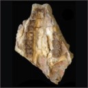
The middle Palaeocene of São José de Itaboraí (State of Rio de Janeiro, Brazil) has yielded a very rich and diverse snake fauna which includes Madtsoiidae, Aniliidae s.l., Boidae, Tropidophiidae s.l., Booidea incertae sedis, and Russellophiidae. The present article (part I) deals with Madtsoiidae and Aniliidae s.l. Madtsoiidae are represented by many vertebrae and a few skull bones. They comprise one new species assigned to the genus Madtsoia (M. camposi sp. nov.). However, the definition of the genus Madtsoia is unsatisfactory and the generic allocation might be provisional. A few elements, vertebrae only, belong to the Aniliidae s. l. Two taxa are referred to this latter group: Coniophis cf. C. precedens and Hoffstetterella brasiliensis gen. et sp. nov. The forthcoming part II will deal with Boidae.
PV article infos
Published in Vol. 27, Fasc. 3-4 (1998)
|
PDF
|

|
Rongeurs muroidés du Néogène supérieur d'Afghanistan, évolution, biogéographie, corrélations
Published online: 30/09/1981
Keywords:
Afghanistan; Muroidea; Neogene
https://doi.org/10.18563/pv.11.4.133-179
Abstract
The rodent faunas of five afghan localities found in 1976 and 1977 (Sherullah, Ghazgay, Pul-e Charkhi, Dawrankhel 14 and 15) are studied.
The rodents (Muridae, Cricetidae and Rhizomyidae) represent 8 genera and 10 species. The detailed description of the 2 new genera and 7 species diagnosed in 1979 is given. An other species is created : Pseudomeriones crapouilloti n. sp. These faunas precise the origin and diversification of Muridae and Cricetidae. A phyletic lineage known in Afghanistan is represented in East Africa by a ramus or a collateral lineage. The five localities are dated from Lower Turolian to Ruscinian. They constitute the frame of a chronologie scale for the Upper Continental Neogene of Afghanistan.
The study of afghan material brings new data to the biogeography of Old Word's rodents during the Upper Neogene; from Pakistan to Europe and Africa, a rather warm and damp province would have existed till Upper Miocene; after which (in the mio-pliocene epoch) it would have divided into 3 parts, by aridification of the central area.
PV article infos
Published in Vol. 11, Fasc. 4 (1981)
|
PDF
|

|
Nouveaux Mammifères Eocènes du Sahara Occidental
Published online: 01/11/1979
Keywords:
Eocene; Mammals; Occidental Sahara
https://doi.org/10.18563/pv.9.3.83-115
Abstract
The fossil mammals collected from the Eocene of Hammada du Dra (northwest Sahara. Algeria) and two fragmentary teeth from the Lutetian of M'Bodione Dadere (Senegal) are described.
The fossils from the northwest Sahara come from a lacustrian deposit dated by charophytes (Raskyella aff. pecki, Raskyella n. sp.. Maedleriella lavocati, Maedleriella sp. et ? Peckichara sp.) as Middle Eocene or perhaps Lower Eocene (Gevin, Feist and Mongereau, 1974). Several hyracoids (3 or 4) identified from this formation extends the age of the family Pliohyracidae Osborn in Africa. Three forms appear to belong in the genera Megalohyrax, Titanohyrax and perhaps Bunohyrax which have been know until now only from the lower Oligocene of the Fayum (M. gevini n. sp. ; T. mongereaui n. sp.. ? Bunohyrax or Megalohyrax indet.). Another hyracoidof small size is referred to a new genus, Microhyrax (M. lavocati n. sp.).
Helioseus insolitus n. g. n. sp. is described without ordinal assignment. Azibius (Sudre, 1975) which has been the subject of questions and interpretations is reviewed.
Only one tooth from the Lutetian of M'Bodione Dadere is complete enough to interpret. lt probably belongs to a condylarth and demonstrates for the first time, the presence of the order in Africa. The second tooth is too fragmentary for comment.
In conclusion., the paleobiogeographic role of Africa at the end of the cretaceous and the beginning of the Cenozoic is discussed.
PV article infos
Published in Vol. 09, Fasc. 3 (1979)
|
PDF
|
|
New Late Miocene plecotine bats (Chiroptera, Vespertilionidae: Plecotini) from Gritsev, Ukraine
Published online: 07/03/2019
Keywords:
Barbastella; bats; late Neogene; Mammalia; Plecotus
https://doi.org/10.18563/pv.42.1.e2
Abstract
The Late Miocene site of Gritsev (MN 9, Ukraine) has yielded a very rich bat fauna, the remains of which are well preserved. Compared to other Neogene bat assemblages of Europe, the Gritsev bat community is unique in preserving plecotine bats, which are rare from Neogene sites. Some peculiar and new bat species, including a large plecotin Otonycteris, already were described from the Gritsev mammal site. Here we report new records of small plecotin bats from Gritsev, including a new taxon, Barbastella maxima nov. sp. This is the earliest reliable fossil record of this genus and it differs from more recent species of Barbastella in being considerably larger. The evolutionary patterns in the odontology within the tribe Plecotini, supported by biostratigraphical distribution of fossil records of Plecotus are discussed. The morphological peculiarities of the new fossils of plecotine bats from Gritsev are discussed in connection with its possible taxonomical affinity.
PV article infos
Published in Vol 42-1 (2019)
|
PDF
|

|
Un giraffidae dans le pliocène de Montpellier ?
Published online: 31/10/1986
Keywords:
Artiodactyla; France; Giraffidae; Mammalia; Montpellier; Ruscinian
https://doi.org/10.18563/pv.16.3.185-189
Abstract
An upper giraffid premolar without any indication about its origin is preserved at the Montpellier University among numerous fossils from the ruscinian formation of Montpellier. It can be related to Samotherium, of the Upper Miocene in Eastern Europe, North Africa and Asia, or more probably to Bramatherium or Hydaspitherium of the Pliocene of South East Asia. The sedimentological study of the matrix shows a calcareous background, which may indicate that this tooth does not come from the Montpellier formation.
PV article infos
Published in Vol. 16, Fasc. 3 (1986)
|
PDF
|

|
Une nouvelle espèce de Steneosaurus (Thalattosuchia, Teleosauridae) dans le Callovien du Poitou (France) et la systématique des Steneosaurus longirostres du Jurassique moyen d'Europe Occidentale.
Published online: 15/09/1998
Keywords:
middle Jurassic; nov. sp.; phylogenetic relationships; skulls; Steneosaurus pictaviensis; Systematics; thalattosuchian crocodile
Abstract
The study of all the available skulls allows us to review the systematic relationships of the longirostrine Steneosaurus from the Middle Jurassic of western Europe. Up to now, Aalenian and Bajocian deposits have not yielded any significant Steneosaurus remain. In the Bathonian, the only valid longirostrine species, S. megistorhynchus, is known in the Britain-Normandy Basin, the Poitou and the Lorraine. In the Callovian, most of the longirostrine Steneosaurus remains can be attributed to the species S. leedsi. Nevertheless, some remains from the Middle Callovian of Poitou (France) show important differences with S. leedsi. A new Steneosaurus species, only known in Poitou, is created and named S. pictaviensis. The specific characters are carried by the skull (preorbital pit well marked, orbit and ptetygoid fossae shapes), by the mandible (symphysis shape) and by the teeth (ornamentation). S. megistorhynchus is probably situated near the stem of the Callovian species but remains from the Bathonian and Lower Callovian are very scarce and it is very difficult to precise the phylogenetic relationships between the longirostrine species of the Middle Jurassic.
PV article infos
Published in Vol. 27, Fasc. 1-2 (1998)
|
PDF
|
|
Two new scyliorhinid shark species (Elasmobranchii, Carcharhiniformes, Scyliorhinidae), from the Sülstorf Beds (Chattian, Late Oligocene) of the southeastern North Sea Basin, northern Germany.
Published online: 30/04/2014
Keywords:
Chattian; Elasmobranchii; North Sea Basin; Scyliorhinidae; Scyliorhinus
https://doi.org/10.18563/pv.38.1.e1
Abstract
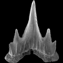
Based on isolated teeth two new scyliorhinid shark species, Scyliorhinus biformis nov. sp. and Scyliorhinus suelstorfensis nov. sp., are described from the Sülstorf Beds, early-middle Chattian, of Mecklenburg, northeastern Germany. They form part of a speciose assemblage of necto-benthic sharks and batoids which populated the warm-temperate to subtropical upper shelf sea of the south-eastern North Sea Basin.
PV article infos
Published in Vol.38-1 (2014)
|
PDF
|

|
Rongeurs de l'Oligocène moyen provenant de nouvelles fouilles dans les phosphorites du Quercy
Published online: 15/09/1969
Keywords:
Oligocene; Quercy Phosphorites; Rodents; Theridomys
https://doi.org/10.18563/pv.2.5.209-239
Abstract
A recent campaign of excavations (1965-68) undertaken by the Laboratoire de Paléontologie of Montpellier in pockets of the Quercy phosphorites, has permitted the dating of several localities thanks to the analysis of their micromammalian fauna.
The rodents of localities belonging to the middle Oligocene (La Sauvetat zone) are the object of this study. This rodent fauna has been compared to the forms coming from three stratified localities belonging to the biochronologic zone of La Sauvetat : Balm, Montalban and Lovagny.
The abundance of material, notably of theridomyids and cricetids, permitted a summary statistical study to be effected and gave some precise details on the variability of the species (Theridomys varians in particular). Other details were provided on certain groups, notably on the upper teeth of Scíuromys cayluxi and on the milk teeth of theridomyids. Some specimens of «Sciurus» sp. and of Plesispermophilus angustidens were collected. By this means, it has been possible to establish, for the first time, a precise upper limit to the epoch of their appearance in Europe.
PV article infos
Published in Vol. 02, Fasc. 5 (1969)
|
PDF
|

|
Eggshell microstructure and porosity of the Nicobar scrubfowl (Megapodius Nicobariensis, great Nicobar island, India)
Published online: 16/12/2008
Keywords:
conductance porosity; eggshell microstructure; incubation conditions; Megapodes
https://doi.org/10.18563/pv.36.1-4.75-88
Abstract
The eggshell of Nicobar scrubfowl (Megapodius nicobariensis) is described for the first time. Its egg porosity is calculated and discussed with data from several taxa (another megapode, some extant and fossil reptiles including a titanosaur group) in order to compare incubation types with eggshell structure. Eggshell microstructure reflects first phylogenetic traits and does not seem to have developed major adaptative features due to the incubation conditions, except for the pore canals.
PV article infos
Published in Vol. 36, Fasc. 1-4 (2008)
|
PDF
|
|
A new species of Propalaeotherium (Palaeotheriidae, Perissodactyla, Mammalia) from the Middle Eocene locality of Aumelas (Hérault, France).
Published online: 24/05/2016
Keywords:
Eocene; new species; Palaeotheriidae; Propalaeotherium
https://doi.org/10.18563/pv.40.2.e1
Abstract
A new Propalaeotherium species, clearly distinct from the genus Eurohippus, is described. It is characterized by having a similar size as P. voigti from the German Geiseltal localities (MP 11 to MP 13 reference-level), but differs in several features suggesting a slighty more derived morphology. It presents indeed less brachyodont crowns with less prominent and less elevated cingula, slightly larger relative surface of premolars, and a more marked metaconid splitting on cheek teeth. This new species is unknown from other European localities except the nearby Saint-Martin de Londres locality which has been considered older than the MP 13 level.
PV article infos
Published in Vol.40-2 (2016)
|
PDF
S.I. Data
|
|
New remains of the giant bird Gargantuavis philoinos from the Late Cretaceous of Provence (south-eastern France)
Published online: 27/08/2015
Keywords:
Aves; Gargantuavis; Late Cretaceous; Pelvis; South-eastern France
https://doi.org/10.18563/pv.39.2.e3
Abstract
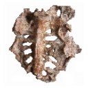
Two incomplete pelves of the giant bird Gargantuavis philoinos are described from Late Cretaceous deposits at Fox-Amphoux (Var, south-eastern France). They consist of synsacra with attached parts of the ilia. One of them has undergone considerable dorsoventral compression, which makes it very similar in appearance to the holotype pelvis of Gargantuavis philoinos from Campagne-sur-Aude (Aude, southern France). The second specimen has suffered some lateral distortion but is uncrushed dorsoventrally. Because of this, its avians characters (including an arched synsacrum and widespread pneumatisation) are especially clear. These new specimens confirm the avian nature of Gargantuavis and reveal new details about its pelvic anatomy, but provide little new evidence about its systematic position within Aves. The geographical distribution and general rarity of Gargantuavis are discussed.
PV article infos
Published in Vol.39-2 (2015)
|
PDF
|

|
Rongeurs (Mammalia, Rodentia) du Miocène de Beni-Mellal
Published online: 15/02/1977
Keywords:
Morocco; Neogene
https://doi.org/10.18563/pv.7.4.91-125
Abstract
The rodent fauna of Beni-Mellal is characterized by the abundance of ctenodactylids and cricetids. The latter are represented by four distinct species, among which a new form. Dakkamys zaiani nov. gen., nov. sp. is described. A detailed morphological analysis shows that, contrary to that which had been established before, « Cricetodon ›› atlasi Lavocat, 1961, is not closely related to any European form known; this species is attributed, in consequence, to the new genus Mellalomys. A simple biometric analysis has shown that the genus Myocricetodon Lavocat, 1952, is represented in this locality by two distinct species. The systematic homogeneity of the Beni-Mellal cricetids is also demonstrated: they can, as a matter of fact, all be referred to the subfamily Myocricetodontinae. The definition of this subfamily is completed. The sciurids and glirids are also reviewed in the light of new systematic and biogeographic information established ln Europe. A new species of Atlantoxevus from the early Pleistocene of Morocco, A. huvelini nov. sp., is described. It is probably the descendant of A. tadlae from Beni-Mellal. Biogeographic analysis leads one to consider this fauna as the result of geographic isolation in the Maghreb since the late Oligocene or the early Miocene. In particular no direct European influence can be discerned. Stratigraphic considerations resulting from the discovery of new localities in North Africa lead to the confirmation of the ante-Vallesian age of this fauna and to its parallelism with the faunas of La Grive in Western Europe and Fort Ternan in East Africa. The peculiar geologic nature of this locality is discussed.
PV article infos
Published in Vol. 07, Fasc. 4 (1977)
|
PDF
|

|
Nouvelles données sur les mammifères du Thanétien et de l'Yprésien du bassin d'Ouarzazate (Maroc) et leur contexte stratigraphique.
Published online: 15/12/1998
Keywords:
early Paleogene; magnetostratigraphy; Mammals; Morocco; North Africa; Ouarzazatz basin; Systematics
Abstract
New faunal and stratigraphical data on the vertebrates localities from the early Paleogene of the Ouarzazate Basin (Adrar Mgorn 1, Adrar Mgorn 1 bis et N'Tagourt 2), Morocco, are presented. A magnetostratigraphical study, the first for such early Paleogene Arabo-African mammal localities, and the discovery of probable remains of the nannofossil Discoaster support the Thanetian age of the Adrar Mgorn 1 site. The magnetostratigraphy suggests a slightly later age than was thought for the Paleogene formations of the local series of Tinerhir and for the vertebrate localities: late or latest Thanetian for Adrar Mgorn 1 and Adrar Mgorn 1 bis, middle Ypresian for N'Tagourt 2. It also indicates a lower position of the KT boundary in the series. Two tons of matrix recovered in the vertebrate sites have vielded new data on the micromammals. A damaged lower molar from N'Tagourt 2 is referable to Khamsaconus bulbosus and supports the proboscidean affinities of this species and especially possible relationships with bunolophodont taxa such as elephantiforms. A lower molar from Adrar Mgorn 1 bis belongs to a new form which can be identified as a plesiadapiform or an euprimate close to Altiatlasius koulchii though significantly larger. A new material from Adrar Mgorn 1 illustrates a new dilambdodont adapisoriculid species which is referable to Garatherium : ?Garatherium todrae n. sp. Another species referred to Garatherium is known in the locality (?Garatherium n. sp.). Garatherium is a new lineage from the Ouarzazate basin which crosses the Paleocene-Eocene boundary together with Palaeoryctes, Didelphodontinae gen. and sp. 2, Todralestes, and Afrodon, and it is the first Paleocene-Eocene lineage identified outside of this basin (Garatheríum is based on a species from El Kohol, Algeria). Among the Paleocene-Eocene lineages from the Ouarzazate basin, it should be also mentioned a new possible carnassial form (carnivoran or creodont; Adrar Mgorn 1), and an upper molar of Cimolestes cf. incisus (Adrar Mgorn 1 bis). The upper molar THR 168 previously reported as from an indeterminate didelphodontine is here identified as the M1/ of Afrodon chleuhi. The micromammal faunas from the Ouarzazate basin are positioned in the global chronological framework of the mammal localities from the Paleogene of the Arabo-African domain.
PV article infos
Published in Vol. 27, Fasc. 3-4 (1998)
|
PDF
|

|
Données nouvelles sur le genre Stehlinia (Vespertilionoidea, Chiroptera) du Paléocène d'Europe
Published online: 01/12/1974
Keywords:
Chiroptera; Palaeocene; Vespertilionoidea
https://doi.org/10.18563/pv.6.3-4.253-272
Abstract
Cet article présente une étude détaillée du genre Stehlinia, un chiroptère du Paléogène d'Europe, basé sur un matériel abondant issu notamment du gisement d'Escamps (Quercy). L'analyse révèle que Stehlinia possède un mélange de caractères primitifs (comme une denture tribosphénique complète et des prémaxillaires soudés) et évolués (notamment dans le squelette post-crânien), le rapprochant des vespertilionoïdes actuels, en particulier des Kerivoula.
PV article infos
Published in Vol. 06, Fasc. 3-4 (1975)
|
PDF
|

|
The Gliridae (Mammalia) from the oligocene (MP24) of Gröben 3 in the folded molasse of southern Germany
Published online: 28/12/2001
Keywords:
Biostratigraphy; Cyrena Beds; folded molasse; Germany; Gliridae; level MP 24; Mammals; Oligocene; Palaeoecology
Abstract
This study describes four taxa of Gliridae from the Oligocene mammal locality Gröben 3: Gliravus tenuis BAI-ILO, 1975, Bransatoglis micio (MISONNE, 1957), B. planus (BAHLO, 1975) and B. heissigi n. sp. Gliravus tenuis from Gröben 3 is somewhat more advanced than the type population found in Heimersheim. This confirms previous research suggesting that Gröben 3 should be dated earlier than Heimersheim (MP 24). The first documented occurrence of B. mício around level MP 24 was found in Gröben 3. An abundance of tooth material from B. planus in Gröben 3 makes it possible, for the first time, to observe evolutionary stages within this species from MP 21 until MP 28. B. heissigi n. sp. is restricted to level MP 24. This species is located between B. mísonnei (MP 20 - 23) and Microdyromys praemurinus (MP 25 - 28). Within the lineage Bransatoglis bahloi - B. misonnei - B. heissigi, a decrease in size is noticeable.
PV article infos
Published in Vol. 30, Fasc. 3-4 (2001)
|
PDF
|

|
The skull of Arsinoitherium (Mammalia, Embrithopoda) and the higher order interrelationships of ungulates
Published online: 17/12/1992
Keywords:
Arsinoitherium; PHYLOGENY; Skull; Ungulate
https://doi.org/10.18563/pv.22.1.1-43
Abstract
Detailed anatomical description of arsinoithere cranial remains from the Lower Oligocene, Fayum Depression, Egypt, provides the basic data for a systematic investigation. All cranial and some postcranial features are assessed from a phylogenetic standpoint. Several soft tissue characters are then added to a cladistic analysis based on 54 derived ungulate morphological characters. The resulting phylogenetic hypothesis implies that perissodactyls, sirenians, proboscideans and arsinoitheres constitute a monophyletic unit (5 synapomorphies). However, increasing the tree length by 3 steps reveals a closer association between hyraxes and perissodactyls. Nevertheless, 13 synapomorphies link proboscideans, sirenians and arsinoitheres to the exclusion of all other ungulates. Form of the sphenopalatine and ethmoid foramina, recurved posttympanic process, absence of a fenestra rotundum in the petrosal, vestigial paroccipital process of the exoccipital and the highly unusual absence of a hypoglossal foramen in the skull, imply a robust sister-group relationship between arsinoitheres and proboscideans. In this analysis artiodactyls share only one derived character with all other ungulates studied. Monophyly of Ungulata, including Artiodactyla, is therefore only weakly supported. It is argued that pedal anatomy of hyraxes is non-homologous with that of Tethytheria. Arsinoitherium should now be classified within Tethytheria, sharing a sister-group relationship with Proboscidea. Hyraxes are excluded, thus refuting the concept of Paenungulata. However, monophyly of the wider concept, Pantomesaxonia, containing hyraxes, perissodactyls, sirenians, proboscideans and now, arsinoitheres, is supported by this study.
PV article infos
Published in Vol. 22, Fasc. 1 (1992)
|
PDF
|

|
Contributions à l'étude des micromammifères du gisement Miocène supérieur de Montredon (Hérault). 1- Le gisement
Published online: 30/06/1982
Keywords:
Hérault; Late Miocene; Micromammals; Montredon
https://doi.org/10.18563/pv.12.3.75-79
Abstract
La localité fossilifère du Puech de Montredon, désignée plus communément sous le nom de Montredon, est située sur la commune de Montouliers (Hérault) à quelques 300 mètres de la limite avec le département de l'Aude. Elle a été découverte en 1845 par Narbonne, Directeur des Mines de La Caunette, et de très nombreux restes de vertébrés continentaux y ont été extraits. La plus ancienne mention de ce gisement dans la littérature semble être celle de Lartet (1859) qui signale que "M. Jourdan, de Lyon, a constaté à Montredon, près de Bize (Aude), l'association des restes de Dinotherium avec l'Hipparion".
PV article infos
Published in Vol. 12, Fasc. 3 (1982)
|
PDF
|
