
|
First evidence of an early Miocene marine teleostean fish fauna (otoliths) from la Paillade.(Montpellier,France)
Published online: 15/06/1999
Keywords:
Aquitanian; Biostratigraphy; La Paillade; marine deposits; Miocene; otoliths; Palaeoecology; Palaeogeography; Southern France; Teleostei
Abstract
A fossil fish fauna, based on 5533 otoliths, from the La Paillade locality at Montpellier is described and figured. The otolith-bearing marls correlate to mammal zone MN l (Aguilar, 1982), and thus represent the earliest Miocene. The fish fauna consists of 30 taxa belonging to 20 families. Two species are new: Dussumieria sittigi and Liza gaudanti. The predominant faunal element is the Lesueurigobius vicínalis-species complex, composing 73% of all investigated otoliths. The palaeoecological analysis reveals a marine to euryhaline fish fauna living under tropical to subtropical conditions in the transition zone littoral - sublittoral. Water depth probably was more than 10 m. The scarcity of pelagic físhes suggests that the habitat was either a sheltered bay and/or far away from the open sea. Furthermore, some genera represented in the La Paillade fish fauna presently live exclusively in the Indopacific realm. Their presence strongly supports a broad connection between the Indian Ocean, the Mediterranean, and the Paratethys Seas during the Early Miocene (Aquitanian). From a palaeobiogeographical point of view, faunal relationships were found between the La Paillade fish fauna and both the Paratethys fish fauna and the fish fauna from the deposits in the Upper Rhinegraben and the Mayence and Hanau Basins (Germany).
PV article infos
Published in Vol. 28, Fasc. 1 (1999)
|
PDF
|

|
Contributions à l'étude des micromammifères du gisement Miocène supérieur de Montredon (Hérault). 3- Les insectivores
Published online: 30/06/1982
Keywords:
Hérault; Insectivora; Late Miocene; Micromammals; Montredon
https://doi.org/10.18563/pv.12.3.119-131
Abstract
This paper presents a preliminary list of insectivores from the Vallesian beds at Montredon (France). The associated rodent fauna has established a Vallesian age for the fauna. Eleven species belonging to the Soricidae, Talpidae, Erinaceidae, and Dimylidae are identified of which four only are referred with certainty to forms already named.
PV article infos
Published in Vol. 12, Fasc. 3 (1982)
|
PDF
|

|
Anatomie du membre antérieur chez un chiroptère Molossidé (Tadarida sp.) du Stampien de Cereste (Alpes-de-Haute-Provence).
Published online: 01/01/1971
Keywords:
Chiroptera; Molossidae; Oligocene
https://doi.org/10.18563/pv.4.1.1-38
Abstract
The present study describes in detail the anterior limb osteology of a molossid chiropteran of the genus Tadarida, from Céreste, a Stampian locality in the Apt-Forcalquier Oligocene basin already known for its fishes, plants and insects.
A comparision with the Miocene forms T. srehlini from Saint-Géraud localities and T. sp. from Württemberg, also with the recent forms T. teniotis and Eumops perotis, does not show any clear morphological differences between the Tertiary and Recent Tadarida, indicating a rather noticeable anatomical stability, not exceptionnal indeed among Chiropterans. The Céreste fossil exhibits however slightly primitive wing proportions if compared to the Saint Gérand Aquitanian species.
Several remarks deal with the peculiar relationships between the ecology of the molossids and their kind of fossilisation, frequently associated with sedimentary facies of the lacustrine type.
PV article infos
Published in Vol. 04, Fasc. 1 (1971)
|
PDF
|
|
New datation of the Tafna Basin (Algeria): A combination between biochronological and magnetostratigraphical data
Published online: 11/03/2015
Keywords:
correlations; Late Miocene; North Africa; Rodentia
https://doi.org/10.18563/pv.39.1.e1
Abstract
The Tafna Basin corresponds to the lowlands, which are located in front of Tessala and Traras ranges, below the Tlemcen mountains, Algeria. This basin displays a complete sedimentary cycle dominated by lagoonal-fluvial and marine deposits. The continental formations located at the base of these deposits are mainly composed of alternating sandstones and clays. An early late Miocene age has been previously attributed to them, based on direct correlations with marine deposits. Search for micromammal fossils led to the discovery of three different rodent species from a single level of the Djebel Guetaf section, located at the bottom of these deposits. The rodent assemblage indicates a late Miocene age. Combined magnetostratigraphical and biostratigraphical investigations were carried out to provide a more accurate age control of these continental deposits. Sixty-four oriented rock samples were collected for a magnetostratigraphic study along a 92 meters thick section including the fossiliferous layer. Rock magnetic investigations indicate the presence of both high and low coercivity minerals. Specimens subjected to progressive thermal demagnetization procedures show that the samples exhibit a high temperature magnetization component and display a normal polarity. Based on biostratigraphic constraints, the Guetaf section is correlated with Chron C4An, indicating an age ranging from
9.1 to 8.7 Myr.
PV article infos
Published in Vol.39-1 (2015)
|
PDF
|

|
The eosimiid and amphipithecid primates (Anthropoidea) from the Oligocene of the Bugti hills (Balochistan, Pakistan): new insight into early higher primate evolution in South Asia.
Published online: 15/10/2006
Keywords:
Amphipithecidae; anthropoid phylogney; Bugti Hills; Early Oligocene; Eosimiidae; Pakistan
https://doi.org/10.18563/pv.34.e15
Abstract
Eosimiid and amphipithecid primates document a long and significant history of primate evolution throughout the Eocene in Southeast Asia. Despite the absence of a comprehensive post-Eocene fossil record, it was generally hypothesized that both families left no descendant in Asia. Recently, two new small-bodied taxa, Bugtipithecus and Phileosimias, have been recovered in early Oligocene coastal deposits from the Bugti Hills (Balochistan, central Pakistan) and referred to the families Amphipithecidae and Eosimiidae, respectively, on the basis of dental fossil remains. In this paper, we provide more exhaustive description, comparison, and discussion of these taxa. As for tarsiid and sivaladapid primates, the persistence of eosimiids and amphipithecids into the Oligocene clearly demonstrates that low latitudes of South Asia provided a continuous access to tropical refugia during the climatic deterioration characterizing the late Eocene-early Oligocene interval, which was seemingly lethal for primate communities elsewhere across the Holarctic continents. As a contribution to the ongoing phylogenetic debates regarding the position of eosimiids and amphipithecids on the primate family tree, we have performed a cladistic analysis in a high-level primate systematic context in order to assess the position and the role of these new taxa in that phylogenetic issue. Our results support the view according to which eosimiids and amphipithecids (and by extension Phileosimias and Bugtipithecus, respectively) are stem anthropoids. These fossils from Pakistan document an unsuspected Oligocene phase of the evolutionary history of anthropoid primates in southern Asia, which clearly enhances the extent of the anthropoid radiation in this province during the Paleogene. Several phylogenetic and paleobiogeographic aspects are discussed, notably the intra- and inter-relationships between Paleogene Asian and Afro-Arabian anthropoids, and the resulting potential dispersal models between both land-masses during the Paleogene.
PV article infos
Published in Vol. 34, Fasc. 1-2 (2006)
|
PDF
|
|
Nouvelles données sur les Ichnites de dinosaures d'El Bayadh (Crétacé Inférieur, Algérie)
Published online: 16/12/2008
Keywords:
Algeria; Brezina; El Bayadh; Ichnites; Lower Cretaceous; Sauropoids; Theropoids
https://doi.org/10.18563/pv.36.1-4.7-35
Abstract
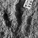
Evidence of 350 Lower Cretaceous Dinosaur footprints is pointed out in El Bayadh area. Their preliminary study allow to distinguish four trackway assemblages which reveal vertebrate bipedal presence forms of tri-and tetradactylous Dinosauroïds (Assemblages 1-3) and quadrupidal Sauropoïd (Assemblage 4).
The analysis of their footprint biometric features will attribute the quadrupidal Sauropoïd form to Brontopodus ichnogenus which is weIl known in the Jurassic and Cretaceous periods. In retum and despite their age, the dinosauroïd forms were approached, temporarily, to Grallator and Eubrontes types.
The occurrence of the dinosaur traces (Theropoïd and Sauropoïd) constitutes, in the Lower Cretaceous, an important first step of the knowlege of the marshy Reptilian fauna which takes over, from the begining of the Secondary Era, a wide paleogeographie area on the Southem Tethyan margin.
PV article infos
Published in Vol. 36, Fasc. 1-4 (2008)
|
PDF
|

|
Mammals and stratigraphy of the continental mammal-bearing Quarternary of South America
Published online: 16/12/1984
Keywords:
Geochronology; Mammalia; Quaternary; South America; Stratigraphy
https://doi.org/10.18563/pv.14.ext
Abstract
Previous chronological arrangements of South American Quaternary land mammal faunas are appraised on the basis of current geological and paleontological data. Three South American late Pliocene-Pleistocene land mammal ages are conventionally recognized, from oldest to youngest, the Uquian, Ensenadan, and Lujanian ; all are defined on Argentine faunas.
The Uquian is based fundamentally and historically on the fauna from the Uquía Formation in Jujuy Province, northwestern Argentina. Important known formations in Argentina yielding Uquian Age faunas include the sub-surface Puelche Formation (or Puelchense) near the city of Buenos Aires, and the Barranca de Los Lobos and Vorohué Formations between Mar del Plata and Miramar, Buenos Aires Province. A tentative subdivision is propos-ed for the Uquian into three subages based on knowledge of the Mar del Plata-Miramar sequence, from oldest to youngest, the Barrancalobian, Vorohuean, and Sanandresian. In Argentina the Uquian is presently marked by the first known record of Scelidodon, Hydrochoeropsis, Ctenomys, Canidae, Ursidae, Gomphotheriidae, Equidae, Tapiridae, Camelidae, Cervidae, and the last known record of Thylatheridium, Thylophorops, Dankomys, Eumysops, Pithanotomys, Eucoelophorus, Hegetotheriidae, Sparassocynidae, and Microtragulidae.
The Ensenadan Age is based on the fauna from the Ensenada Formation near the city of Ensenada, Buenos Aires Province. In Argentina the Ensenadan is marked by the first known record of Lomaphorus, Neothoracophorus, Plaxhaplous, Cavia, Lyncodon, Lutra, Galera, Smilodon, Dicotyles, Lama, Vicugna, the last known record of Orthomyctera, and the only known record of Brachynasua.
Typícal beds of late Lujanian Age in Argentina consist of fluvial deposits occupying stream channels, and shallow basins, often incised into beds of early Lujanian (i.e. Bonaerian of early workers) and Ensenadan Age. The Lujanian Age is based on a fauna from beds along the Rio Luján, about 65 km west of the city of Buenos Aires, Buenos Aires Province. The Lujanian in Argentina is marked by the first record of Equus, Chlamyphorus, and Holochilus, and the last record of Megatherioidea, Glyptodontoidea, Arctodus (=Arctotherium), Smilodon, Litopterna, Notoungulata, Proboscidea, Equidae, Morenelaphus, and Palaeolama.
These land mammal ages are often difficult to recognize in other South American countries. The compositions of South American Pleistocene faunas vary with the environment. Some taxa were widely distributed in fossil deposits throughout the continent, but their occurrences need not reflect synchroneity. This is a result of changing climates and habitats in time. Consequently, proposed intracontinental correlations need confirmation based on magnetostratigraphy and a radioisotope time scale. Paleontologic characterizations of these land mammal ages (i.e. first and last record, and guide fossils) are useful for much of Argentina, but extensions to most of the other parts of South America are at best tenuous.
The majority of known non-Argentine Pleistocene faunas are believed to be Lujanian in age. Possible non Argentine early Pleistocene (Uquian) faunas include Ayo Ayo and Anzaldo in Bolivia, and Cocha Verde in southern Columbia. A possible middle Pleistocene (Ensenadan or early Lujanian) fauna is the Chichense of Ecuador. Paleomagnetic and radioisotopic date (MacFadden et al., 1983) clearly indicate that the greater part of the Tarija fauna (Bolivia) is Ensenadan in age.
The end of the Pleistocene and beginning of the Holocene in South America is marked by extinction of nearly all large mammalian herbivores and their specialized large predators. Radiocarbon age determinations suggest that large scale extinctions of megafauna occurred between 15,000 and 8,000 yrs. B.P. (years before present).
PV article infos
Published in Vol. 14, Ext (1984)
|
PDF
|
|
Difficulties with the origin of dinosaurs: a comment on the current debate
Published online: 01/07/2020
Keywords:
dinosaur anatomy; dinosaur evolution; Ornithoscelida; palaeobiogeography; Triassic Period
https://doi.org/10.18563/pv.43.1.e3
Abstract
The origin and early evolutionary history of the dinosaurs is a topic that has recently gone through a period of renewed interest and academic debate. For 130 years, one way of classifying the various dinosaur subgroups persisted as the accepted model, with increasing levels of research in the past quarter-century also providing evidence for the hypothesis that dinosaur origination occurred in the Southern Hemisphere, particularly in South America. It is, after all, from within the Late Triassic strata of countries like Argentina and Brazil that we get some of the very best early dinosaur specimens; many of these specimens are the earliest known representatives of some of the major dinosaur subgroups, such as the theropods and sauropodomorphs. However, some recent analyses have brought about a shift in terms of what is currently accepted and what is now disputed regarding the origin of dinosaurs – the Southern Hemisphere origination hypothesis was questioned (although this was based upon observations and not with quantitative analysis techniques), as has the shape of the dinosaur tree. Responses to the new hypothesis were numerous; many further supported a Southern Hemisphere point of origin. Whilst the interrelationships between the major dinosaur clades remains to be resolved, the current data does seem to comprehensively answer the question of where the dinosaurs first originated. However, it is arguable whether the current data that is being used in such palaeobiogeographical analyses is sufficient to provide an answer to the question of where specifically the dinosaur clade first appeared. This short communication urges a degree of caution about the current consensus and what steps may need to be taken to ensure that more meaningful results are produced in the future.
PV article infos
Published in Vol 43-1 (2020)
|
PDF
|

|
A new rodent from Quaternary deposits of the Canary Islands and its relationships with Neogène and recent murids of Europe and Africa.
Published online: 15/12/1988
Keywords:
Canary Islands; Holocene; Island evolution; Muridae; PHYLOGENY; Rodents; Spain
https://doi.org/10.18563/pv.18.4.241-262
Abstract
A peculiar new rodent, Malpaisomys insularis nov. gen., nov. sp., is described from subfossil deposits of the eastern Canary Islands. The species shows some highly specialized skull features although its molars exhibit a mixture of primitive and derived characters among which a partial stephanodonty is most notable. A comparison of the new rodent with several Miocene to Holocene Muridae shows that Malpaisomys possibly shares a common ancestor with Acomys and Uranomys.
PV article infos
Published in Vol. 18, Fasc. 4 (1988)
|
PDF
|

|
Ostéologie de la tête de Richardus excavans Lavocat,1988
Published online: 30/10/1989
Keywords:
Africa; anatomy; Bathyergidae; Miocene; Rodents
https://doi.org/10.18563/pv.19.2.73-80
Abstract
Remarkable association of a small infraorbital foramen, similar to that in recent Heterocephalus, and of a strong muscular print on the dorsal anterior part of the zygomatic plate and on the premaxillary. Several anatomical structures to be compared with those of Heterocephalus suggest relationships with this genus. Richardus supports the ancestrality of the hystricomorph character in the bathyergids
PV article infos
Published in Vol. 19, Fasc. 2 (1989)
|
PDF
|

|
Contributions à l'étude du gisement Miocène supérieur de Montredon (Hérault). Les grands mammifères. 7 - Les proboscidiens Deinotheriidae
Published online: 15/11/1988
Keywords:
allometry; Astaracian; Deinotherium; Montredon; Systematics; taphonomy; Vallesian
https://doi.org/10.18563/pv.18.ext.135-175
Abstract
Some complete tooth rows and about one hundred isolated teeth enabled the identification of the deinothere of the Vallesian site Montredon (Hérault) as Deinotherium giganteum KAUP 1829, mainly by comparisons with the likewise Vallesian sample of the type locality Eppelsheim (Rheinhessen, F.R.G.).
Scatterdiagrams of the teeth show the importance of allometry during the phyletic size increase of the European deinotheres.
Some taphonomic problems of the Montredon deinothere are briefly mentioned.
PV article infos
Published in Vol. 18, Ext (1988)
|
PDF
|

|
Un crane de Chalicothere (Mammalia, Perissodactyla) du Miocène supérieur de Macédoine (Grèce) : remarque sur la phylogénie des Chalicotheiinae
Published online: 14/06/1995
Keywords:
Chalicotheriidae; Cladistics; Greece; Miocene; Perissodactyla; PHYLOGENY
Abstract
The discovery in the Turolian (Late Miocene) of Dytiko 3 (Macedonia, Greece) of a complete skull with mandibles and cervical vertebrae, atlas and epistropheus, is a very important contribution to the knowledge of the subfarnily Chalicotheríinae. After the description, the comparison with other specimens of Miocene chalicotheres permits the revival of the generic name Macrotherium with a new species M. macedonicum. This genus is mainly characterized by a short snout and an inflated cerebral skull. It coexists during the Miocene with Chalicotherium. A cladistic analysis leads to conclusion that the species which has been described from the Early Middle Miocene of Rusinga must be identified as the type-species of a new genus: Butleria.
PV article infos
Published in Vol. 24, Fasc. 1-2 (1995)
|
PDF
|

|
Morphotypes dentaires actuels et fossiles des Chiroptères Vespertilioninés. 1e partie: Etude des morphologies dentaires
Published online: 30/09/1985
Keywords:
bats; Dental morphology; fossils; PHYLOGENY; recent; Systematics
https://doi.org/10.18563/pv.15.2.71-128
Abstract
The classifications of the recent vespertilionine bats were made wihtout taking in account the teeth morphology; this resulted in a reduction of the possibilities of comparison with the available fossils. The generalized use of dental formulae was abusive: this contributed to the admission of artificial genera. These conditions have long delayed the consideration of characters able to frame the phylogeny of the sub-family. In the first part of the study, the teeth morphologies are described and analysed. morphological reference types are established for each upper and lower tooth: they should make an easier elaboration of criteria for the differentiation at generic level. The position of the species in view of these criteria allows one to group them into homogeneous genera, and to appreciate the degree of relationship that the latter have between them. The second part of the study (next publicationà will develop inferences dealing with systematics and phylogeny
PV article infos
Published in Vol. 15, Fasc. 2 (1985)
|
PDF
|

|
Les oiseaux aquatiques (Gaviiformes à Anseriformes) du gisement Aquitanien de Saint-Gerand-le-Puy (Allier, France): Révision systématique.
Published online: 01/11/1984
Keywords:
Aves; Early Miocene; Osteology; Palaeoecology; Systematics
https://doi.org/10.18563/pv.14.2.33-115
Abstract
Six orders of birds adapted to aquatic life are represented among the numerous avifauna of "Saint-Gérand-le-Puy": Gaviiformes, Procellariiformes, Pelecaniformes, Ciconiiformes, Phoenicopteriformes, and Anseriformes. The present study of this avifauna proposes several changes in systematics:- Procellariiformes: Puffinus arvernensis does not belong in Procellariidae but in Diomodeidae, and it is transferred to the fossil genus Plotornis previously described in the Middle Miocene of France. - Pelecaniformes: Phalacrocorax littoralis remains in Phalacrocoracidae; P. míocaenus is different from the modern species, and is transferred to the new genus Nectornis. Empheresula arvernensis, described in the Oligocene deposits of Gannat, seems to be present in Saint-Gérand-le-Puy too. Pelecanus gracilis shows many differences from the modern species, and belongs to the new genus Miopelecanus, - Ciconiiformes: Ardea formosa nom. oblit. is a synonym of Proardeola walkeri. - Anseriformes: a new species closely related to swans is described, and belongs to the fossil genus Cygnopterus, of the Middle Oligocene of Europe; this species is called C. alphonsi. The ecology of each species is suggested by comparison with that of its nearest living relatives, and by study of osteological adaptations.
PV article infos
Published in Vol. 14, Fasc. 2 (1984)
|
PDF
|

|
Les mammifères Montiens de Hainin (Paléocène moyen de Belgique) Part1: Multituberculés.
Published online: 01/11/1979
Keywords:
Belgium; Hainin; Mammals; multituberculates; Paleocene
https://doi.org/10.18563/pv.9.4.117-131
Abstract
The Montian locality of Hainin (Hainaut, Belgium) yielded about twenty teeth of Multituberculates. They are very peculiar forms, showing no affinities, at the generic level, with those hitherto known from North America, Asia and Europe. They are referred to the new taxa Boffius splendidus nov. gen., nov. sp., Hainina belgica nov. gen., nov. sp., and H. godfriauxi nov. gen., nov. sp. They expose some common features, such as the advanced type of first upper molar. possessing at least three complete rows of cusps. Because of this, and also of the upper premolar reduction, Boffius splendidus appears as the most specialized form within the Ptilodontoidea suborder.
Several other characters of Hainina seem to be less advanced, such as the great number of upper premolars and the simple cusp-formula of the first lower molar.
Till now, only H. godfriauxi has been recovered within the Thanetian fauna from Cemay-les-Reims, where it is very poorly documented.
PV article infos
Published in Vol. 09, Fasc. 4 (1979)
|
PDF
|

|
Le genre Plagiolophus (Palaeotheriidae, Perissodactyla, Mammalia): révision systématique, morphologie et histologie dentaires, anatomie crânienne, essai d'interprétation fonctionnelle
Published online: 15/12/2004
Keywords:
New taxa; Paléogène; perissodactyls; skull anatomy; tooth histology
Abstract
The genus Plagiolophus is documented, almost solely in Western Europe, from the middle Eocene up to the mid Oligocene (MP 12 to MP 25), i.e. more than for 15 MY. Seventeen species are now recorded whose two of them are new, P. ringeadei nov. sp. and P. mamertensis nov. sp. Some anatomical variations and the deflection of certain evolutionary trends justify the distinction of three subgenera, Paloplotherium, Fraasiolophus nov. and Plagiolophus s.s. The genus displays a wide range in size and weight (between 10 and 150 kg). The detailed description of the skull of several species is here given for the first time.
Despite important evolutionary drifts during this long time span, the dentition shows a great structural homogeneity, which renders difficult the determination of fragmentary specimens or isolated teeth. It is characterized by a great heterodonty; premolars are little molarized and present a certain regression through time with paradoxically some progress in the molarization. The hypsodonty increases: the first Plagiolophus are hardly less brachyodont than Propalaeotherium, and the last ones are nearly as hypsodont as Merychippus from the early Miocene. The upper molars change from a wide crown pattern, with an open occlusal surface, lightly oblique transverse lophs and rounded internal cusps, to a narrower pattern, with a frontally constricted occlusal surface and internal lophs aligned parallel to the ectoloph. The M3/3 become always longer.
The dental enamel displays horizontal Schreger-bands with imprecise limits occupying only the middle part of the enamel layer. The dentine is remarkable by its high rate of pericanalicular dentine. The crown cementum, lacking in earlier forms, increases to the point where it fills the occlusal valleys of the
teeth.
The masticatory musculature shows a increasing prominence of the temporal, with probably an important role devoted to the pterygoid muscles in lateral movements related to a two-phase type of chewing.
The evolution of the dentition, of the masticatory musculature and of the repartition of masticatory forces indicate that the Plagiolophus have known different diets through their long evolutionary history; at first browsers they became mixed feeders and finally grazers. Their relatively long neck allowed these animals to reach different vegetal layers. The strength of the nuchal crests also suggests that they were able to have strong backwards movements of the head to pull up their food.
This evolution of diet seems related to the slow degradation of environmental conditions attested during this period in western Europe, with the generalization of more open landscapes, increasing aridity and more marked seasons.
Besides, a remodeling of the face is ontogenetically and along time observed, in relation with the evolution of the masticatory apparatus and especially with that of the mandibular lever arm. The postcanine diastemata become longer in the course of evolution; the free extremities of the nasals are always relatively long which contradicts the hypothesis according to which Paloplotherium may have had a trunk. At last the lineage Fraasiolophus can be distinguished by the presence of a deep malar fossa, probably related to a strong development of the maxillo-labialis superior muscle.
The orbit is always large and tends to increase in size, which indicates a good development of the vision and its increasing role in the life relations. A peculiar type of epitympanic sinus could have been used as a resonance chamber insuring a certain amplification of sounds before their transmission to the eardrum. The endocranial cast reveals a relatively large brain with an advanced degree of gyrencephaly. Beside the role eventually played in food research and social relations, these neurophysiological abilities, also related to an advance in cursorial fitness, could have contributed to the survival of these animals facing the predation pressure of the first fissipede carnivores and the competition with new immigrant herbivores after the "Grande Coupure".
On the basis of some shared apomorphies with the Pachynolophinae, which prevent from considering the latter as Equidae (molarization of the premolars, reduction of the premaxilla dorsal apophysis, peculiar epitympanic sinus, splitting of the jugular process), the hypothesis of an autochthonous origin of Plagiolophus issued from a form near Propalaeotherium, is once again proposed and discussed. Finally, intra-generic relationships are taken into consideration.
PV article infos
Published in Vol. 33, Fasc. 1-4 (2004)
|
PDF
|
|
Contributions à l'étude de l'anatomie crânienne des rongeurs. 1- Principaux types de cricétodontinés
Published online: 25/09/1967
Keywords:
Cricetodon; Cricetodontinae; Miocene
https://doi.org/10.18563/pv.1.2.47-64
Abstract
Description, for the first time, of the skull of Ruscinomys Depéret on the basis of a nearly complete specimen, and description of a new facial part of a Megacricetodon Fahlbusch skull (material from upper Miocene, Spain). New description of the skull (facial part) of " Cricetodon" incertum Schlosser on the basis of the specimen from the Oligocene of Quercy phosphorites already published by S. Schaub.
Comparison of the skull of “ C. ” incertum with that of the asiatic genus Cricetops Matthew and Granger and that of the North-American Eumys Leidy. These three genera of similar age display very distinct characteristics; their common origin must go back to the Eocene, as Wood believed.
Comparisons of the auditory regions of Ruscínomys and of Megacricetodon where important differences are noted. Comparison of the Cricetodontines from European Miocene with contemporary Cricetines. Contrary of the opinion held by Schaub, their anatomical differences do not seem suflicient to warrant the contention that none of the Cricetines derive from a form close to one of the known Cricetodontines.
PV article infos
Published in Vol. 01, Fasc. 2 (1967)
|
PDF
|

|
Rongeurs du Miocène inférieur et moyen en Languedoc. Leur apport pour les correlations Marin-Continental et la Stratigraphie.
Published online: 31/03/1980
Keywords:
Languedoc; Miocene; Rodents; Southern France
https://doi.org/10.18563/pv.9.6.155-203
Abstract
The rodents (Cricetidae, Gliridae, Sciuridae) found in lacustrine, brackish marine and karstic sediments of Miocene age in Languedoc, assign the position of the different localities in the scale of "niveaux repères" used by mammalogists. Some detailed stratigraphical studies bring several correlations between this continental biochronological scale and the marine scale ; the most important results are the Aquitanian age of the "niveaux repères" of Coderet and Paulhiac, the Burdigalian age of Laugnac, Estrepouy, Vieux-Collonges, La Romieu and Sansan and the Langhian or Lower Serravallian age of La Grive M. The correlations between the Tethys and the Central Paratethys for the Lower Neogene profit also of these results, since the locality of Neudorf Spalte 1, 2 (Czechoslovakia) is shown to be younger than Sansan (France). The paleontological study has also several geological inferences for the Miocene of Languedoc ; with the calibration of this Miocene, we know quite precisely that the Lower Miocene is chiefly a time lacustrine sedimentation, and also that the marine Miocene sedimentation ends early in the Miocene Period, in Langhian or lower Serravallian times.
PV article infos
Published in Vol. 09, Fasc. 6 (1980)
|
PDF
|
|
Batoids (Rajiformes, Torpediniformes, Myliobatiformes) from the Sülstorf Beds (Chattian, Late Oligocene) of Mecklenburg, northeastern Germany: a revision and description of three new species
Published online: 24/06/2015
Keywords:
Batoids; Chattian; Elasmobranchii; North Sea Basin; Oligocene
https://doi.org/10.18563/pv.39.2.e2
Abstract
Bulk-sampling of fossil-rich tempestites from the Chattian Sülstorf Beds of
Mecklenburg, north-eastern Germany, yielded a rich selachian fauna in which batoids
predominate by the abundance of teeth but are subordinate by the number of taxa. Thirteen
taxa are identified, among which rajiform batoids are the most diverse (six species). One
genus and three species are newly described: Raja thiedei sp. nov., Oligoraja pristina gen. et
sp. nov., and Torpedo chattica sp. nov. Two species are reallocated: Atlantoraja cecilae
(Steurbaut & Herman, 1978) new comb., and Dipturus casieri (Steurbaut & Herman, 1978)
new comb. Ontogenetic heterodonty is documented for the first time in the dental pattern of
Myliobatis sp. Stratigraphical ranges of batoid taxa in the period from Rupelian to Langhian
are presented and partly discussed in context with the palaeoclimatic evolution and
palaeogeographic situation of the North Sea Basin.
PV article infos
Published in Vol.39-2 (2015)
|
PDF
|

|
Les vertébrés fossiles de Colombie et les problèmes posés par l'isolement du Continent sud-Américain.
Published online: 20/01/1969
Keywords:
Columbia; Cretaceous; Fauna; Quaternary; South America
https://doi.org/10.18563/pv.2.2.77-94
Abstract
A general view is given of the vertebrate faunas, Cretaceous to Quaternary of age, found in Columbia and of their principal characteristics. This view leads to the discussion of the isolation of the South American continent and of the role played by the Bolivar syncline with respect to North American immigrants during the Oligocene. The absence of marine deposits of Oligocene age in the north and northwest of Columbia suggests the possibility of a communication with Central America. This communication would have permitted the passage of hystricomorph rodents, of platyrrhine monkeys, and of colubrids. The non-occupation, until then, of the ecologie niches of these groups would have favored their installation beside the indigenous fauna. In this hypothesis it would no longer be necessary to admit that these vertebrates arrived as «island hoppers ››. The eco-biologic conditions would explain the absence of large-sized forms of North American origin.
PV article infos
Published in Vol. 02, Fasc. 2 (1969)
|
PDF
|
