
|
La poche à phosphate de Sainte-Néboule (Lot) et sa faune de vertébrés du Ludien supérieur. 13-Rongeurs
Published online: 25/09/1978
Keywords:
Eocene; Quercy Phosphorites
https://doi.org/10.18563/pv.8.2-4.313-318
Abstract
Sainte-Néboule has yielded only 4 species of Rodents. But the Theridomyids (Blainvillimys rotundidens and Patriotheridomys altus) are very significative of the age of the locality: Ste-Néboule is lower than the marker level of Escamps
PV article infos
Published in Vol. 08, Fasc. 2-4 (1978)
|
PDF
|

|
Données nouvelles sur le genre Stehlinia (Vespertilionoidea, Chiroptera) du Paléocène d'Europe
Published online: 01/12/1974
Keywords:
Chiroptera; Palaeocene; Vespertilionoidea
https://doi.org/10.18563/pv.6.3-4.253-272
Abstract
Cet article présente une étude détaillée du genre Stehlinia, un chiroptère du Paléogène d'Europe, basé sur un matériel abondant issu notamment du gisement d'Escamps (Quercy). L'analyse révèle que Stehlinia possède un mélange de caractères primitifs (comme une denture tribosphénique complète et des prémaxillaires soudés) et évolués (notamment dans le squelette post-crânien), le rapprochant des vespertilionoïdes actuels, en particulier des Kerivoula.
PV article infos
Published in Vol. 06, Fasc. 3-4 (1975)
|
PDF
|

|
Les nouvelles faunes de rongeurs proches de la limite mio-pliocène en Roussillon. Implications biostratigraphiques et biogéographiques
Published online: 29/04/1991
Keywords:
Arvicolidae; Cricetidae; Gliridae; Miocene; Muridae; Pliocene; Rodents; Southern France
https://doi.org/10.18563/pv.20.4.147-174
Abstract
Three new fossiliferous localities, two of karstic origin, Castelnou 3 and Font Estramar, respectively Late Upper Miocene and Lower Pliocene, and one of lacustrine origin, Thuir, Lower Pliocene, add data about the transition between Miocene and Pliocene faunas of rodents in southern France. An unexpected association of taxa was present in the late Upper Miocene, including between others, Myocricetodon, Hispanomys, Ruscinomys, Cricetus barrierei, Promimomys and a new species of Stephanomys, S. dubari nov. sp. Myocricetodon is still known in the Lower Pliocene. It is shown that the large field-mice known since the Late Upper Miocene belong to two different lineages, on one side, A. jeanteti, on the other side, A. gudrunae followed by A. gorafensis. Biochronological and biogeographical implications are discussed.
PV article infos
Published in Vol. 20, Fasc. 4 (1991)
|
PDF
|

|
Rongeurs (Mammalia, Rodentia) du Miocène de Beni-Mellal
Published online: 15/02/1977
Keywords:
Morocco; Neogene
https://doi.org/10.18563/pv.7.4.91-125
Abstract
The rodent fauna of Beni-Mellal is characterized by the abundance of ctenodactylids and cricetids. The latter are represented by four distinct species, among which a new form. Dakkamys zaiani nov. gen., nov. sp. is described. A detailed morphological analysis shows that, contrary to that which had been established before, « Cricetodon ›› atlasi Lavocat, 1961, is not closely related to any European form known; this species is attributed, in consequence, to the new genus Mellalomys. A simple biometric analysis has shown that the genus Myocricetodon Lavocat, 1952, is represented in this locality by two distinct species. The systematic homogeneity of the Beni-Mellal cricetids is also demonstrated: they can, as a matter of fact, all be referred to the subfamily Myocricetodontinae. The definition of this subfamily is completed. The sciurids and glirids are also reviewed in the light of new systematic and biogeographic information established ln Europe. A new species of Atlantoxevus from the early Pleistocene of Morocco, A. huvelini nov. sp., is described. It is probably the descendant of A. tadlae from Beni-Mellal. Biogeographic analysis leads one to consider this fauna as the result of geographic isolation in the Maghreb since the late Oligocene or the early Miocene. In particular no direct European influence can be discerned. Stratigraphic considerations resulting from the discovery of new localities in North Africa lead to the confirmation of the ante-Vallesian age of this fauna and to its parallelism with the faunas of La Grive in Western Europe and Fort Ternan in East Africa. The peculiar geologic nature of this locality is discussed.
PV article infos
Published in Vol. 07, Fasc. 4 (1977)
|
PDF
|

|
Schmelzmikrostruktur in den inzisiven alt-und neuweltlicher histricognather nagetiere
Published online: 15/12/1992
Keywords:
Africa; Caviomorpha; Ctenodactyloidea; Deseadan; Enamel microstructure; Hunter-Schreger bands; Hystricognathi; Incisors; Ischyromyoidea; multiserial; Paleobiogeography; pauciserial; Phiomorpha; Rodentia; South America
https://doi.org/10.18563/pv.21.ext.1-168
Abstract
Enamel microstructure in the incisors of Old- and New World hystricognath rodents:
The incisor enamel microstructure in more than 100 genera of fossil and Recent hystricognath and sciurognath rodents was studied. A multiserial schmelzmuster is present in the Hystricognathi, the Ctenodactylidae, advanced Chapattimyidae, and in Pedetes. A redefinition of pauciserial and multiserial HSB is given that makes the two enamel types unambiguously distinguishable which apparently represent well defined evolutionary levels. In the pauciserial Schmelzmuster the IPM is thicker than in the multiserial one. In pauciserial HSB the IPM always surrounds each prism, and the crystallites of the IPM run parallel to prism direction; transition zones between HSB are lacking; the inclination of the HSB is normally very low and the prism cross sections are not flattened but somewhat irregular. The number of prisms per HSB is no good distinctive character for pauciserial and multiserial HSB, since there exists a wide overlap. The pauciserial schmelzmuster is primitive, the multiseiial derived because: 1. the pauciseiial schmelzmuster appears earlier in the fossil record in the most primitive rodents (Paramyids s.l. and Ctenodactyloids); 2. the Eocene Ctenodactyloidea show pauciserial HSB but the Oligocene and younger ones are characterized by multiserial HSB; 3. in the outgroup comparison, the Eurymylidae (Mixodontia) show pauciserial HSB; 4. biomechanically, multiserial HSB strenghten the enamel better than pauciserial HSB, since their IPM runs nearly always in an angle of 45° or more to the prisms.
In multiseríal HSB three subtypes can be distinguished which are differentiated by the IPM orientation. Primitive is a (rarely strict) parallel or acute angular, anastomozing IPM, and derived is an interrow sheet-like ("plattenartige") IPM. This evolutionary polarity is indicated by enamel evolution in the Ctenodactylidae which show an acute angular IPM in the Oligocene and a rectangular interrow sheet-like IPM since the Miocene. Among the Caviomorpha a rectangular interrow sheet-like IPM is restricted to the Octodontoidea; therefore they must be considered derived in terms of their enamel structure. The first multiserial HSB in rodent incisors appear in phiomyids or chapatrimyids from the Upper Eocene of Algeria. The IPM is acute angular and anastomozing. The worldwide next younger multiserial HSB are found in Lower Oligocene phiomyids of Fayum, Egypt There already a rectangular interrow sheet like IPM is present (in Metaphiomys) besides the acute angular anastomozing IPM.
The first Caviomorpha from the Deseadan (Oligocene-Miocene) likewise show already acute angular anastomozing IPM (e.g. Scozamys) and rectangular interrow sheet-like IPM (Platypittamys). Therefore the first Caviomorpha cannot be positioned close to a transition from pauciserial to multiserial HSB. In none of the potential caviomorph ancestors from southern North America multiserial HSB or transitional stage between pauciserial and multiserial HSB could be found. The similarities between the enamel types of the Fayum rodents and the rodents from the Deseadan of South America make a derivation of the Caviomorpha from Paleogene North African phiomorph rodents or their direct ancestors most probable. This supports at the same time a descent of the platyrrhine Primates from North African anthropoids.
PV article infos
Published in Vol. 21, Ext (1992)
|
PDF
|

|
Contributions à l'étude des micromammifères du gisement Miocène supérieur de Montredon (Hérault). 3- Les insectivores
Published online: 30/06/1982
Keywords:
Hérault; Insectivora; Late Miocene; Micromammals; Montredon
https://doi.org/10.18563/pv.12.3.119-131
Abstract
This paper presents a preliminary list of insectivores from the Vallesian beds at Montredon (France). The associated rodent fauna has established a Vallesian age for the fauna. Eleven species belonging to the Soricidae, Talpidae, Erinaceidae, and Dimylidae are identified of which four only are referred with certainty to forms already named.
PV article infos
Published in Vol. 12, Fasc. 3 (1982)
|
PDF
|

|
A new rodent from Quaternary deposits of the Canary Islands and its relationships with Neogène and recent murids of Europe and Africa.
Published online: 15/12/1988
Keywords:
Canary Islands; Holocene; Island evolution; Muridae; PHYLOGENY; Rodents; Spain
https://doi.org/10.18563/pv.18.4.241-262
Abstract
A peculiar new rodent, Malpaisomys insularis nov. gen., nov. sp., is described from subfossil deposits of the eastern Canary Islands. The species shows some highly specialized skull features although its molars exhibit a mixture of primitive and derived characters among which a partial stephanodonty is most notable. A comparison of the new rodent with several Miocene to Holocene Muridae shows that Malpaisomys possibly shares a common ancestor with Acomys and Uranomys.
PV article infos
Published in Vol. 18, Fasc. 4 (1988)
|
PDF
|

|
Neolicaphrium recens Frenguelli,1921,the only surviving proterotheriidae (Litopterna, Mammalia) into the south american Pleistocene.
Published online: 30/07/2001
Keywords:
Litopterna; Neolicaphrium recens; Pleistocene; Proterotheriidae; South America
Abstract
The litoptem Proterotheriidae are extinct endemic South American ungulates frequently used as an example of evolutionary convergence with the horses. They were considered to be exclusively Tertiary representatives with the youngest record being in the late Pliocene, before the appearence of the equids and cervids during the Great American Interchange. Two undoubted Pleistocene records in Argentina and the specimen here described from Uruguay, confirm the persistence of the proterotherids into that period. In the Quaternary, these ungulates are found outside the typical pampean region and probably were confined to a few northern and warmer more forested relictual microhabitats.
PV article infos
Published in Vol. 30, Fasc. 1-2 (2001)
|
PDF
|

|
Les mammifères Montiens de Hainin (Paléocène moyen de Belgique) Part1: Multituberculés.
Published online: 01/11/1979
Keywords:
Belgium; Hainin; Mammals; multituberculates; Paleocene
https://doi.org/10.18563/pv.9.4.117-131
Abstract
The Montian locality of Hainin (Hainaut, Belgium) yielded about twenty teeth of Multituberculates. They are very peculiar forms, showing no affinities, at the generic level, with those hitherto known from North America, Asia and Europe. They are referred to the new taxa Boffius splendidus nov. gen., nov. sp., Hainina belgica nov. gen., nov. sp., and H. godfriauxi nov. gen., nov. sp. They expose some common features, such as the advanced type of first upper molar. possessing at least three complete rows of cusps. Because of this, and also of the upper premolar reduction, Boffius splendidus appears as the most specialized form within the Ptilodontoidea suborder.
Several other characters of Hainina seem to be less advanced, such as the great number of upper premolars and the simple cusp-formula of the first lower molar.
Till now, only H. godfriauxi has been recovered within the Thanetian fauna from Cemay-les-Reims, where it is very poorly documented.
PV article infos
Published in Vol. 09, Fasc. 4 (1979)
|
PDF
|
|
Batoids (Rajiformes, Torpediniformes, Myliobatiformes) from the Sülstorf Beds (Chattian, Late Oligocene) of Mecklenburg, northeastern Germany: a revision and description of three new species
Published online: 24/06/2015
Keywords:
Batoids; Chattian; Elasmobranchii; North Sea Basin; Oligocene
https://doi.org/10.18563/pv.39.2.e2
Abstract
Bulk-sampling of fossil-rich tempestites from the Chattian Sülstorf Beds of
Mecklenburg, north-eastern Germany, yielded a rich selachian fauna in which batoids
predominate by the abundance of teeth but are subordinate by the number of taxa. Thirteen
taxa are identified, among which rajiform batoids are the most diverse (six species). One
genus and three species are newly described: Raja thiedei sp. nov., Oligoraja pristina gen. et
sp. nov., and Torpedo chattica sp. nov. Two species are reallocated: Atlantoraja cecilae
(Steurbaut & Herman, 1978) new comb., and Dipturus casieri (Steurbaut & Herman, 1978)
new comb. Ontogenetic heterodonty is documented for the first time in the dental pattern of
Myliobatis sp. Stratigraphical ranges of batoid taxa in the period from Rupelian to Langhian
are presented and partly discussed in context with the palaeoclimatic evolution and
palaeogeographic situation of the North Sea Basin.
PV article infos
Published in Vol.39-2 (2015)
|
PDF
|

|
Rongeurs muroidés du Néogène supérieur d'Afghanistan, évolution, biogéographie, corrélations
Published online: 30/09/1981
Keywords:
Afghanistan; Muroidea; Neogene
https://doi.org/10.18563/pv.11.4.133-179
Abstract
The rodent faunas of five afghan localities found in 1976 and 1977 (Sherullah, Ghazgay, Pul-e Charkhi, Dawrankhel 14 and 15) are studied.
The rodents (Muridae, Cricetidae and Rhizomyidae) represent 8 genera and 10 species. The detailed description of the 2 new genera and 7 species diagnosed in 1979 is given. An other species is created : Pseudomeriones crapouilloti n. sp. These faunas precise the origin and diversification of Muridae and Cricetidae. A phyletic lineage known in Afghanistan is represented in East Africa by a ramus or a collateral lineage. The five localities are dated from Lower Turolian to Ruscinian. They constitute the frame of a chronologie scale for the Upper Continental Neogene of Afghanistan.
The study of afghan material brings new data to the biogeography of Old Word's rodents during the Upper Neogene; from Pakistan to Europe and Africa, a rather warm and damp province would have existed till Upper Miocene; after which (in the mio-pliocene epoch) it would have divided into 3 parts, by aridification of the central area.
PV article infos
Published in Vol. 11, Fasc. 4 (1981)
|
PDF
|
|
New remains of the giant bird Gargantuavis philoinos from the Late Cretaceous of Provence (south-eastern France)
Published online: 27/08/2015
Keywords:
Aves; Gargantuavis; Late Cretaceous; Pelvis; South-eastern France
https://doi.org/10.18563/pv.39.2.e3
Abstract
Two incomplete pelves of the giant bird Gargantuavis philoinos are described from Late Cretaceous deposits at Fox-Amphoux (Var, south-eastern France). They consist of synsacra with attached parts of the ilia. One of them has undergone considerable dorsoventral compression, which makes it very similar in appearance to the holotype pelvis of Gargantuavis philoinos from Campagne-sur-Aude (Aude, southern France). The second specimen has suffered some lateral distortion but is uncrushed dorsoventrally. Because of this, its avians characters (including an arched synsacrum and widespread pneumatisation) are especially clear. These new specimens confirm the avian nature of Gargantuavis and reveal new details about its pelvic anatomy, but provide little new evidence about its systematic position within Aves. The geographical distribution and general rarity of Gargantuavis are discussed.
PV article infos
Published in Vol.39-2 (2015)
|
PDF
|

|
La poche à phosphate de Ste-Néboule (Lot) et sa faune de vertebres du Ludien supérieur. 7- Didelphides (Marsupiaux)
Published online: 25/09/1978
Keywords:
Eocene; Quercy Phosphorites
https://doi.org/10.18563/pv.8.2-4.231-242
Abstract
The family Didelphidae is represented by three species in the Sainte-Néboule site, phosphorites of Quercy (lower Oligocene, San Cugat's nivel): Amphiperatherium minutum (Aymard), Amphiperatherium sp. and Peratherium cuvieri (Fischer). Only the first and third species are abundant (88 and 97 pieces). This two populations are described. The marsupial fauna of the european lower Oligocene is not recognized in its entirety in this site.
PV article infos
Published in Vol. 08, Fasc. 2-4 (1978)
|
PDF
|

|
Données et hypothèses sur la radiation initiale des rongeurs.
Published online: 01/10/1980
Keywords:
Diversification scheme; Radiation; Rodents
https://doi.org/10.18563/pv.9.ext.285-302
Abstract
About the early radiation of Rodents, we have now from the early tertiary of Asia, a new fossil record, and we can do new interpretations. First the problem of the origin of Rodents is studied : considered as a sister group of Lagomorpha, it is possible to find their ancestors between the Mixodontia. Second the new facts about the origin of modern groups of Rodents are reviewed. A general scheme of this diversification can be proposed.
PV article infos
Published in Vol. 9, Ext (1980)
|
PDF
|

|
Contributions à l'étude du gisement Miocène supérieur de Montredon (Hérault). Les grands mammifères. 7 - Les proboscidiens Deinotheriidae
Published online: 15/11/1988
Keywords:
allometry; Astaracian; Deinotherium; Montredon; Systematics; taphonomy; Vallesian
https://doi.org/10.18563/pv.18.ext.135-175
Abstract
Some complete tooth rows and about one hundred isolated teeth enabled the identification of the deinothere of the Vallesian site Montredon (Hérault) as Deinotherium giganteum KAUP 1829, mainly by comparisons with the likewise Vallesian sample of the type locality Eppelsheim (Rheinhessen, F.R.G.).
Scatterdiagrams of the teeth show the importance of allometry during the phyletic size increase of the European deinotheres.
Some taphonomic problems of the Montredon deinothere are briefly mentioned.
PV article infos
Published in Vol. 18, Ext (1988)
|
PDF
|
|
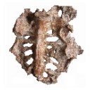
A pangolin (Manidae, Pholidota, Mammalia) from the French Quercy phosphorites (Pech du Fraysse, Saint-Projet, Tarn-et-Garonne, late Oligocene, MP 28)
Published online: 14/09/2015
Keywords:
Oligocene; Pangolin; Pech du Fraysse; Quercy Phosphorites
https://doi.org/10.18563/pv.39.2.e4
Abstract
Pangolins have never shown a high taxic diversity and their fossil record is scarce. We report here the first discovery of a partial humerus from late Oligocene deposits in Pech du Fraysse (MP28, France). The new specimen from Pech du Fraysse is described and compared to various extant and extinct species of pangolins. It shows a suite of morphological features very similar to the humeri discovered in Saint-André (MP 26), Solnhofen (Burdigalien), and Saulcet (Aquitanian), attributed here to Necromanis franconica. The description of the specimen from Pech du Fraysse allowed us to discuss the systematics of Paleogene and Neogene pholidotans. The differences between PFY 4051 and N. franconica on the one side, and N. quercyi on the other side, might be sufficiently important to justify a generic distinction. A comparison with extant species showed that N. franconica was likely terrestrial and fossorial based on its humeral morphology.
PV article infos
Published in Vol.39-2 (2015)
|
PDF
|

|
The eosimiid and amphipithecid primates (Anthropoidea) from the Oligocene of the Bugti hills (Balochistan, Pakistan): new insight into early higher primate evolution in South Asia.
Published online: 15/10/2006
Keywords:
Amphipithecidae; anthropoid phylogney; Bugti Hills; Early Oligocene; Eosimiidae; Pakistan
https://doi.org/10.18563/pv.34.e15
Abstract
Eosimiid and amphipithecid primates document a long and significant history of primate evolution throughout the Eocene in Southeast Asia. Despite the absence of a comprehensive post-Eocene fossil record, it was generally hypothesized that both families left no descendant in Asia. Recently, two new small-bodied taxa, Bugtipithecus and Phileosimias, have been recovered in early Oligocene coastal deposits from the Bugti Hills (Balochistan, central Pakistan) and referred to the families Amphipithecidae and Eosimiidae, respectively, on the basis of dental fossil remains. In this paper, we provide more exhaustive description, comparison, and discussion of these taxa. As for tarsiid and sivaladapid primates, the persistence of eosimiids and amphipithecids into the Oligocene clearly demonstrates that low latitudes of South Asia provided a continuous access to tropical refugia during the climatic deterioration characterizing the late Eocene-early Oligocene interval, which was seemingly lethal for primate communities elsewhere across the Holarctic continents. As a contribution to the ongoing phylogenetic debates regarding the position of eosimiids and amphipithecids on the primate family tree, we have performed a cladistic analysis in a high-level primate systematic context in order to assess the position and the role of these new taxa in that phylogenetic issue. Our results support the view according to which eosimiids and amphipithecids (and by extension Phileosimias and Bugtipithecus, respectively) are stem anthropoids. These fossils from Pakistan document an unsuspected Oligocene phase of the evolutionary history of anthropoid primates in southern Asia, which clearly enhances the extent of the anthropoid radiation in this province during the Paleogene. Several phylogenetic and paleobiogeographic aspects are discussed, notably the intra- and inter-relationships between Paleogene Asian and Afro-Arabian anthropoids, and the resulting potential dispersal models between both land-masses during the Paleogene.
PV article infos
Published in Vol. 34, Fasc. 1-2 (2006)
|
PDF
|

|
Additions of the Geiseltal mammalian faunas, Middle Eocene: Didelphidae, Nyctitheriidae, Myrmecophagidae.
Published online: 04/12/1989
Keywords:
Edentata; Geiseltalian; German Democratic Republic; Lipotyphla; Marsupialia; MP 11-13
https://doi.org/10.18563/pv.19.3.95-114
Abstract
New and hitherto unpublished mammals from the stratigraphical levels Unterkohle, Untere Mittelkohle and Obere Mittelkohle of the Geiseltal near Halle, GDR, are described (= biochronological levels MP 11-13, Geiseltalian sensu Franzen & Haubold 1986a, b). The marsupial taxa Amphiperatherium aff. maximum (MP 12), A. goethei (MP 12), and Peratherium aff. monspeliense (MP 12 and 13) are recorded for the first time. A lectotype for Amphiperatherium giselense is designated, and the alleged primate Microtarsioides voigzi is assigned to Marsupialia, incertae sedis. A new insectivore species, Saturninia ceciliensis n. sp., is described (MP 13). The anteater Eurotamandua joresi is recorded for the first time outside its type locality, Grube Messel, FRG (MP 11). The present humerus and ulna display the autapomorphic features of the myrmecophagids.
PV article infos
Published in Vol. 19, Fasc. 3 (1989)
|
PDF
|

|
Eggshell microstructure and porosity of the Nicobar scrubfowl (Megapodius Nicobariensis, great Nicobar island, India)
Published online: 16/12/2008
Keywords:
conductance porosity; eggshell microstructure; incubation conditions; Megapodes
https://doi.org/10.18563/pv.36.1-4.75-88
Abstract
The eggshell of Nicobar scrubfowl (Megapodius nicobariensis) is described for the first time. Its egg porosity is calculated and discussed with data from several taxa (another megapode, some extant and fossil reptiles including a titanosaur group) in order to compare incubation types with eggshell structure. Eggshell microstructure reflects first phylogenetic traits and does not seem to have developed major adaptative features due to the incubation conditions, except for the pore canals.
PV article infos
Published in Vol. 36, Fasc. 1-4 (2008)
|
PDF
|

|
Révision des Rhombodontidae (Neoselachii Batomorphii) des bassins à phosphate du Maroc
Published online: 20/05/1994
Keywords:
Batomorphii; Maastrichtian; Morocco; New taxa; Phosphate; Rbombodontidae
https://doi.org/10.18563/pv.23.1-4.1-49
Abstract
The revision of the Rhombodontidae from the Maastrichtian of Morocco led us to the description of a new species: Rhombodus andriesi.
The biometrical study of populations of R. binkhorsti and R. microdon, species sometimes considered as synonymous, supports the conclusion that they represent two distinct species and not young and adult specimens of a single species. The stratigraphical range of these two species confirms this
result.
The reexarnination of type-series studied by C. Ararnbourg led us to revise the generic status of Rhombodus bondoni which is now ranged, because his dental features, in the new genus Dasyrhombodus showing a less derived dentition than Rhombodus. The stratigraphical range of each species is clarified within the phosphatic series.
PV article infos
Published in Vol. 23, Fasc. 1-4 (1994)
|
PDF
|
