|
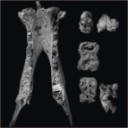
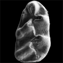
A new species of hippopotamine (Cetartiodactyla, Hippopotamidae) from the late Miocene Baynunah Formation, Abu Dhabi, United Arab Emirates
Published online: 07/04/2017
Keywords:
Arab Peninsula; Hippopotamidae; Hippopotamine event; Systematics
https://doi.org/10.18563/pv.41.1.e2
Abstract
The discovery of new hippopotamid material from the late Miocene Baynunah Formation (Abu Dhabi, United Arab Emirates) has prompted the revision of the existing material of this as yet unnamed fossil taxon. The Baynunah hippopotamid appears to be distinct from all other contemporary and later species in having a relatively more elongate symphysis, a feature similar to the earlier (and more primitive) Kenyapotamus. Yet, the Baynunah hippopotamid presents a dentition typical of the Hippopotaminae. It is therefore a distinct species attributed to the later subfamily, described and named in this contribution. This species provides further evidence for a ca. 8 Ma evolutionary event (termed “Hippopotamine Event”) that initiated the spread and ecological significance of the Hippopotaminae into wet habitats across Africa and Eurasia. The morphological affinities of the new species from Abu Dhabi suggest that the Arabian Peninsula was not a dispersal route from Africa toward southern Asia for the Hippopotamidae at ca. 7.5 Ma to 6.5 Ma.
PV article infos
Published in Vol 41-1 (2018)
|
PDF
S.I. Data
|

|
Les poissons crétacés et tertiaires du bassin des Iullemmeden (République du Niger)
Published online: 15/09/1972
Keywords:
Actinopterygians; Cenozoic; Cretaceous; Dipnoans; Selachians
https://doi.org/10.18563/pv.5.5.179-251
Abstract
The present work is devoted to the study of the Cretaceous and Tertiary fishes (teeth of Selachians, Actinopterygians and Dipnoans) collected during a recent expedition in Niger. The Maestrichtian localities have yielded a new genus and a new subspecies of Selachian: Igdabatis sigmodon nov. gen., nov. sp. and Lamna biauriculata nigeriana nov. subsp. The locality of Sessao, which has been attributed to the Thanetian by means of the study of the fish, has furnished by screen-washing an interesting fauna wherein six new species are described: Raja Iouisi, Dasyatis sessaoensis, D. sudrei, D. russelli, Hypolophites thaleri and Ceratodus casieri. Comparison of these faunas with contemporary faunas of Africa has brought out a certain endemism in the Iullemmeden Basin during the late Cretaceous and the early Tertiary.
PV article infos
Published in Vol. 05, Fasc. 5 (1972)
|
PDF
|
|
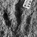
Nouvelles données sur les Ichnites de dinosaures d'El Bayadh (Crétacé Inférieur, Algérie)
Published online: 16/12/2008
Keywords:
Algeria; Brezina; El Bayadh; Ichnites; Lower Cretaceous; Sauropoids; Theropoids
https://doi.org/10.18563/pv.36.1-4.7-35
Abstract
Evidence of 350 Lower Cretaceous Dinosaur footprints is pointed out in El Bayadh area. Their preliminary study allow to distinguish four trackway assemblages which reveal vertebrate bipedal presence forms of tri-and tetradactylous Dinosauroïds (Assemblages 1-3) and quadrupidal Sauropoïd (Assemblage 4).
The analysis of their footprint biometric features will attribute the quadrupidal Sauropoïd form to Brontopodus ichnogenus which is weIl known in the Jurassic and Cretaceous periods. In retum and despite their age, the dinosauroïd forms were approached, temporarily, to Grallator and Eubrontes types.
The occurrence of the dinosaur traces (Theropoïd and Sauropoïd) constitutes, in the Lower Cretaceous, an important first step of the knowlege of the marshy Reptilian fauna which takes over, from the begining of the Secondary Era, a wide paleogeographie area on the Southem Tethyan margin.
PV article infos
Published in Vol. 36, Fasc. 1-4 (2008)
|
PDF
|
|
New Late Miocene plecotine bats (Chiroptera, Vespertilionidae: Plecotini) from Gritsev, Ukraine
Published online: 07/03/2019
Keywords:
Barbastella; bats; late Neogene; Mammalia; Plecotus
https://doi.org/10.18563/pv.42.1.e2
Abstract
The Late Miocene site of Gritsev (MN 9, Ukraine) has yielded a very rich bat fauna, the remains of which are well preserved. Compared to other Neogene bat assemblages of Europe, the Gritsev bat community is unique in preserving plecotine bats, which are rare from Neogene sites. Some peculiar and new bat species, including a large plecotin Otonycteris, already were described from the Gritsev mammal site. Here we report new records of small plecotin bats from Gritsev, including a new taxon, Barbastella maxima nov. sp. This is the earliest reliable fossil record of this genus and it differs from more recent species of Barbastella in being considerably larger. The evolutionary patterns in the odontology within the tribe Plecotini, supported by biostratigraphical distribution of fossil records of Plecotus are discussed. The morphological peculiarities of the new fossils of plecotine bats from Gritsev are discussed in connection with its possible taxonomical affinity.
PV article infos
Published in Vol 42-1 (2019)
|
PDF
|
|
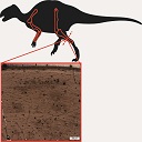
Comparative bone histology of rhabdodontid dinosaurs
Published online: 17/11/2014
Keywords:
bone histology-based ontogeny; Mochlodon; Rhabdodon; skeletal maturation; Zalmoxes
https://doi.org/10.18563/pv.38.2.e1
Abstract
A comparative bone histological study of the three known genera of the endemic European ornithopod dinosaur family, Rhabdodontidae, is presented here in an ontogenetic context. Investigated specimens were assigned to different ontogenetic stages based exclusively on the histological indicators of osteologic maturation during diametrical bone growth; an entirely size-independent method as opposed to most previous studies. Qualitative comparison of bone histology of corresponding ontogenetic stages and elements among the three valid rhabdodontid genera, Mochlodon, Zalmoxes, and Rhabdodon, revealed some consistent patterns. Genus specific histological differences within Rhabdodontidae are most expressed between Rhabdodon and the Mochlodon-Zalmoxes clade. These indicate a prolonged phase of fast growth and a less constrained cyclicity in the growth dynamics of Rhabdodon, as opposed to the slower and more regulated growth strategy reflected in the bones of Mochlodon and Zalmoxes. These genus specific differences are consistent with the phylogenetic interrelation of the genera and are most probably related to the pronounced differences in body size. However, when compared to other ornithopods, most detected histological features in rhabdodontids do not seem to reliably reflect either phylogenetic relations or body size. A notable common feature of all rhabdodontid genera irrespective of body size is the ontogenetically early onset of cyclical growth and secondary remodelling; a pattern that more resembles the condition found in derived ornithopods than that described in more basal taxa which are closer relatives of rhabdodontids. The recognition of taxon-specific histological patterns as well as patterns indicative of ecological and thereby functional traits clearly requires more accurate, preferably quantitative evaluations.
PV article infos
Published in Vol.38-2 (2014)
|
PDF
|

|
Anatomie du membre antérieur chez un chiroptère Molossidé (Tadarida sp.) du Stampien de Cereste (Alpes-de-Haute-Provence).
Published online: 01/01/1971
Keywords:
Chiroptera; Molossidae; Oligocene
https://doi.org/10.18563/pv.4.1.1-38
Abstract
The present study describes in detail the anterior limb osteology of a molossid chiropteran of the genus Tadarida, from Céreste, a Stampian locality in the Apt-Forcalquier Oligocene basin already known for its fishes, plants and insects.
A comparision with the Miocene forms T. srehlini from Saint-Géraud localities and T. sp. from Württemberg, also with the recent forms T. teniotis and Eumops perotis, does not show any clear morphological differences between the Tertiary and Recent Tadarida, indicating a rather noticeable anatomical stability, not exceptionnal indeed among Chiropterans. The Céreste fossil exhibits however slightly primitive wing proportions if compared to the Saint Gérand Aquitanian species.
Several remarks deal with the peculiar relationships between the ecology of the molossids and their kind of fossilisation, frequently associated with sedimentary facies of the lacustrine type.
PV article infos
Published in Vol. 04, Fasc. 1 (1971)
|
PDF
|

|
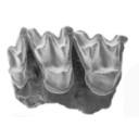
Un crane de Chalicothere (Mammalia, Perissodactyla) du Miocène supérieur de Macédoine (Grèce) : remarque sur la phylogénie des Chalicotheiinae
Published online: 14/06/1995
Keywords:
Chalicotheriidae; Cladistics; Greece; Miocene; Perissodactyla; PHYLOGENY
Abstract
The discovery in the Turolian (Late Miocene) of Dytiko 3 (Macedonia, Greece) of a complete skull with mandibles and cervical vertebrae, atlas and epistropheus, is a very important contribution to the knowledge of the subfarnily Chalicotheríinae. After the description, the comparison with other specimens of Miocene chalicotheres permits the revival of the generic name Macrotherium with a new species M. macedonicum. This genus is mainly characterized by a short snout and an inflated cerebral skull. It coexists during the Miocene with Chalicotherium. A cladistic analysis leads to conclusion that the species which has been described from the Early Middle Miocene of Rusinga must be identified as the type-species of a new genus: Butleria.
PV article infos
Published in Vol. 24, Fasc. 1-2 (1995)
|
PDF
|
|
New datation of the Tafna Basin (Algeria): A combination between biochronological and magnetostratigraphical data
Published online: 11/03/2015
Keywords:
correlations; Late Miocene; North Africa; Rodentia
https://doi.org/10.18563/pv.39.1.e1
Abstract
The Tafna Basin corresponds to the lowlands, which are located in front of Tessala and Traras ranges, below the Tlemcen mountains, Algeria. This basin displays a complete sedimentary cycle dominated by lagoonal-fluvial and marine deposits. The continental formations located at the base of these deposits are mainly composed of alternating sandstones and clays. An early late Miocene age has been previously attributed to them, based on direct correlations with marine deposits. Search for micromammal fossils led to the discovery of three different rodent species from a single level of the Djebel Guetaf section, located at the bottom of these deposits. The rodent assemblage indicates a late Miocene age. Combined magnetostratigraphical and biostratigraphical investigations were carried out to provide a more accurate age control of these continental deposits. Sixty-four oriented rock samples were collected for a magnetostratigraphic study along a 92 meters thick section including the fossiliferous layer. Rock magnetic investigations indicate the presence of both high and low coercivity minerals. Specimens subjected to progressive thermal demagnetization procedures show that the samples exhibit a high temperature magnetization component and display a normal polarity. Based on biostratigraphic constraints, the Guetaf section is correlated with Chron C4An, indicating an age ranging from
9.1 to 8.7 Myr.
PV article infos
Published in Vol.39-1 (2015)
|
PDF
|

|
Mammals and stratigraphy : the Paleocene of Europe
Published online: 01/12/1982
Keywords:
Europe; Mammalia; Mammalian biochronology; Paléogène; Stratigraphy
https://doi.org/10.18563/pv.12.ext
Abstract
The mammalian faunas of the Paleogene of Europe and their localities are reviewed with comments on problems of European stratigraphy (epoch, stage and substage limits) and on the possibilities of faunal migrations. Radiometric dating is discussed. A stratigraphic scale for the Paleogene is presented, as well as a refined system of sequential faunal levels.
PV article infos
Published in Vol. 12, Ext (1982)
|
PDF
|

|
Mammals and stratigraphy of the continental mammal-bearing Quarternary of South America
Published online: 16/12/1984
Keywords:
Geochronology; Mammalia; Quaternary; South America; Stratigraphy
https://doi.org/10.18563/pv.14.ext
Abstract
Previous chronological arrangements of South American Quaternary land mammal faunas are appraised on the basis of current geological and paleontological data. Three South American late Pliocene-Pleistocene land mammal ages are conventionally recognized, from oldest to youngest, the Uquian, Ensenadan, and Lujanian ; all are defined on Argentine faunas.
The Uquian is based fundamentally and historically on the fauna from the Uquía Formation in Jujuy Province, northwestern Argentina. Important known formations in Argentina yielding Uquian Age faunas include the sub-surface Puelche Formation (or Puelchense) near the city of Buenos Aires, and the Barranca de Los Lobos and Vorohué Formations between Mar del Plata and Miramar, Buenos Aires Province. A tentative subdivision is propos-ed for the Uquian into three subages based on knowledge of the Mar del Plata-Miramar sequence, from oldest to youngest, the Barrancalobian, Vorohuean, and Sanandresian. In Argentina the Uquian is presently marked by the first known record of Scelidodon, Hydrochoeropsis, Ctenomys, Canidae, Ursidae, Gomphotheriidae, Equidae, Tapiridae, Camelidae, Cervidae, and the last known record of Thylatheridium, Thylophorops, Dankomys, Eumysops, Pithanotomys, Eucoelophorus, Hegetotheriidae, Sparassocynidae, and Microtragulidae.
The Ensenadan Age is based on the fauna from the Ensenada Formation near the city of Ensenada, Buenos Aires Province. In Argentina the Ensenadan is marked by the first known record of Lomaphorus, Neothoracophorus, Plaxhaplous, Cavia, Lyncodon, Lutra, Galera, Smilodon, Dicotyles, Lama, Vicugna, the last known record of Orthomyctera, and the only known record of Brachynasua.
Typícal beds of late Lujanian Age in Argentina consist of fluvial deposits occupying stream channels, and shallow basins, often incised into beds of early Lujanian (i.e. Bonaerian of early workers) and Ensenadan Age. The Lujanian Age is based on a fauna from beds along the Rio Luján, about 65 km west of the city of Buenos Aires, Buenos Aires Province. The Lujanian in Argentina is marked by the first record of Equus, Chlamyphorus, and Holochilus, and the last record of Megatherioidea, Glyptodontoidea, Arctodus (=Arctotherium), Smilodon, Litopterna, Notoungulata, Proboscidea, Equidae, Morenelaphus, and Palaeolama.
These land mammal ages are often difficult to recognize in other South American countries. The compositions of South American Pleistocene faunas vary with the environment. Some taxa were widely distributed in fossil deposits throughout the continent, but their occurrences need not reflect synchroneity. This is a result of changing climates and habitats in time. Consequently, proposed intracontinental correlations need confirmation based on magnetostratigraphy and a radioisotope time scale. Paleontologic characterizations of these land mammal ages (i.e. first and last record, and guide fossils) are useful for much of Argentina, but extensions to most of the other parts of South America are at best tenuous.
The majority of known non-Argentine Pleistocene faunas are believed to be Lujanian in age. Possible non Argentine early Pleistocene (Uquian) faunas include Ayo Ayo and Anzaldo in Bolivia, and Cocha Verde in southern Columbia. A possible middle Pleistocene (Ensenadan or early Lujanian) fauna is the Chichense of Ecuador. Paleomagnetic and radioisotopic date (MacFadden et al., 1983) clearly indicate that the greater part of the Tarija fauna (Bolivia) is Ensenadan in age.
The end of the Pleistocene and beginning of the Holocene in South America is marked by extinction of nearly all large mammalian herbivores and their specialized large predators. Radiocarbon age determinations suggest that large scale extinctions of megafauna occurred between 15,000 and 8,000 yrs. B.P. (years before present).
PV article infos
Published in Vol. 14, Ext (1984)
|
PDF
|

|
Nouveaux Mammifères Eocènes du Sahara Occidental
Published online: 01/11/1979
Keywords:
Eocene; Mammals; Occidental Sahara
https://doi.org/10.18563/pv.9.3.83-115
Abstract
The fossil mammals collected from the Eocene of Hammada du Dra (northwest Sahara. Algeria) and two fragmentary teeth from the Lutetian of M'Bodione Dadere (Senegal) are described.
The fossils from the northwest Sahara come from a lacustrian deposit dated by charophytes (Raskyella aff. pecki, Raskyella n. sp.. Maedleriella lavocati, Maedleriella sp. et ? Peckichara sp.) as Middle Eocene or perhaps Lower Eocene (Gevin, Feist and Mongereau, 1974). Several hyracoids (3 or 4) identified from this formation extends the age of the family Pliohyracidae Osborn in Africa. Three forms appear to belong in the genera Megalohyrax, Titanohyrax and perhaps Bunohyrax which have been know until now only from the lower Oligocene of the Fayum (M. gevini n. sp. ; T. mongereaui n. sp.. ? Bunohyrax or Megalohyrax indet.). Another hyracoidof small size is referred to a new genus, Microhyrax (M. lavocati n. sp.).
Helioseus insolitus n. g. n. sp. is described without ordinal assignment. Azibius (Sudre, 1975) which has been the subject of questions and interpretations is reviewed.
Only one tooth from the Lutetian of M'Bodione Dadere is complete enough to interpret. lt probably belongs to a condylarth and demonstrates for the first time, the presence of the order in Africa. The second tooth is too fragmentary for comment.
In conclusion., the paleobiogeographic role of Africa at the end of the cretaceous and the beginning of the Cenozoic is discussed.
PV article infos
Published in Vol. 09, Fasc. 3 (1979)
|
PDF
|

|
Reflections on some Russian eotheriodonts (Reptilia, Synapsida, Therapsida)
Published online: 28/02/1972
Keywords:
Reptilia; Russia; Synapsida; Therapsida
https://doi.org/10.18563/pv.5.3.79-109
Abstract
As a result of the enrichment of eotheriodont material by one of us (P.K.T.), these specimens (essentially Biarmosuchur and Eotitanosuchur) are reexamined and refigured. A reevaluation of their particularities supports the distinction of two families, for which new diagnoses are proposed. This leads us to discuss the affinities of these families, with respect to the sphenacodonts on one hand, and to the South African primitive theriodonts on the other (gorgonopsids and ictidorhinids). This study contains inherent paleogeographic consequences which are considered in conclusion.
PV article infos
Published in Vol. 05, Fasc. 3 (1972)
|
PDF
|

|
Agriotherium intermedium (Stach 1957) from a Pliocene fissure filling of Xiaoxian County (Anhuei Province, China) and the phylogenetic position of the genus.
Published online: 30/09/1983
Keywords:
Carnivora; China; PHYLOGENY; Pliocene; skull anatomy; Ursidae
https://doi.org/10.18563/pv.13.3.65-81
Abstract
A fragmentary mandible and maxilla of a small sized Agriotherium of a young individual discovered from a Pliocene fissure filling in Xiaoxian county (Anhuei Province, China) are described. Judging from the morphology of the dentition and its dimensions the new material can be identified as Agriotherium inlermedium (STACH l957). Hendey's proposition (1980) that the Agriotherium species are derived from Indarctos is reconsídered on the basis of the new documents. As a result of a more general phylogenetic discussion it can be stated, that: 1. the supposed size increase as well as other trends, leading from Indarctos to Agriotherium are untenable ; 2. there are no positive indications to assume a phylogenetic transition of these two genera. 3. there are no real arguments in favor of an adaptational reversal in the evolution of Agriotherium. Hence, many features of that genus supposed by Hendey to be derived are plesiomorphic ; 4. regardless of the previous points it is methodologícally impossible to establish direct ancestor - descendant relationships between Indarctos and Agriotherium species, as Hendey did. Based on the data available and especially on the characters of the new material from China it is more likely that Agriotherium and Indarctos are two genera which developed independently. While advanced Agriotherium species, e.g. A. africanum, resemble in some respects Indarctos by adaptational analogies, more primitive species, e.g. Agriotherium intermedium, are quite dissimilar to lndarctos. While Indarctos might be derived from an Ursavus like forerunner, Agriotherium has its roots more likely somewhere in between Ursavus and the Hemicyon-group.
PV article infos
Published in Vol. 13, Fasc. 3 (1983)
|
PDF
|

|
Un giraffidae dans le pliocène de Montpellier ?
Published online: 31/10/1986
Keywords:
Artiodactyla; France; Giraffidae; Mammalia; Montpellier; Ruscinian
https://doi.org/10.18563/pv.16.3.185-189
Abstract
An upper giraffid premolar without any indication about its origin is preserved at the Montpellier University among numerous fossils from the ruscinian formation of Montpellier. It can be related to Samotherium, of the Upper Miocene in Eastern Europe, North Africa and Asia, or more probably to Bramatherium or Hydaspitherium of the Pliocene of South East Asia. The sedimentological study of the matrix shows a calcareous background, which may indicate that this tooth does not come from the Montpellier formation.
PV article infos
Published in Vol. 16, Fasc. 3 (1986)
|
PDF
|

|
L'occlusion dentaire chez Peradectes, Amphiperatherium et Peratherium, Marsupiaux du tertiaire d'Europe.
Published online: 01/10/1980
Keywords:
Didelphidae; Eocene; Mastication; Oligocene; Wear facets
https://doi.org/10.18563/pv.9.ext.79-89
Abstract
The general principles guiding the study of wear facets which develop during mastication in mammals possessing tribosphenic molars are named. The application of this method of study to the molars of European Tertiary Didelphidae shows that the lineage of this family as represented by the species Peratherium cuvieri (Upper Eocene), P. elegans (Lower-middle Oligocene) and P. antiquum (Upper Oligocene) has propessively evolved toward a more carnivorous diet.
PV article infos
Published in Vol. 9, Ext (1980)
|
PDF
|

|
Die Ohr-Region der Paulchoffatiidae (Multituberculata, Ober-Jura).
Published online: 15/11/1988
Keywords:
Multituberculata; Ober-Jura; Paulchoffatiidae; Petrosum; Portugal
https://doi.org/10.18563/pv.18.3.155-185
Abstract
The petrosal of the Paulchoffatiidae HAHN, 1969 is described and compared with that of younger multituberculates and of other Mesozoic mammals. The "Morrison petrosal", described by Prothero (1983), is also discussed; it probably belongs to the multituberculates. The reconstruction of the ventral side of the Paulchoffatiinae-skull, given by Hahn in 1987, is completed by addition of the otic and the occipital region.
PV article infos
Published in Vol. 18, Fasc. 3 (1988)
|
PDF
|

|
Nouvelles données sur les mammifères du Thanétien et de l'Yprésien du bassin d'Ouarzazate (Maroc) et leur contexte stratigraphique.
Published online: 15/12/1998
Keywords:
early Paleogene; magnetostratigraphy; Mammals; Morocco; North Africa; Ouarzazatz basin; Systematics
Abstract
New faunal and stratigraphical data on the vertebrates localities from the early Paleogene of the Ouarzazate Basin (Adrar Mgorn 1, Adrar Mgorn 1 bis et N'Tagourt 2), Morocco, are presented. A magnetostratigraphical study, the first for such early Paleogene Arabo-African mammal localities, and the discovery of probable remains of the nannofossil Discoaster support the Thanetian age of the Adrar Mgorn 1 site. The magnetostratigraphy suggests a slightly later age than was thought for the Paleogene formations of the local series of Tinerhir and for the vertebrate localities: late or latest Thanetian for Adrar Mgorn 1 and Adrar Mgorn 1 bis, middle Ypresian for N'Tagourt 2. It also indicates a lower position of the KT boundary in the series. Two tons of matrix recovered in the vertebrate sites have vielded new data on the micromammals. A damaged lower molar from N'Tagourt 2 is referable to Khamsaconus bulbosus and supports the proboscidean affinities of this species and especially possible relationships with bunolophodont taxa such as elephantiforms. A lower molar from Adrar Mgorn 1 bis belongs to a new form which can be identified as a plesiadapiform or an euprimate close to Altiatlasius koulchii though significantly larger. A new material from Adrar Mgorn 1 illustrates a new dilambdodont adapisoriculid species which is referable to Garatherium : ?Garatherium todrae n. sp. Another species referred to Garatherium is known in the locality (?Garatherium n. sp.). Garatherium is a new lineage from the Ouarzazate basin which crosses the Paleocene-Eocene boundary together with Palaeoryctes, Didelphodontinae gen. and sp. 2, Todralestes, and Afrodon, and it is the first Paleocene-Eocene lineage identified outside of this basin (Garatheríum is based on a species from El Kohol, Algeria). Among the Paleocene-Eocene lineages from the Ouarzazate basin, it should be also mentioned a new possible carnassial form (carnivoran or creodont; Adrar Mgorn 1), and an upper molar of Cimolestes cf. incisus (Adrar Mgorn 1 bis). The upper molar THR 168 previously reported as from an indeterminate didelphodontine is here identified as the M1/ of Afrodon chleuhi. The micromammal faunas from the Ouarzazate basin are positioned in the global chronological framework of the mammal localities from the Paleogene of the Arabo-African domain.
PV article infos
Published in Vol. 27, Fasc. 3-4 (1998)
|
PDF
|

|
La poche à phosphate de Ste-Néboule (Lot) et sa faune de vertébres du Ludien Supérieur. 2- Amphibiens. Etude Preliminaire
Published online: 25/09/1978
Keywords:
Eocene; Quercy Phosphorites
https://doi.org/10.18563/pv.8.2-4.175-179
Abstract
The Caudata are known by two Salamandridae ; one of them is attributed to the genus Megalotriton. The Pelobatidae form the major part of the Anura ; a few bones indicate also the presence of Neobatrachia.
PV article infos
Published in Vol. 08, Fasc. 2-4 (1978)
|
PDF
|

|
Ostéologie de la tête de Richardus excavans Lavocat,1988
Published online: 30/10/1989
Keywords:
Africa; anatomy; Bathyergidae; Miocene; Rodents
https://doi.org/10.18563/pv.19.2.73-80
Abstract
Remarkable association of a small infraorbital foramen, similar to that in recent Heterocephalus, and of a strong muscular print on the dorsal anterior part of the zygomatic plate and on the premaxillary. Several anatomical structures to be compared with those of Heterocephalus suggest relationships with this genus. Richardus supports the ancestrality of the hystricomorph character in the bathyergids
PV article infos
Published in Vol. 19, Fasc. 2 (1989)
|
PDF
|

|
Les nouvelles faunes de rongeurs proches de la limite mio-pliocène en Roussillon. Implications biostratigraphiques et biogéographiques
Published online: 29/04/1991
Keywords:
Arvicolidae; Cricetidae; Gliridae; Miocene; Muridae; Pliocene; Rodents; Southern France
https://doi.org/10.18563/pv.20.4.147-174
Abstract
Three new fossiliferous localities, two of karstic origin, Castelnou 3 and Font Estramar, respectively Late Upper Miocene and Lower Pliocene, and one of lacustrine origin, Thuir, Lower Pliocene, add data about the transition between Miocene and Pliocene faunas of rodents in southern France. An unexpected association of taxa was present in the late Upper Miocene, including between others, Myocricetodon, Hispanomys, Ruscinomys, Cricetus barrierei, Promimomys and a new species of Stephanomys, S. dubari nov. sp. Myocricetodon is still known in the Lower Pliocene. It is shown that the large field-mice known since the Late Upper Miocene belong to two different lineages, on one side, A. jeanteti, on the other side, A. gudrunae followed by A. gorafensis. Biochronological and biogeographical implications are discussed.
PV article infos
Published in Vol. 20, Fasc. 4 (1991)
|
PDF
|
