
Abstract book of the 18th Conference of the EAVP
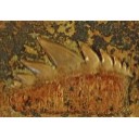
Pterosaurs from Coahuila
Pliocene-Pleistocene large mammals from Le Riège and Saint-Palais
Les sélaciens du Miocène de la région de Montpellier
Muridae du Pliocène supérieur d'Espagne et du midi de la France.
Contribution à l'étude des genres Gliravus et Microparamys.
Eocene (57) , Quercy Phosphorites (38) , Systematics (32) , Rodents (29) , Mammalia (27) , Rodentia (25) , Miocene (24)
|
First Neogene Otonycteris (Chiroptera: Vespertilionidae) from Ukraine: its biostratigraphic and paleogeographic significance.Valentina V. RosinaPublished online: 19/03/2015Keywords: bats; East Europe; Gritsev; Late Miocene; Mammalia https://doi.org/10.18563/pv.39.1.e2 Abstract A new species, Otonycteris rummeli nov. sp., is described from the Late Miocene site Gritsev (MN 9) in the Ukraine. Otonycteris rummeli nov. sp. differs from those of most vespertilionids, except recent Otonycteris, Antrozous and Early Miocene Karstala silva, in having a well-developed entocingulid at the foot of the trigonid valley in the lower molars. The morphological resemblance of Otonycteris, Antrozous and Karstala is apparently a case of convergence in the evolution of the Old and New Worlds bat faunas. From at least the Middle Miocene the range of Otonycteris distribution spread to the whole of Central Europe and such a situation continued during the whole Late Miocene. This indicates a more arid climate in Europe during the Upper Miocene compared to the Quaternary. The reduction of the distribution range of Otonycteris and its extinction in most of the territory of Europe could have been caused by the global climatic cooling and increasing glacial cycle amplitude during the onset of the Quaternary. PV article infos Published in Vol.39-1 (2015) |
|

|
A survey of Cretaceous tribosphenic mammals from middle Asia (Uzbekistan, Kazakhstan and Tajikistan), of their geological setting, age and faunal environmentLev A. Nessov, Denise Sigogneau-Russell and Donald E. RussellPublished online: 20/05/1994Keywords: Cretaceous; Environment; Middle Asia; Sharks; Tribosphenic mammals https://doi.org/10.18563/pv.23.1-4.51-92 Abstract This paper is an English concentrate of various Russian publications by the senior author presenting the mammaIian taxa from the Cretaceous (Albian through Santonian) of the region termed Middle Asia by Soviet geographers. The diagnoses are the unmodified, literal translation of the original version, but are followed with short complementary remarks; most of the species are illuslrated anew with SEM photographs, others are by normal photography. The fossiliferous formations are cited and arguments for their dating are given. Finally, the main vertebrate groups accompanying mammaIs are listed and the environment and climate at the time of deposition are suggested. In conclusion, an hypothesis on the origin and high diversity of tribosphenic mammals on the Cretaceous coastal plains of southwest Asia is proposed. In appendix the taxon Khuduklestes bohlini novo gen. novo sp. is formally defined. PV article infos Published in Vol. 23, Fasc. 1-4 (1994) |
|
|
Latest Early-early Middle Eocene deposits of Algeria (Glib Zegdou, HGL50), yield the richest and most diverse fauna of amphibians and squamate reptiles from the Palaeogene of AfricaJean-Claude Rage
Published online: 08/02/2021 |
|
|
Révision systématique des Anchilophini (Palaeotheriidae, Perissodactyla, Mammalia).Jean-Albert RemyPublished online: 15/10/2012Keywords: Anchilophus; Eocene; new genus; new species; Palaeotheriidae; Paranchilophus; Perissodactyla; Systematics https://doi.org/10.18563/pv.37.1-3.1-165 Abstract The knowledge of the Anchilophini has been lately renewed by the discovery of a rather large amount of new material still largely unpublished. This new material offers the opportunity of a systematic revision of this tribe gathering those of European Eocene Equoidea which bear no mesostyle on upper check teeth and display a heavy trend to the molarization of premolars. PV article infos Published in Vol. 37, Fasc. 1-3 (2012) |
|

|
Arvicolinae (Rodentia) du Pliocène terminal et du quaternaire ancien de France et d'Espagne.Jacques MichauxPublished online: 30/10/1971Keywords: Arvicolinae; France; Pleistocene; Pliocene; Spain https://doi.org/10.18563/pv.4.5.137-214 Abstract Two steps can be distinguished in the history of the first invasion of western and south western Europe by the arvicolines. The first step corresponds to the installation of these rodents with the immigration of Promimonys inxuliferus Kowalski, then of Mimomys stehlini Kormos and of Mimomys gracilis (Kretzoi). The second is characterized by the establishment of a geographic differentiation in the arvicoline fauna between the south of France and Spain, from where are described new species of Mimomys (Mimamys cappettai, Mimomys septimanus, Mimonys medasensis), and the rest of France, where are found only elements already known from central Europe or England (Mimomys polonicus Kowalski, Mimomys pliocaenicus F. Major, Mimomys reidi Hinton, or forms very close to the latter). This geographic differentiation, which is very certainly the consequence of the division of Europe into distinct climatic provinces, one of them being the southern province comprising at least Spain and southern France, could result from a cladogenetic evolution of Mimomys stehlini and Mimomys gracilis after their immigration. The present work is also a contribution to the search for correlations between the diverse micromammal localities of the latest Pliocene (or early Villafranchian) and of the early Quaternary of Europe. PV article infos Published in Vol. 04, Fasc. 5 (1971) |
|

|
Les Dipodidae (Mammalia, Rodentia) d'Europe occidentale au Paléogène et au Néogène inférieur: origine et évolution.Marguerite Hugueney and Monique Vianey-Liaud
Published online: 01/10/1980 |
|
|
Fallen in a dead ear: intralabyrinthine preservation of stapes in fossil artiodactylsMaeva J. Orliac
Published online: 09/03/2016 |
S.I. Data |

|
Les Pseudosciuridae (Mammalia, Rodentia) de l'Eocène moyen de Bouxwiller, Egerkingen et Lissieu.Jean-Louis HartenbergerPublished online: 30/10/1969Keywords: Bouxwiller; cranium; Egerkingen; Middle Eocene; Rodents https://doi.org/10.18563/pv.3.2.27-64 Abstract The description of new material from three classic middle Eocene localities of western Europe permits the addition of details to the systematics of primitive Pseudosciurids. The cranial anatomy of Protadelomys cartieri (STEHLIN and SCHAUB) from Egerkingen is described here and compared to that of the Adelomyines from the late Eocene, until now the only ones known. The morphologic and biometric study of the dentition of P. cartieri compared to that of P. alsaticus n. sp. from Bouxwiller and P. Iugdunensis n. sp. from Lissieu, forms respectively older and younger than P. cartieri, permits the evolutionary tendencies of the group to be demonstrated and shows that notable differences in age exist between these localities. This ensemble of forms can constitute a valuable guide lineage in the establishment of a fine stratigraphy of the period. Other less well known lineages are present at Egerkingen along with P. cartieri. They can be related to genera that have been noted int he late Eocene. In conclusion, a criticism of recent zonation proposals, made by divers authors, completes this article. PV article infos Published in Vol. 03, Fasc. 2 (1969) |
|

|
The Pleistocene vertebrate fauna of Robinson Cave, Overton County, TennesseeJ. E. Guilday, H. W. Hamilton and A. D. Mc CradyPublished online: 20/01/1969Keywords: Fauna; Mammalia; Pleistocene; Tennessee https://doi.org/10.18563/pv.2.2.25-75 Abstract A late Pleistocene deposit of 60 species of vertebrates and 12 of invertebrates is described from Robinson Cave, Overton County, Tennessee, U.S.A. Forty-eight species of mammals are represented by at least 2,483 individuals; 10 % are extinct, 10 % occur in the state only as boreal relicts in the Great Smoky Mountains; 23 % no longer occur as far south as Tennessee; 57 % occur at or near the site today. Nínety-one percent of the Recent mammal species can be found living today in the Minnesota-Wisconsin area, approximately 10 degrees farther north. Fluorine analysis suggests a long period of accumulation. The following 10 mammalian species are recorded from Tennessee for the first time. Sorex arcticus, Microsorex hoyi, Citellus tridecemlineatus, Clethrionomys gapperi, Microtus pennsylvanicus, Synaptomys cooperi, Synaptomys borealis, Zapus nudsonius, Napaeozapus insignis, Martes americana. Six additional species are present as boreal relicts in the Great Smoky Mountains of eastern Tennessee but not at the site today : Sorex cinereus, Sorex dispar, Sorex palustris, Parascalops breweri, Glaucomys sabrinus, Mustela nivalis. Six forms are extinct: Canis dirus, Ursus americanus amplidens, Sangamona furtiva, Dasypus bellus, Mammut americanus,Megalonyx jeffersoni. Twenty-six additional species of mammals, all of the snails, birds, reptiles, and amphibians recovered from the fauna still inhabit the area today: The fauna is indicative of a cold-temperate climatic episode associated with the Wisconsin glaciation, but may be chronologically mixed. PV article infos Published in Vol. 02, Fasc. 2 (1969) |
|

|
Les gisements de Robiac (Eocène supérieur) et leurs faunes de Mammifères.Jean SudrePublished online: 05/04/1969Keywords: Fauna; Late Eocene; Mammalia; Robiac https://doi.org/10.18563/pv.2.3.95-156 Abstract Designated the type-locality of a late Eocene paleomammal zone, Robiac has recently been the object of important excavations. The first results of the new collecting, as well as a revision of the material in old collections, are given in this work. PV article infos Published in Vol. 02, Fasc. 3 (1969) |
|
|
Late Campanian theropod trackways from Porvenir de Jalpa, Coahuila, MexicoHéctor E. Rivera-Sylva
Published online: 29/11/2017 |
|
|
Les rongeurs de Chéry-Chartreuve et Rocourt-Saint-Martin (est du bassin de Paris; Aisne, France). Leur place parmi les faunes de l'Eocène Moyen d'EuropeBernard Comte, Maurice Sabatier and Monique Vianey-Liaud
Published online: 15/12/2012 |
|

|
The late Miocene percrocutas (Carnivora,Mammalia) of Madedonia, Greece.George D. KoufosPublished online: 14/06/1995Keywords: Biochronology; Carnivora; Comparisons; Dinocrocuta; Greece; Late Miocene; Mammalia Abstract Some new material of percrocutas from the late Miocene of Axios valley (Macedonia, Greece) is studied. They have been found in the locality of "Pentalophos 1" (PNT). The material has been described and compared with the known late Miocene percrocutas of Eurasia. This comparison indicates that it can be identified as Dinocrocuta gigantea (SCHLOSSER, 1903). A maxilla of a percrocuta, named ”Hyaena" salonicae, was found in the same area (Andrews, 1918). "Hyaena" salonicae is smaller than the PNT material. It is also compared with other material from Eurasia while its taxonomic and age problems are discussed. It belongs to Dinocrocuta and shows close relationships with D. robusta and D. senyureki; its age can be considered as late Vallesian-early Turolian. The age of the locality PNT is also discussed and a possible Vallesian age is proposed for it. PV article infos Published in Vol. 24, Fasc. 1-2 (1995) |
|
|
Designating a lectotype for Mesacanthus pusillus (Gnathostomata: Acanthodii)Matthew G. Baron
Published online: 03/03/2021 |
S.I. Data |
|
Enamel hypoplasia on rhinocerotoid teeth: Does CT-scan imaging detect the defects better than the naked eye?Manon Hullot
Published online: 03/01/2022 |
S.I. Data |

|
The Ctenodactylidae (Rodentia) from the Oligocene of Ulantatal (inner Mongolia, China)Monique Vianey-Liaud
Published online: 15/12/2006 |
|
|
Discovery of the most ancient Notidanodon tooth (Neoselachii: Hexanchiformes) in the Late Jurassic of New Zealand. New considerations on the systematics and range of the genus
|
|

|
Study of the Turolian hipparions of the lower Axios valley (Macedonia, Greece). 4. Localities of Dytiko.George D. KoufosPublished online: 15/12/1988Keywords: Equidae; Greece; Hipparion; Lower Axios Valley; Macedonia; Mammalia; Turolian https://doi.org/10.18563/pv.18.4.187-239 Abstract The hipparions from the Dytiko localities of the lower Axios valley (Macedonia, Greece) are studied. The material comes from three localities Dytiko-l, 2, 3 (DTK, DIT, DKO), which are situated near the village of Dytiko, about 60 km northwest to Thessaloniki. Three species have been determined, the medium-sized H. mediterraneum, the small-sized H. matthewi and the very small-sized H. periafricanum. The determined Hipparion species, their morphological characters and their comparison with the other Axios valley material indicate a Late Turolian age for the Dytiko localities. PV article infos Published in Vol. 18, Fasc. 4 (1988) |
|
|
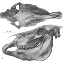
Old world hemiones and new world slender species (Mammalia, Equidae)Véra Eisenmann, John Howe and Mario PichardoPublished online: 16/12/2008Keywords: Amerhippus; biometry; Equus; Holocene; New World; Old World; Osteology; Pleistocene; Pliocene https://doi.org/10.18563/pv.36.1-4.159-233 Abstract Morphological and biometrical description of skulls, teeth, and limb bones of extant and fossil Old World herniones (including E. hydruntinus) and of New World 'stilt-Iegged' and other slender species from Blancan to Holocene. An Appendix presents ways in which the approximate size of some missing bones or dimensions may be deduced from available ones. PV article infos Published in Vol. 36, Fasc. 1-4 (2008) |
|

|
Les traces de pas d'amphibiens, de dinosaures et autres reptiles du Mesozoïque Français : inventaire et interprétations.Georges Gand, Georges Demathieu and Christian MontenatPublished online: 15/12/2007Keywords: Footprints; France; Inventory; Mesozoic; palaeontology; palaeovenvironments; Stratigraphy https://doi.org/10.18563/pv.35.1-4.1-149 Abstract Since the 19th century, thousands of footprints were observed in the geological series of the French Mesozoic. All are located in the Triassic and Jurassic. After a promising beginning, in France, it is only a few papers which will be published in the first half 20th century, unlike the USA and of others countries of Western Europe. One ought to wait about 1950 for a revival and now they are nearly 200 papers which were devoted to the ichnofossils. The literature abundance and the renewed interest of the naturalists for the palichnologic studies decided to us to write a synthesis work. This one begins with a stratigraphic inventory in which, localisation, age and paleontological contents of about 180 fossiliferous sites are specified. After having pointed out the followed methods, the footprints paleontological interpretation is then approached in detail and the results obtained are replaced in stratigraphy to deduce the fauna evolution during the Mesozoic. So, it appears that Ichnologic data, more varied and rich in the Triassic and Liassic than those relating to the bones, very rare for the considered periods, are very informative. The middle Triassic (Anisian-Ladinian), thus reveals Cotylosauria, Lepidosauria, Crurotarsi with Rauisuchia, Ornithosuchidae, Crocodylia and Dinosauromorpha more the "Prodinosauria": Dinosamiforme whose skeletons are known in Argentina but only in Ladinian. The rather fast domination of Dinosaurs during Norian is also as well shown. The almost exclusive presence of their footprints, up to fifty cm long, in the Lower Hettangian indicates their supremacy in the environments. Footprints characterise not very deep life places located between inter-supratidal limits and often out of water. Sedimentologic and Palaeontologic studies showed that they were great coastal spaces during Middle Triassic, flood-plain with sebkhas while Upper Triassic, and a large !!coastal marsh!! in Grands-Causses during Liassic in which, mainly, fine stromatolithic layers were deposited. During the same periad, bay beaches spread in Vendée. During the Middle Jurassic, they are also brackish to lacustrine environments and recifallagoons in- the Upper Jurassic. Numerous measurements of the footprints and trackways directions showed that the animaIs moved there in weil defined directions, for long periods. They seem due to the palaeotopography of the life environments relatively stable. Also, the discovery of vegetal radicular networks and small footprints far away from the continental borderlands has suggested that the animals continuously lived in these palaeoenvironnements, belonging to large ecosystems, where the sedimentation rate was weak. This explains that thebadies could not fossilize there but only their footprints through the cyanobacterian action in main cases. From the vertical distribution of different ichnospecies, defined with adapted statistical methods, explained in this work, a palichnostratigraphy was established for the Middle Triassic. Although the footprints are also abundant in Hettango-Sinemurian of "Grands-Causses" and the Vendée, it was not possible, up to now, to establish any zonation in this series; Probably because the palichnofauna is too little diversified there, currently reduced to a majority of Theropods II-IV tridactyl traces. PV article infos Published in Vol. 35, Fasc. 1-4 (2007) |
|