
|
Le genre Plagiolophus (Palaeotheriidae, Perissodactyla, Mammalia): révision systématique, morphologie et histologie dentaires, anatomie crânienne, essai d'interprétation fonctionnelle
Published online: 15/12/2004
Keywords:
New taxa; Paléogène; perissodactyls; skull anatomy; tooth histology
Abstract
The genus Plagiolophus is documented, almost solely in Western Europe, from the middle Eocene up to the mid Oligocene (MP 12 to MP 25), i.e. more than for 15 MY. Seventeen species are now recorded whose two of them are new, P. ringeadei nov. sp. and P. mamertensis nov. sp. Some anatomical variations and the deflection of certain evolutionary trends justify the distinction of three subgenera, Paloplotherium, Fraasiolophus nov. and Plagiolophus s.s. The genus displays a wide range in size and weight (between 10 and 150 kg). The detailed description of the skull of several species is here given for the first time.
Despite important evolutionary drifts during this long time span, the dentition shows a great structural homogeneity, which renders difficult the determination of fragmentary specimens or isolated teeth. It is characterized by a great heterodonty; premolars are little molarized and present a certain regression through time with paradoxically some progress in the molarization. The hypsodonty increases: the first Plagiolophus are hardly less brachyodont than Propalaeotherium, and the last ones are nearly as hypsodont as Merychippus from the early Miocene. The upper molars change from a wide crown pattern, with an open occlusal surface, lightly oblique transverse lophs and rounded internal cusps, to a narrower pattern, with a frontally constricted occlusal surface and internal lophs aligned parallel to the ectoloph. The M3/3 become always longer.
The dental enamel displays horizontal Schreger-bands with imprecise limits occupying only the middle part of the enamel layer. The dentine is remarkable by its high rate of pericanalicular dentine. The crown cementum, lacking in earlier forms, increases to the point where it fills the occlusal valleys of the
teeth.
The masticatory musculature shows a increasing prominence of the temporal, with probably an important role devoted to the pterygoid muscles in lateral movements related to a two-phase type of chewing.
The evolution of the dentition, of the masticatory musculature and of the repartition of masticatory forces indicate that the Plagiolophus have known different diets through their long evolutionary history; at first browsers they became mixed feeders and finally grazers. Their relatively long neck allowed these animals to reach different vegetal layers. The strength of the nuchal crests also suggests that they were able to have strong backwards movements of the head to pull up their food.
This evolution of diet seems related to the slow degradation of environmental conditions attested during this period in western Europe, with the generalization of more open landscapes, increasing aridity and more marked seasons.
Besides, a remodeling of the face is ontogenetically and along time observed, in relation with the evolution of the masticatory apparatus and especially with that of the mandibular lever arm. The postcanine diastemata become longer in the course of evolution; the free extremities of the nasals are always relatively long which contradicts the hypothesis according to which Paloplotherium may have had a trunk. At last the lineage Fraasiolophus can be distinguished by the presence of a deep malar fossa, probably related to a strong development of the maxillo-labialis superior muscle.
The orbit is always large and tends to increase in size, which indicates a good development of the vision and its increasing role in the life relations. A peculiar type of epitympanic sinus could have been used as a resonance chamber insuring a certain amplification of sounds before their transmission to the eardrum. The endocranial cast reveals a relatively large brain with an advanced degree of gyrencephaly. Beside the role eventually played in food research and social relations, these neurophysiological abilities, also related to an advance in cursorial fitness, could have contributed to the survival of these animals facing the predation pressure of the first fissipede carnivores and the competition with new immigrant herbivores after the "Grande Coupure".
On the basis of some shared apomorphies with the Pachynolophinae, which prevent from considering the latter as Equidae (molarization of the premolars, reduction of the premaxilla dorsal apophysis, peculiar epitympanic sinus, splitting of the jugular process), the hypothesis of an autochthonous origin of Plagiolophus issued from a form near Propalaeotherium, is once again proposed and discussed. Finally, intra-generic relationships are taken into consideration.
PV article infos
Published in Vol. 33, Fasc. 1-4 (2004)
|
PDF
|

|
Un gisement à mammifères dans la formation lacustre d'âge Miocène moyen du Collet Redon près de St-Cannat (Bouches-du-Rhone). Implications stratigaphiques
Published online: 10/04/1979
Keywords:
France; Neogene; Rodentia
https://doi.org/10.18563/pv.8.5.327-341
Abstract
The new fauna of Collet Redon (Bouches-du-Rhône, France) is dated by three rodents: Megacricetodon aff. bavaricus, Democricetodon affinis mutilus and Peridyromys cf. hamadryas. They correlate this locality with Oggenhof and Ohningen in Bavaria (Western Germany). As the radiometric age of Ohningen is estimated between 14 and 13 M.Y., these three localities are of Serravallian age. This datation brings a complete readjusment of the stratigraphy of the section of Collet Redon formerly described by Collot and Combaluzier. The marine deposits with underly the continental formation with the mammal fauna, are Burdigalian. The angular unconformity between the marine and the continental deposits gives evidence of an episode of emersion on the margin of a sedimentary basin, with deformation and erosion. Owing to the newly discovered fauna, this geodynamical event is clearly settled within the regional geographical and chronological context. Lacustrine and continental deposits of such an age were up to now unsuspected in this area.
PV article infos
Published in Vol. 08, Fasc. 5 (1979)
|
PDF
|

|
La plus ancienne faune de mammifères du Quercy : Le Bretou
Published online: 01/12/1974
Keywords:
Le Bretou; Quercy Phosphorites
https://doi.org/10.18563/pv.6.3-4.177-196
Abstract
Redécouvertes en 1968 dans le bois du Bretou, les deux poches à phosphorites qui ont livré cette faune sont parmi les plus méridionales du plateau quercynois. De faible profondeur, huit mètres, les cavités sont de petite taille et font partie d'un complexe de sept fissures réparties dans les bois du Bretou. Les autres trous n'ont livré aucun fossile, soit parce qu'ils avaient été intégralement vidés de leur sédiment, soit que des venues d'eau en interdisent l'accès.
Dans les deux poches fossilifères, proches de quelques mètres, le sédiment que l'on rencontre en placage sur les parois présente un aspect particulier. Il s'agit en effet d'une brèche très indurée, où la calcite domine, et contenant assez peu de sédiments sidérolithiques. Les ossements contenus dans cette brèche sont assez nombreux, très souvent brisés. Les dents isolées y sont relativement rares. Exploitées séparément, les deux poches ont livré des faunes identiques, aucune différence chronologique ne pouvant être décelée entre elles. Les formes rencontrées dans une seule des cavités sont rares mais la poche la plus riche est celle dénommée Le Bretou 2.
L'intérêt de la faune récoltée réside dans son ancienneté. On sait en effet que H.G. Stehlin avait signalé dans les collections du Quercy des restes attribuables à Lophiodon lautricense, mais la provenance exacte de ces fossiles reste inconnue. Au Bretou si nous n'avons pas rencontré à ce jour le Lophiodon, il n'en demeure pas moins que l'âge de la faune récoltée doit être proche de celui de la faune de Robiac.
PV article infos
Published in Vol. 06, Fasc. 3-4 (1975)
|
PDF
|

|
The Quaternary avifauna of Crete, Greece.
Published online: 01/09/1988
Keywords:
Avifauna; Crete; Quaternary; Systematics
https://doi.org/10.18563/pv.18.1.1-94
Abstract
Pleistocene bird fossils have been studied from nine localities on Crete. Part of this material was described earlier by the author (Weesie, 1982) and will not be treated here in extenso, the results will be incorporated. More than one third of the over 10,000 fossil bird bones available could be identified ; they were found to represent at least 65 bird species. The following species of the Pleistocene Cretan avifauna are new to the fauna of Crete : Branta ruficollis, Haliaeetus albicilla, Gyps melitensis, Aquila chrysaetos simurgh n. ssp., Ketupa zeylomensis, Aegolius funereus, Dendrocopos leucotos, Zoothera dauma, Turdus iliacus and Pyrrhula pyrrhula. The Pleistocene Cretan avifauna differs less from comparable mainland avifaunas than (fossil) avifaunas from oceanic islands do. Still, the Pleistocene Cretan avifauna has two qualities that are characteristic of island avifaunas : the almost complete absence of a group of birds (the Galliformes) and the presence of two endemic (sub)species : the giant eagle Aquila chrysaetos simurgh n. ssp. and the long-legged owl Athene cretensis (Weesie, 1982). The new subspecies is described in the present study.
These endemic birds of prey were found in association with their supposedly principal prey species (now extinct as well) : endemic mice for the owl and endemic deer for the eagle. Endemic mammals have been found in association with endemic birds of prey on many islands, not only in the Mediterranean. There is evidence that the size of endemic birds of prey becomes optimally adapted to their feeding on certain endemic mammals, especially rodents. Another characteristic of the Pleistocene Cretan avifauna is the great number of species of birds of prey. This appears to be a common characteristic of fossil avifaunas from caves on Mediterranean islands as well as from caves on the European mainland. However, we think that ecological conditions on Pleistocene Crete (especially the abundant presence of mice) helped to account for the high representation of birds of prey. Furthemore, the fossil avifauna enables us to draw some conclusions about the climate and vegetation on Pleistocene Crete : it is concluded that the climate was cooler than today and that Crete was largely covered with forests. Finally, the reasons for the extinction or disappearance from Crete of some bird species of the Pleistocene Cretan avifauna are discussed.
PV article infos
Published in Vol. 18, Fasc. 1 (1988)
|
PDF
|

|
Contributions à l'étude du gisement Miocène supérieur de Montredon (Hérault). Les grands mammifères. 4 - Les artiodactyles Suidae.
Published online: 15/11/1988
Keywords:
Artiodactyla; France; Mammalia; Montredon; Upper Miocene
https://doi.org/10.18563/pv.18.ext.57-64
Abstract
There is only one suid known in the Upper Miocene of Montredon (Hérault): Microstonyx (Limnostonyx nov. subgen.) antiquus (KAUP). It is differenciated from Microstonyx major by the presence of upper and lower canines which are considerably longer and biger. Its presence at Montredon corroborates the palustrine habitat for the species.
PV article infos
Published in Vol. 18, Ext (1988)
|
PDF
|

|
Contributions à l'étude du gisement Miocène supérieur de Montredon (Hérault). Les grands mammifères. 2 - les carnivores
Published online: 15/11/1988
Keywords:
anatomy; Carnivora; France; Montredon; Systematics; Upper Miocene
https://doi.org/10.18563/pv.18.ext.15-42
Abstract
The locality of Montredon has provided 8 species of camivores, often little documented, that are discribed and depicted. All the remains are fragmentary and generally badly preserved which lowers very much the possible precision of the taxonomic study; this one has however allowed the creation of a new subspecies. The most richlypdocumented forms are an ursid (Indarctos) and a felid (Machairodus). Issuing often from a westem Europe evolution, the carnivores are well inserted between those of the better known faunas of the "Classical Pontian" and of the lower Vallesian and this situation fits also well with their stratigraphic level, that cannot however be more accurately defined with them alone.
PV article infos
Published in Vol. 18, Ext (1988)
|
PDF
|

|
An Australian Miocene Brachipposideros (Mammalia, Chiroptera) related to Miocene representatives from France
Published online: 15/12/1982
Keywords:
Australia; bats; Chiroptera; Miocene
https://doi.org/10.18563/pv.12.5.149-172
Abstract
A new middle Miocene hipposiderid bat is described from a limestone deposit on Riversleigh Station in north-western Queensland. Hipposideros (Brachipposideros) nooraleebus n. sp. is the first record of this subgenus from anywhere in the world outside of France. The palaeoecological setting of the fossil bats appears to have been a relatively quiet, sunny lime-enriched tropical pool that contained tortoises, crocodiles and fish. It is possible that the bats were washed into the pool from an adjacent cave.
The Riversleigh bat most closely resembles the French Burdigalian (early middle Miocene) bat H. (B.) aguilari. It is also possible that it may have been closely related to the original Australian hipposiderid stock that ultimately gave rise to the endemic monotÿpic Rhinonycteris aurantius. The disjunct distribution of species of H. (Brachipposideros) suggests that representatives of this subgenus will be found in at least tropical southern Asia.
PV article infos
Published in Vol. 12, Fasc. 5 (1982)
|
PDF
|
|
Dortokid turtle remains from the Upper Cretaceous of Cruzy (Hérault, southern France) and phylogenetic implications
Published online: 14/11/2022
Keywords:
Cruzy; Dortoka vasconica; France; Late Cretaceous; Turtle
https://doi.org/10.18563/pv.45.2.e3
Abstract
An isolated right costal 1 from the Late Cretaceous Massecaps locality (Cruzy, Hérault, southern France) is assigned to Dortoka vasconica (Dortokidae). This find adds a new element to the Late Cretaceous turtle fauna of Cruzy and further supports the hypothesis that two distinct lineages of Dortokidae were present in Europe during the Late Cretaceous-Paleogene due to geographical isolation.
PV article infos
Published in 45-2 (2022)
|
PDF
|

|
Contribution à l'étude des Cricétidés oligocènes d'Europe occidentale
Published online: 20/01/1972
Keywords:
Cricetidae; Europe; Oligocene
https://doi.org/10.18563/pv.5.1.1-44
Abstract
Of the ten cricetid species from the Oligocene of Western Europe, attributed until now to the genus Eucricetodon, only four prove to be utilizable - E. atavus, E. huberi, E. praecursor, E. collatum - to which it is possible to add two forms newly described: E. huerzeleri and E. quercyi. The evolullon of the genus Pseudocricetodon is also the subject of new observations. The study of the dental morphology allows us to distinguish in these two genera three lineages beginning in the middle Oligocene:
- Lineage P. montalbenensis-P. thaleri (from Montalban to Goderet), of small size, without
increase in size.
- LineageE. atavus-E. infralactorensis (from Hoogbulsel to Estrepouy), of middle size, with a regular increase in size.
- Lineage E. huerzeleri-E. haslachense (level of Montalban to that of Estrepouy), of large size, with an increase in size.
Two other forms are equally represented in these loealitles: Heterocricetodon aff. helbengi and Melissiodon quercyi. It has been possible to attribute a precise age (zone of "Cournon") to the last species, which has been defined by Schaub (l925) from material in the Old Quercy collections. The genera Pseudocricetodon, Eucricetodon, ? "Cricetodon", Leydimys, Eumys, differentiated at the beginning of the middle Oligocene in Europe, Asia and North America, seem to derive from a common ancestral group. The place of origin of this group could be situated in Asia.
PV article infos
Published in Vol. 05, Fasc. 1 (1972)
|
PDF
|

|
Rongeurs nouveaux de l'Oligocène Moyen d'Espagne.
Published online: 15/09/1969
Keywords:
Cricetidae; Oligocene; Pseudocricetodon; Rodents; Theridomys
https://doi.org/10.18563/pv.2.5.191-207
Abstract
Description of four new rodents from a recently discovered locality at Montalban. Theridomys crusafonti nov. sp. is considered as the ancestry of T. Iembronicus. Theridomys varian: nov. sp. includes «Theridomys» morphotypes and «Blainvilllimys» morphotypes; it could be ancestral to B. blainvillei. Pseudoltinomys nanus nov. sp. represents a new lineage paralleling in evolution that of P. gaillardi (which is equally found at Montalban). Pseudocricetodon montalbanensis nov. gen., nov. sp. designates a lineage of very small Cricetidae accompanying Eucricetodon. With these well defined new species and six others present in the locality, Montalban appears as the best faunal reference point within the biochronologic zone of La Sauvetat.
As an annex, discussion of two rodent specimens from the classic localíty of Tárrega, close in age to that of Montalban.
PV article infos
Published in Vol. 02, Fasc. 5 (1969)
|
PDF
|
|
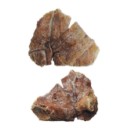
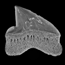
New Squalicorax species (Neoselachii: Lamniformes) from the Lower Maastrichtian of Ganntour phosphate deposit, Morocco
Published online: 05/12/2014
Keywords:
Anacoracidae; Chondrichthyes; Maastrichtian; Morocco; New taxa
https://doi.org/10.18563/pv.38.2.e3
Abstract
Two new Squalicorax species, S. benguerirensis nov. sp. and S. microserratus nov. sp. are described from the Lower Maastrichtian of the Benguérir phosphate open mine, Ganntour deposit, Morocco. The species S. benguerirensis nov. sp. was classically assigned to S. yangaensis since Arambourg (1952) and has been also recognized in coeval deposits from eastern USA to Mid-East. The species S. microserratus nov. sp. correspond to the lateral teeth of S. kaupi as reported by Arambourg (1952) and which is now referred in fact to S. bassanii. The comparison of these two new species with other Anacoracids, known in Moroccan or elsewhere, allows highlighting the great taxonomic and ecological diversities of this family during the Cretaceous.
PV article infos
Published in Vol.38-2 (2014)
|
PDF
|

|
Compléments sur les Chiroptères de l'Eocène moyen d'Europe. Les genres Palaeochiropteryx et Cecilionycteris.
Published online: 01/10/1980
Keywords:
Chiroptera; Geiseltal; Messel; Middle Eocene
https://doi.org/10.18563/pv.9.ext.91-126
Abstract
New dental and skeletal material referable to Palaeochiropteryx tupaiodon from the Middle Eocene locality of
Messel (G.F.R.) is studied, which provides additions to the previously gained knowledge of this european genus. Dental specimens from Geiseltal (G.D.R.), also of Middle Eocene age, allow us to analyze Cecilionycteria prisca. Some of these are separated to establish a new genus, Matthesia, and two new species, M. germanica and M. ? insolita.
PV article infos
Published in Vol. 9, Ext (1980)
|
PDF
|
|
A new species of Propalaeotherium (Palaeotheriidae, Perissodactyla, Mammalia) from the Middle Eocene locality of Aumelas (Hérault, France).
Published online: 24/05/2016
Keywords:
Eocene; new species; Palaeotheriidae; Propalaeotherium
https://doi.org/10.18563/pv.40.2.e1
Abstract
A new Propalaeotherium species, clearly distinct from the genus Eurohippus, is described. It is characterized by having a similar size as P. voigti from the German Geiseltal localities (MP 11 to MP 13 reference-level), but differs in several features suggesting a slighty more derived morphology. It presents indeed less brachyodont crowns with less prominent and less elevated cingula, slightly larger relative surface of premolars, and a more marked metaconid splitting on cheek teeth. This new species is unknown from other European localities except the nearby Saint-Martin de Londres locality which has been considered older than the MP 13 level.
PV article infos
Published in Vol.40-2 (2016)
|
PDF
S.I. Data
|

|
Deux nouveaux primates dans l'Oligocène inférieur de Taqah (Sultanat d'Oman): premiers Adapiformes (?Anchomomyini) de la péninsule arabique?
Published online: 15/11/1993
Keywords:
Adapids; Afro-Arabian plate; Early Oligocene; New taxa; Primates; Trans-Tethyan dispersals
https://doi.org/10.18563/pv.22.4.141-196
Abstract
Two new species, Omanodon minor n. g., n. sp. and Shizarodon dhofarensis n. g., n. sp., known from fifteen isolated teeth, are described here as the first adapiform primates (?Anchomomyini) recognizable to date in the Taqah material (early Oligocene of Sultanate of Oman).
Omanodon minor n. g., n. sp. displays special morphological similarity to the adapid tribe Anchomomyini from the Eocene of Europe, and especially to the Anchomomys lineage. Resemblances with the extant lemurifonn Microcebus are also noticeable and could be regarded as supporting Schwartz & Tattersall (1983) hypothesis of special relationships between the anchomomyine adapids and the cheirogaleid lemuriformes. However, these morphological affmities can be interpreted, altematively, as the results of parallelisms: important differences in upper molars indicate that the resemblances of cheirogaleids and Omanodon minor n. g., n. sp. are indeed probably due to parallelisms. Phyletic relationship of O. minor n. g., n. sp. to Anchomomyini is finally the most likely hypothesis.
Shizarodon dhofarensis n. g., n. sp., although much more poorly known, is closely related to Omanodon minor n. g., n. sp., at least at a familial level. The general morphology of this species suggests
also a close link with adapid Anchomomyini, although precise relationships within this tribe remain obscure. Interesting resemblances of Shizarodon dhofarensis n. g., n. sp. to Djebelemur martinezi lower molars (early Eocene of Tunisia) are also noticeable. These resemblances are even stronger than those betwen Omanodon minor and Djebelemur martinezi. However the very bunodont upper molars referred to D. martinezi are unusual for adapids, and there are moreover some notable differences in their lower molars. Thus resemblances in Djebelemur and Shizarodon are probably due to paralellisms.
Because of the fragmentary nature of the material and of possible parallelisms, the systematic position of Omanodon and Shizarodon within adapiformes cannot however yet be established definitively.
PV article infos
Published in Vol. 22, Fasc. 4 (1993)
|
PDF
|

|
First evidence of an early Miocene marine teleostean fish fauna (otoliths) from la Paillade.(Montpellier,France)
Published online: 15/06/1999
Keywords:
Aquitanian; Biostratigraphy; La Paillade; marine deposits; Miocene; otoliths; Palaeoecology; Palaeogeography; Southern France; Teleostei
Abstract
A fossil fish fauna, based on 5533 otoliths, from the La Paillade locality at Montpellier is described and figured. The otolith-bearing marls correlate to mammal zone MN l (Aguilar, 1982), and thus represent the earliest Miocene. The fish fauna consists of 30 taxa belonging to 20 families. Two species are new: Dussumieria sittigi and Liza gaudanti. The predominant faunal element is the Lesueurigobius vicínalis-species complex, composing 73% of all investigated otoliths. The palaeoecological analysis reveals a marine to euryhaline fish fauna living under tropical to subtropical conditions in the transition zone littoral - sublittoral. Water depth probably was more than 10 m. The scarcity of pelagic físhes suggests that the habitat was either a sheltered bay and/or far away from the open sea. Furthermore, some genera represented in the La Paillade fish fauna presently live exclusively in the Indopacific realm. Their presence strongly supports a broad connection between the Indian Ocean, the Mediterranean, and the Paratethys Seas during the Early Miocene (Aquitanian). From a palaeobiogeographical point of view, faunal relationships were found between the La Paillade fish fauna and both the Paratethys fish fauna and the fish fauna from the deposits in the Upper Rhinegraben and the Mayence and Hanau Basins (Germany).
PV article infos
Published in Vol. 28, Fasc. 1 (1999)
|
PDF
|

|
Les mammifères Montiens de Hainin (Paléocène moyen de Belgique) Part III : Marsupiaux
Published online: 30/09/1983
Keywords:
Belgium; Marsupials; Paleobiogeography; Paleocene
https://doi.org/10.18563/pv.13.3.51-64
Abstract
The oldest european marsupials are described from some specimens (isolated upper molars) recently found from the Hainin sediment (Middle Paleocene of Belgium). These fossils document a new species of the Peradectes genus. They give evidence of a much older occurrence of the marsupials in Europe than it was assumed. They allow us to postulate a didelphid dispersal from South America towards the western-holarctic area operating in two phases : the first one of the Peradectes genus at the end of the Cretaceous; the second one of the Didelphíni tribe at the end of the Paleocene. A central american crossing is likely for the first one, whereas a transafrican way is tentatively argued for the second one.
PV article infos
Published in Vol. 13, Fasc. 3 (1983)
|
PDF
|

|
Les Amphibiens et les reptiles du Pliocène supérieur de Balaruc II (Herault, France)
Published online: 15/09/1989
Keywords:
amphibians; Europe; France; Pliocene; Reptiles
https://doi.org/10.18563/pv.19.1.7-28
Abstract
The late Pliocene site (MN 16) of Balaruc II (Hérault, France) has provided remains of the following amphibians and reptiles: Chelotriton pliocenicus nov. sp. and Triturus marmoratus (Salamandridae), cf. Rana (Ranidae), cf. Blanus (Amphisbaenidae), cf. Agama (Agamidae), Gekkonidae indet., Lacerta s.l. (Lacertidae), "Ophisaurus" sp. (Anguidae), Michauxophis occitanus (Aniliidae), Erycinae indet. (Boidae), Elaphe cf. E. longissima and Malpolon sp. (Colubridae), cf. Naja (Elapidae) and Vipera sp. (Viperidae). The salamandrid Chelotriton pliocenicus and the aniliid Michauxophis occitanus constitute, up to now, the only records of these groups in the European Pliocene. The fauna is indicative of a warm, dry
subtropical climate.
PV article infos
Published in Vol. 19, Fasc. 1 (1989)
|
PDF
|

|
Pantolestidae nouveaux (Mammalia, Insectivora) de l'Eocène moyen de Bouxwiller (Alsace).
Published online: 31/03/1970
Keywords:
Bouxwiller; Insectivora; Mammalia; Middle Eocene; Pantolestidae
https://doi.org/10.18563/pv.3.3.63-82
Abstract
The Pantolestidae from the middle eocene of Bouxwiller are the subject of a detailed study. Buxolestes hammeli (n. g., n. sp.) is not closely related to any other European or North American form described until now; it presents, however, some characters in common with Pantolestes, a form of the same age from North America. A parallel evolution from a common ancestral form could explain this ressemblance.
Another form (gen. and sp. indet.) accompanies Buxolertes hammeli in the Bouxwiller fauna.
PV article infos
Published in Vol. 03, Fasc. 3 (1970)
|
PDF
|

|
Etude de la Variabilité chez Lophiodon lautricense Noulet
Published online: 28/02/1971
Keywords:
Cheek teeth; Eocene; Lophiodon; variability
https://doi.org/10.18563/pv.4.3.67-95
Abstract
The biometric and morphologie variability of the cheek teeth in the end-of-the-phylum species Lophiodon lautricense Noulet studied in this note, reposes on the observation of about 800 teeth. These were revealed to be little variable in absolute dimensions. The considerable morphologie variability in the upper premolars permitted the problem of the molarization process to be taken up. An hypothesis concerning the order of eruption of the cheek teeth is formulated based on an examination of a large number of milk dentitions. In conclusion, it is suggested that reservations be held on the value of dental characters classically used in systematics for the group under consideration.
PV article infos
Published in Vol. 04, Fasc. 3 (1971)
|
PDF
|
|
Difficulties with the origin of dinosaurs: a comment on the current debate
Published online: 01/07/2020
Keywords:
dinosaur anatomy; dinosaur evolution; Ornithoscelida; palaeobiogeography; Triassic Period
https://doi.org/10.18563/pv.43.1.e3
Abstract
The origin and early evolutionary history of the dinosaurs is a topic that has recently gone through a period of renewed interest and academic debate. For 130 years, one way of classifying the various dinosaur subgroups persisted as the accepted model, with increasing levels of research in the past quarter-century also providing evidence for the hypothesis that dinosaur origination occurred in the Southern Hemisphere, particularly in South America. It is, after all, from within the Late Triassic strata of countries like Argentina and Brazil that we get some of the very best early dinosaur specimens; many of these specimens are the earliest known representatives of some of the major dinosaur subgroups, such as the theropods and sauropodomorphs. However, some recent analyses have brought about a shift in terms of what is currently accepted and what is now disputed regarding the origin of dinosaurs – the Southern Hemisphere origination hypothesis was questioned (although this was based upon observations and not with quantitative analysis techniques), as has the shape of the dinosaur tree. Responses to the new hypothesis were numerous; many further supported a Southern Hemisphere point of origin. Whilst the interrelationships between the major dinosaur clades remains to be resolved, the current data does seem to comprehensively answer the question of where the dinosaurs first originated. However, it is arguable whether the current data that is being used in such palaeobiogeographical analyses is sufficient to provide an answer to the question of where specifically the dinosaur clade first appeared. This short communication urges a degree of caution about the current consensus and what steps may need to be taken to ensure that more meaningful results are produced in the future.
PV article infos
Published in Vol 43-1 (2020)
|
PDF
|
