
Abstract book of the 18th Conference of the EAVP
Pterosaurs from Coahuila
Pliocene-Pleistocene large mammals from Le Riège and Saint-Palais
Les sélaciens du Miocène de la région de Montpellier
Muridae du Pliocène supérieur d'Espagne et du midi de la France.
Contribution à l'étude des genres Gliravus et Microparamys.
Eocene (57) , Quercy Phosphorites (38) , Systematics (32) , Rodents (29) , Mammalia (27) , Rodentia (25) , Miocene (24)

|
Eggshell microstructure and porosity of the Nicobar scrubfowl (Megapodius Nicobariensis, great Nicobar island, India)Géraldine Garcia
Published online: 16/12/2008 |
|

|
Sur les empreintes de pas des gros mammifères de l'Eocène supérieur de Garrigues-ste-Eulalie (Gard)Paul EllenbergerPublished online: 01/10/1980Keywords: Eocene; Euzet; Footprints; Ichnofauna https://doi.org/10.18563/pv.9.ext.37-78 Abstract Is hereby described an impressive lchnoiauna belonging to the Lower to Middle Ludian of the Gard (S. France). The slab, already cleaned over a length of 18 m, is located near the top of the Potamides aporoschema lacustrine limestone (Lower Ludian, Euzet zone). It is therefore older than the Célas sandstone deposit, and still more than the Melanoides albigensis and M. acutus marly limestone corresponding to the Upper Levels of the Ludian stage. Although biostratigraphically older than the La Débruge and Montmartre zone, the biotope shows already a sampling of very tall Artiodactyles, Perissodactyles and Carnivorous. One of the most « majestic ›› Artiodactyles, Anopolotheriipus lavocati, nov., points out a huge size type. To mention also among the Ichnotypes described, 10, the big Perissodactyle Palaeotheriipus similimedius, nov., and the big Carnivorous Hyaenodontipus praedator, nov. PV article infos Published in Vol. 9, Ext (1980) |
|

|
The stratigraphic sequence of North American rodent faunasRobert W. WilsonPublished online: 01/10/1980Keywords: North America; Rodents; Stratigraphic sequence https://doi.org/10.18563/pv.9.ext.273-283 Abstract Rodents first appear in the latest Paleocene or earliest Eocene as very fragmentary specimens (Family Paramyidae) known largely from a single locality. After this sparse beginning, rodents are usually abundant in the North American record if proper recovery methods are used. Utilization of rodents for biostratigraphic purposes depends on 1/ extinction, and 2/ replacement by evolution of endemic groups and/or incursions of Old World rodents, and rarely and late by South American kinds. These incursions are separated by relatively long periods of isolation in the Paleogene, but more episodic in the Neogene. At least 10 rodent zones can be characterized by major distinctions, and these zones can be amplified into as many as 16 with little trouble. In general, rodent genera permit as refined a zonation as do genera of large mammals. Distinction at a specific level has not been attempted herein except in the Blancan and Post-Blancan. PV article infos Published in Vol. 9, Ext (1980) |
|
|
A reassessment of the giant birds Liornis floweri Ameghino, 1895 and Callornis giganteus Ameghino, 1895, from the Santacrucian (late Early Miocene) of Argentina.Eric Buffetaut
Published online: 13/12/2016 |
|

|
Rongeurs caviomorphes de l'Oligocène de Bolivie. 1 Introduction au deseadien de BolivieRobert HoffstetterPublished online: 01/08/1976Keywords: Rodentia; South America https://doi.org/10.18563/pv.7.3.1-16 Abstract The Tertiary of the Salla-Luribay basin consists of red beds affected by the second period of the andine compression, of Miocene ending age. The Tertiary layers are exposed at an approximate elevation of 3.500 to 4.000 meters. Two stratigraphic units can be distinguished in them: the Luribay conglomerates, in which vertical clifts result from erosion, and the Salla layers consisting mostly of consolidated clays. These clays are very fossiliferous and have furnished a rich vertebrate fauna which gave to R. Hoffstetter the possibility to establish the Oligocene age of these beds. Sediments of same age has been reported to be present in several other places of Bolivia, particularly near Lacayani, where have been collected highly hypsodont Rodents, different from those found in Salla-Luribay basin. PV article infos Published in Vol. 07, Fasc. 3 (1976) |
|

|
La poche à Phosphate de Ste-Néboule (Lot) et sa faune de vertébres du Ludien supérieur. 5-SquamatesJean-Claude Rage
Published online: 25/09/1978 |
|

|
Première occurrence d'un mégachiroptère ptéropodidé dans le Miocène moyen d'Europe (Gisement de Lo Fournas-II, Pyrénées-Orientales, France).Jean-Pierre Aguilar, Marc Calvet
Published online: 31/10/1986 |
|
|
Contributions à l'étude de l'anatomie crânienne des rongeurs. 1- Principaux types de cricétodontinésJean-Louis HartenbergerPublished online: 25/09/1967Keywords: Cricetodon; Cricetodontinae; Miocene https://doi.org/10.18563/pv.1.2.47-64 Abstract Description, for the first time, of the skull of Ruscinomys Depéret on the basis of a nearly complete specimen, and description of a new facial part of a Megacricetodon Fahlbusch skull (material from upper Miocene, Spain). New description of the skull (facial part) of " Cricetodon" incertum Schlosser on the basis of the specimen from the Oligocene of Quercy phosphorites already published by S. Schaub. PV article infos Published in Vol. 01, Fasc. 2 (1967) |
|

|
Rongeurs Miocènes dans le Valles-Penedes 2 : Les rongeurs de Castell de BarberaJean-Pierre Aguilar, Jordi Agusti
Published online: 20/04/1979 |
|

|
La poche à phosphate de Ste-Néboule (Lot) et sa faune de vertebres du Ludien supérieur. 9- Primates et ArtiodactylesJean SudrePublished online: 25/09/1978Keywords: Eocene; Quercy Phosphorites https://doi.org/10.18563/pv.8.2-4.269-290 Abstract La faune d'artiodactyles de Ste-Néboule, qui comprend neuf espèces, présente de nombreux PV article infos Published in Vol. 08, Fasc. 2-4 (1978) |
|
|
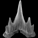
Two new scyliorhinid shark species (Elasmobranchii, Carcharhiniformes, Scyliorhinidae), from the Sülstorf Beds (Chattian, Late Oligocene) of the southeastern North Sea Basin, northern Germany.Thomas ReineckePublished online: 30/04/2014Keywords: Chattian; Elasmobranchii; North Sea Basin; Scyliorhinidae; Scyliorhinus https://doi.org/10.18563/pv.38.1.e1 Abstract Based on isolated teeth two new scyliorhinid shark species, Scyliorhinus biformis nov. sp. and Scyliorhinus suelstorfensis nov. sp., are described from the Sülstorf Beds, early-middle Chattian, of Mecklenburg, northeastern Germany. They form part of a speciose assemblage of necto-benthic sharks and batoids which populated the warm-temperate to subtropical upper shelf sea of the south-eastern North Sea Basin. PV article infos Published in Vol.38-1 (2014) |
|

|
Sur les Condylarthres Cernaysiens Tricuspiodon et Landenodon (Paléocène supérieur de France)Donald E. RussellPublished online: 01/10/1980Keywords: Arctocyonidae; Condylarths; Late Paleocene; Tricuspiodontidae https://doi.org/10.18563/pv.9.ext.127-166 Abstract The numerical importance of the Condylarths in the Cernaysian fauna is discussed. The Condylarth family, Tricuspiodontidae, is reviewed in the light of new material and its close relationships to the Phenacodontidae is suggested ; one new species is recognized : Tricuspiodon sobrinus. European Arctocyonidae are reviewed and the recentclassification of Van Valen is briefly commented on. Also, the arctocyonine Landenodon is described for the first time in Thanetian (Late Paleocene) sediments ; two new species are proposed : T. lavocati and T. phelizoni. PV article infos Published in Vol. 9, Ext (1980) |
|

|
Les rongeurs de l' Eocène inférieur et moyen d'Europe Occidentale; Systématique, phylogénie, biochronologie et paléobiogéographie des niveaux-repères MP 7 à MP 14.Gilles Escarguel
Published online: 15/12/1999 |
|

|
Prospection paléontologique de la région de Torralba de Ribota (Burdigalien du bassin de Calatayud, prov. de Zaragoza, Espagne)Edouard Boné, Maria T. Alberdi
Published online: 01/10/1980 |
|
|
Rongeurs Miocènes dans le valles-Penedes 1 : Les rongeurs de Can Ponsic 1Jean-Louis Hartenberger and Miquel Crusafont i PairóPublished online: 20/04/1979Keywords: Can Ponsic 1; Miocene; Rodents; Valles-Penedes https://doi.org/10.18563/pv.9.1.1-15 Abstract The rodents from the spanish locality of Can Ponsic 1 bring new data about some rodents species of the beginning of the Upper Miocene in South-West Europe. The criticims made by Mein and Freudenthal about the validity of the species Hispanomys thaleri from Can Llobateres are not justiíied. The study of the anatomy of the skull of Rotundomys from Can Ponsic 1 gives accurate information about the affinity of this genus with Cricetulus, and shows that the hypothesis, according to which Rotundomys is an ancestral form of the Arvicolids, is unlikely. The systematics of Heteroxerus and the phylogeny of the mio-pliocene Muscardinus species are also discussed. The Can Ponsic 1 locality is a little older than Can Llobateres. PV article infos Published in Vol. 09, Fasc. 1 (1979) |
|

|
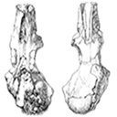
Revision der Equoidea aus den Eozänen Braunkohlen des Geiseltales bel Halle (DDR).Jens L. Franzen and Hartmut HauboldPublished online: 15/04/1986Keywords: Eocene; Europe; Mammalia; Perissodactyla; Stratigraphy; taxonomy https://doi.org/10.18563/pv.16.1.1-34 Abstract The dentitions as well as one complete and several partial skeletons of Equoids from the Eocene lignite beds of the Geiseltal locality are revised. Instead of 13 species distinguished up to now 3 chronoclines with 5 species and 3 separate species are recognized (text. fig. 1). Propalaeotherium hassiacum HAUPT, 1925 is evolving into Propalaeotherium isselanum (CUVIER, 1824) between the levels of the « obere Unterkohle ›› and the « untere Mittelkohle ›› of the Geiseltal section. Propalaeotherium argentonicum GERVAIS, 1849 is shown to be present in the « untere Unterkohle ››, whereas Lophiotherium pygmaeum (DEPERET,1901) occurs in the « obere Mittelkohle ›› and in the « oberes Hauptmittel ››. Plagiolophus cartieri STEHLIN, 1904 appears during the transition from the « Mittelkohle ›› into the « Oberkohle ›› as the earliest true Palaeothere. Therefore the « Oberkohle ›› is already regarded as Upper Eocene. This is corroborated by the occurrence of a phyletic descendant of Propalaeatherium parvulum (Propalaeotherium n.sp.) in the middle and upper "Oberkohle " because this species appears otherwise for the first time at the mammal level of Lissieu. On the other hand Propachynolophus gaudryz (LEMOINE, 1878) described by Matthes (1977) from the « untere Unterkohle ›› turns out te be in fact a Phenacodont. Thus the decisive argument for classifying the « untere Unterkohle ›› as Lower Eocene has to be dropped. Biostratigraphically the « Unterkohle ›› and the «Basishauptrnittel ›› correspond with the lower Middle Eocene (mammal level of Messel), whereas the «unteres Hauptmittel ›› and the « untere Mittelkohle ›› are equivalent to the middle part of the middle Eocene (mammal level of lssel), and the « obere Mittelkohle ›› together with the « oberes Hauptmittel ›› coincide with the upper Middle Eocene (mammal level of Bouxwiller). PV article infos Published in Vol. 16, Fasc. 1 (1986) |
|

|
The Gliridae (Mammalia) from the oligocene (MP24) of Gröben 3 in the folded molasse of southern GermanyUndine UhligPublished online: 28/12/2001Keywords: Biostratigraphy; Cyrena Beds; folded molasse; Germany; Gliridae; level MP 24; Mammals; Oligocene; Palaeoecology Abstract This study describes four taxa of Gliridae from the Oligocene mammal locality Gröben 3: Gliravus tenuis BAI-ILO, 1975, Bransatoglis micio (MISONNE, 1957), B. planus (BAHLO, 1975) and B. heissigi n. sp. Gliravus tenuis from Gröben 3 is somewhat more advanced than the type population found in Heimersheim. This confirms previous research suggesting that Gröben 3 should be dated earlier than Heimersheim (MP 24). The first documented occurrence of B. mício around level MP 24 was found in Gröben 3. An abundance of tooth material from B. planus in Gröben 3 makes it possible, for the first time, to observe evolutionary stages within this species from MP 21 until MP 28. B. heissigi n. sp. is restricted to level MP 24. This species is located between B. mísonnei (MP 20 - 23) and Microdyromys praemurinus (MP 25 - 28). Within the lineage Bransatoglis bahloi - B. misonnei - B. heissigi, a decrease in size is noticeable. PV article infos Published in Vol. 30, Fasc. 3-4 (2001) |
|

|
Fossil snakes from the Palaeocene of Sao José de Itaborai, Brazil.Part 1 Madtsoiidae, Aniliidae.Jean-Claude Rage
Published online: 15/12/1998 |
|

|
Une nouvelle espèce de Steneosaurus (Thalattosuchia, Teleosauridae) dans le Callovien du Poitou (France) et la systématique des Steneosaurus longirostres du Jurassique moyen d'Europe Occidentale.Patrick Vignaud
Published online: 15/09/1998 |
|

|
Hexanchiforme nouveau (Neoselachii) du Crétacé inférieur du Sud de la FranceHenri Cappetta
Published online: 18/12/1990 |
|