
Abstract book of the 18th Conference of the EAVP
Pterosaurs from Coahuila
Pliocene-Pleistocene large mammals from Le Riège and Saint-Palais
Les sélaciens du Miocène de la région de Montpellier
Muridae du Pliocène supérieur d'Espagne et du midi de la France.
Contribution à l'étude des genres Gliravus et Microparamys.
Eocene (57) , Quercy Phosphorites (38) , Systematics (32) , Rodents (29) , Mammalia (27) , Rodentia (25) , Miocene (24)

|
Les traces de pas d'amphibiens, de dinosaures et autres reptiles du Mesozoïque Français : inventaire et interprétations.Georges Gand, Georges Demathieu and Christian MontenatPublished online: 15/12/2007Keywords: Footprints; France; Inventory; Mesozoic; palaeontology; palaeovenvironments; Stratigraphy https://doi.org/10.18563/pv.35.1-4.1-149 Abstract Since the 19th century, thousands of footprints were observed in the geological series of the French Mesozoic. All are located in the Triassic and Jurassic. After a promising beginning, in France, it is only a few papers which will be published in the first half 20th century, unlike the USA and of others countries of Western Europe. One ought to wait about 1950 for a revival and now they are nearly 200 papers which were devoted to the ichnofossils. The literature abundance and the renewed interest of the naturalists for the palichnologic studies decided to us to write a synthesis work. This one begins with a stratigraphic inventory in which, localisation, age and paleontological contents of about 180 fossiliferous sites are specified. After having pointed out the followed methods, the footprints paleontological interpretation is then approached in detail and the results obtained are replaced in stratigraphy to deduce the fauna evolution during the Mesozoic. So, it appears that Ichnologic data, more varied and rich in the Triassic and Liassic than those relating to the bones, very rare for the considered periods, are very informative. The middle Triassic (Anisian-Ladinian), thus reveals Cotylosauria, Lepidosauria, Crurotarsi with Rauisuchia, Ornithosuchidae, Crocodylia and Dinosauromorpha more the "Prodinosauria": Dinosamiforme whose skeletons are known in Argentina but only in Ladinian. The rather fast domination of Dinosaurs during Norian is also as well shown. The almost exclusive presence of their footprints, up to fifty cm long, in the Lower Hettangian indicates their supremacy in the environments. Footprints characterise not very deep life places located between inter-supratidal limits and often out of water. Sedimentologic and Palaeontologic studies showed that they were great coastal spaces during Middle Triassic, flood-plain with sebkhas while Upper Triassic, and a large !!coastal marsh!! in Grands-Causses during Liassic in which, mainly, fine stromatolithic layers were deposited. During the same periad, bay beaches spread in Vendée. During the Middle Jurassic, they are also brackish to lacustrine environments and recifallagoons in- the Upper Jurassic. Numerous measurements of the footprints and trackways directions showed that the animaIs moved there in weil defined directions, for long periods. They seem due to the palaeotopography of the life environments relatively stable. Also, the discovery of vegetal radicular networks and small footprints far away from the continental borderlands has suggested that the animals continuously lived in these palaeoenvironnements, belonging to large ecosystems, where the sedimentation rate was weak. This explains that thebadies could not fossilize there but only their footprints through the cyanobacterian action in main cases. From the vertical distribution of different ichnospecies, defined with adapted statistical methods, explained in this work, a palichnostratigraphy was established for the Middle Triassic. Although the footprints are also abundant in Hettango-Sinemurian of "Grands-Causses" and the Vendée, it was not possible, up to now, to establish any zonation in this series; Probably because the palichnofauna is too little diversified there, currently reduced to a majority of Theropods II-IV tridactyl traces. PV article infos Published in Vol. 35, Fasc. 1-4 (2007) |
|
|
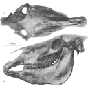
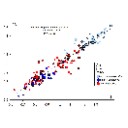
Old world hemiones and new world slender species (Mammalia, Equidae)Véra Eisenmann, John Howe and Mario PichardoPublished online: 16/12/2008Keywords: Amerhippus; biometry; Equus; Holocene; New World; Old World; Osteology; Pleistocene; Pliocene https://doi.org/10.18563/pv.36.1-4.159-233 Abstract Morphological and biometrical description of skulls, teeth, and limb bones of extant and fossil Old World herniones (including E. hydruntinus) and of New World 'stilt-Iegged' and other slender species from Blancan to Holocene. An Appendix presents ways in which the approximate size of some missing bones or dimensions may be deduced from available ones. PV article infos Published in Vol. 36, Fasc. 1-4 (2008) |
|

|
New remains of the very small cuckoo, Chambicuculus pusillus (Aves, Cuculiformes, Cuculidae) from the late Early/early Middle Eocene of Djebel Chambi, TunisiaCécile Mourer-Chauviré
Published online: 15/02/2016 |
|
|
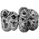
A new study of the anthracotheres (Mammalia, Artiodactyla) from pondaung formation, Myanmar: systematics implicationsAung N. SoePublished online: 16/12/2008Keywords: Anthracohyus; Anthracokeryx; Anthracotherium; Pondaung Formation; sexual dimorphism; Siamotherium; South East Asia; taxonomy https://doi.org/10.18563/pv.36.1-4.89-157 Abstract Anthracotheres from the Pondaung Formation, Myanmar, are considered as one of the most primitive artiodactyl groups and they represent the oldest known record in the world. Thus, the understanding of this group has numerous implications for evolutionary biology and biochronological correlations. However, the systematlcs of these mammals has been interpreted in different ways, and the main debate focuses on the number of taxa represented in the Pondaung Formation. The revised taxonomy proposed here is mainly based on the relative development of the upper molar W-shaped ectoloph, system of crests and stylar cusps, and on body size. On the basis of these characters, they are classified into four genera including six different species. Two well-known genera, Anthracotherium and Anthracokeryx, are validated and more precisely diagnosed. Anthracokeryx possesses a better developed W-shaped ectoloph, system of crests and stylar cusps than Anthracotherium, which displays notable differences with the more derived representatives of this genus. Both of these Pondaung genera show evidence for sexual dimorphism. However, the incompleteness of fossil material fueled a debate concerning the status of two additional Pondaung anthracotheres, Siamotherium and Anthracohyus. The latter genus is of uncertain affinities, but it has been considered as a hippopotamid ancestor. Despite new material attributed to these two forms, additional discoveries are still required to establish their taxonomic status. The hypothesis that Southeast Asia was the centre of origin of Anthracotheriidae is supported by the retention of numerous primitive dental characters in these taxa and by the antiquity of the Pondaung Formation, to which an age of 37 My is now generally accepted. PV article infos Published in Vol. 36, Fasc. 1-4 (2008) |
|
|
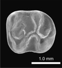
A new and primitive species of Protophiomys (Rodentia, Hystricognathi) from the late middle Eocene of Djebel el Kébar, Central TunisiaLaurent Marivaux
Published online: 02/06/2014 |
|

|
First record of dinosaur eggshells and teeth from the north-west african Maastrichtian (Morocco).Géraldine Garcia
Published online: 15/12/2003 |
|

|
Observations sur l'anatomie crânienne du genre Palaeotherium (Perissodactyla, Mammalia): mise en évidence d'un nouveau sous-genre, FranzenitheriumJean-Albert RemyPublished online: 01/12/1992Keywords: Palaeotherium; Paléogène; Perissodactyla; skull anatomy; Systematics https://doi.org/10.18563/pv.21.3-4.203-224 Abstract The skull remains referred to the genus Palaeotherium are recorded and described. Biometrical tests are made to elucidate intrageneric allometric relationships and to allow comparisons with various other perissodactyls. Apart from the well known shortness of post canine diastems and deepness of the narial opening, the genus is characterized by a great lengthening of the splanchnocranium, owing to a spreading of the post-orbital facial region, by a reduced area of the eye-socket and by the prevalence of the temporal muscle with regard to the masseter; this original shape of the masticatory apparatus needs to be related to the morphology of the jugal teeth and particularly to their asymmetrical semi-hypsodonty. PV article infos Published in Vol. 21, Fasc. 3-4 (1992) |
|

|
Rongeurs du Miocène inférieur et moyen en Languedoc. Leur apport pour les correlations Marin-Continental et la Stratigraphie.Jean-Pierre AguilarPublished online: 31/03/1980Keywords: Languedoc; Miocene; Rodents; Southern France https://doi.org/10.18563/pv.9.6.155-203 Abstract The rodents (Cricetidae, Gliridae, Sciuridae) found in lacustrine, brackish marine and karstic sediments of Miocene age in Languedoc, assign the position of the different localities in the scale of "niveaux repères" used by mammalogists. Some detailed stratigraphical studies bring several correlations between this continental biochronological scale and the marine scale ; the most important results are the Aquitanian age of the "niveaux repères" of Coderet and Paulhiac, the Burdigalian age of Laugnac, Estrepouy, Vieux-Collonges, La Romieu and Sansan and the Langhian or Lower Serravallian age of La Grive M. The correlations between the Tethys and the Central Paratethys for the Lower Neogene profit also of these results, since the locality of Neudorf Spalte 1, 2 (Czechoslovakia) is shown to be younger than Sansan (France). The paleontological study has also several geological inferences for the Miocene of Languedoc ; with the calibration of this Miocene, we know quite precisely that the Lower Miocene is chiefly a time lacustrine sedimentation, and also that the marine Miocene sedimentation ends early in the Miocene Period, in Langhian or lower Serravallian times. PV article infos Published in Vol. 09, Fasc. 6 (1980) |
|
|
|
|

|
Physogaleus hemmooriensis (Carcharhinidae, Elasmobranchii), a new shark species from the early to middle Miocene of the north sea basin.Thomas Reinecke and Kristiaan HoedemakersPublished online: 15/10/2006Keywords: Carcharhinidae; Early Miocene; Elasmobranchii; Hemmoorian; new species; North Sea Basin; Physogaleus https://doi.org/10.18563/pv.34.e14 Abstract A new carcharhinid shark species, Physogaleus hemmooriensis sp. nov., is described from the Lower Hemmoorian (Behrendorfian, late Burdigalian, early Miocene) of Werder, Lower Saxony, Germany. P. hemmooriensis also occurs in the Edegem and Antwerpen Sands Members of the Berchem Formation, Belgium, and in the Miste Bed, Aalten Member of the Breda Formation, The Netherlands, which have an early to middle Miocene age. In the Western Atlantic region, the taxon is present in the early Miocene Calvert Formation of Delaware, U.S.A, which is largely contemporaneous with the Hemmoorian. PV article infos Published in Vol. 34, Fasc. 1-2 (2006) |
|

|
Artiodactyla aus den Eozänen Braunkohlen des Geiseltales bei Halle (DDR)Jorg Erfurt and Hartmut HauboldPublished online: 04/12/1989Keywords: Artiodactyles; Eocene; Europe; Paleoecology; Stratigraphy; taxonomy https://doi.org/10.18563/pv.19.3.131-160 Abstract The present study of Artiodactyla from the Middle Eocene of the Geiseltal lignite beds concems systematics, biostratigraphy, and palaeoecology on the basis of 174 specimens: isolated remains to more complete skeletons. Instead of the formerly known five species of two families are now recognized 14 species of the Diacodexeidae, Dichobunidae, Cebochoeridae, and Haplobunodontidae. New species are Aumelasia maniai, Anthracobunodon neumarkensis, Masillabune franzeni. Four species of the Geiseltalfauna are definitely known from elswere, and five species are closely related to those from other European localities. Evidently the faunal situation of artiodactyls during the Middle Eocene of Europe was largely uniform. The distribution of artiodactyls within the sequence of the Geiseltal strata corroborates the biostratigraphical concept of the land mammal age Geiseltalian (Franzen & Haubold l986b) as well as the mammalian reference levels MP 11-13 (Franzen 1987). Reconstructions of the skulls and skeletons allow conclusions on the functional morphology and palaeoecology of the artiodactyls of the European Middle Eocene PV article infos Published in Vol. 19, Fasc. 3 (1989) |
|

|
Osteology of Prolagus sardus, a Quaternary Ochotonid (Mammalia, Lagomorpha).Mary R. DawsonPublished online: 21/06/1969Keywords: Lagomorpha; Ochotonidae; Prolagus https://doi.org/10.18563/pv.2.4.157-190 Abstract Prolagus sardus is the last representative of the diverse lineages of European endemic ochotonids. It is also the most abundant in the collections. The previous studies made of this species have established rather well its dental morphology, its phylogenetic position, its geographic and temporal distribution, and its intraspecific individual variation. On the other hand, no osteologic study has fully utilized the superb material from Corsica and Sardinia collected by Forsyth Major. PV article infos Published in Vol. 02, Fasc. 4 (1969) |
|
|
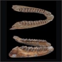
A mandible of the hyracoid mammal Titanohyrax andrewsi in the collections of the Muséum National d'Histoire Naturelle, Paris (France) with a reassessment of the speciesRodolphe Tabuce
Published online: 18/04/2016 |
|
|
Critical comments on the genus Propachynolophus Lemoine, 1891 (Mammalia, Perissodactyla, Equoidea)
|
S.I. Data |

|
Les traces de pas de Dinosaures et autres Archosaures du Lias inférieur des grands Causses, Sud de la FranceGeorges Demathieu, Georges Gand, Jacques Sciau, Pierre Freytet and Jacques GarricPublished online: 15/12/2002Keywords: Dinosauroid footprints; France; Grands-Causses; Hettangian; ichnostratigraphy; paleoenvironments; Sinemurian; statistical results https://doi.org/10.18563/pv.31.1-4.1-143 Abstract The Causses" is a near 3400 km2 large plateau located in the south of France. Here the first dinosaur footprints where found in 1935. After this, this area has yielded an ever-increasing number of ichnites now in excess of 500 specimens. These latter, 15 to 50 cm long, tridactyl or tetradactyl footprints of generally biped animals, were discovered at the surface of Hettangian to lower Sinemurian dolomite layers within 4 distinct stratigraphic units. The 35 sites bearing ichnites are located on the plateau margin. For the first time, morphologic characters studied through descriptive statistic methods with the usual parameters and classical Student and Snédecor tests, allowed us, to divide the whole set of biped traces into 6 ichnospecies. Their definitions are further constrained by multivariate statistical results using Principal Component Analysis (PCA), Factor Analysis of correspondances (FAC) and Discriminant Analysis (DA). All have confirmed the morphologic observations. So that now, the following taxa are identified : Grallator variabilis, G. lescurei, G. sauclierensis, G. minusculus, Eubrontes giganteus, Dilophosauripus williamsi, cf. Moraesichnium, Orníthopus fabrei nov ichnosp. The more immediately visible differences relate to the interdigital II-IV divarication and the digit length ratio. To this panel, we must add Batrachopus deweyi and shapes suggesting Trisauropodichnus and/or Anomoepus. Among all ichnite associations described in the lower Liasic, the New England assemblage presents the most affinities with ours. It shows the ichnotaxa Grallator, Dilophosauripus, Eubrontes, Batrachopus without forgetting Ornithopus fabrei nov. ichnosp. which is close to Ornithopus gallinaceus from the Massachusetts and Connecticut basins. On comparing the present early Jurassic ichnofauna of the Causses with the ones of the Middle and Upper Triassic formations of the eastem border of the Massif Central (France), it appears that tridactyl footprints become more and more numerous and large from Triassic to Early Jurassic. In the Causses, these latest are prevalent but in Quercy (France), Poland, Italy, USA, they are also associated with Omithopoda, Thyreophora and Sauropoda ichnites. Footprint areas considered here were generaly under an arid climate. Animals that passed by were heavy and bulky possible Megalosaur trackmakers, and lighter and slender Coelophysids or Ceratosaurs. For all, these areas were pathways as the orientations of the trackways seem point out. The directions followed by these reptiles were without any important variation during the Hettango-Sinemurian stages. These areas were also used from time to time by Crocodilomorpha and may be tetradactyl (I-IV) bipedal avian Theropods. However, the number of such trackways in sites, sometimes substantial, should not lead us to overestimate the trackmakers populations. These last were probably relatively moderately abondant in this inter-supratidal swamp environment. In the Causses, ichnites are connected with former algo-laminated deposits (Algal mats) which were rapidly hardened by means of calcitisation of cyanobacteria. The result has been a moderate depth of footprints; autopodia disturbing only a few cm of the carbonate substrate. Other fossils have been discovered : invertebrates with thin bivalve and gastropod shells, crustaceans tests and plants. These latter suggest the existence of paleomangroves like environments but also continental vegetation periodically overruning the swamp environment during regression/transgression cycles. At these times, wooded parts of it, could become protecting, feeding, resting and nesting places. PV article infos Published in Vol. 31, Fasc. 1-4 (2002) |
|

|
Nouvelle quantification de l'Hypsodontie chez les Theridomyidae : l'exemple de Theridomys ludensis nov. sp.Monique Vianey-Liaud
Published online: 30/12/1985 |
|

|
Rythme et modalités de l'évolution chez les rongeurs à la fin de l'Oligocène-leurs relations avec les changements de l'environnement.Bernard ComtePublished online: 15/12/2000Keywords: Environment; evolution; Oligocene; Rodents; Systematics Abstract The analysis of oxygene isotope variations as well as paleobotanical data suggest that the Oligocene/Miocene boundary corresponds to a transitional period marked by floristical and climatic variations. During this period, the pyreneo-alpine tectonics has contribued to modify the geography and western Europe landscapes. Faunal changes (appearances, extinctions, migrations) are observed in different mammalian groups, notably in the rodents. A study of the evolutionary trends and patterns in paleogene rodents is involved for the period ranging from level MP 28 of the Late Oligocene to the Early Miocene, including the Oligo-Miocene boundary. PV article infos Published in Vol. 29, Fasc. 2-4 (2000) |
|

|
Contribution à la classification des Pistes de Vertébrés du Trias : les types du Stormberg d'Afrique du Sud (2 ème Partie: le Stormberg supérieur - 1. Le biome de la zone B/1 ou niveau de Moyeni: ses biocénoses).Paul EllenbergerPublished online: 01/12/1974Keywords: biocenosis; Footprints; South Africa; Stormberg; Trias https://doi.org/10.18563/pv.6.ext Abstract Les Pistes de Vertébrés du Stormberg Supérieur ("Trias terminal à Rhétien"), ou Quthingien PV article infos Published in Vol. 6, Ext (1974) |
|

|
Observations sur des remaniements structuraux post-mortem dans des dents de mammifères fossiles provenant des phosphorites du QuercyJean-Albert RemyPublished online: 01/12/1974Keywords: Quercy Phosphorites; rearrangements; Teeth https://doi.org/10.18563/pv.6.3-4.163-176 Abstract Deux types de remaniements post mortem me paraissent caractéristiques de l'état de conservation des dents de mammifères fossiles dans les Phosphorites du Quercy : PV article infos Published in Vol. 06, Fasc. 3-4 (1975) |
|

|
The Quaternary avifauna of Crete, Greece.Peter D. WeesiePublished online: 01/09/1988Keywords: Avifauna; Crete; Quaternary; Systematics https://doi.org/10.18563/pv.18.1.1-94 Abstract Pleistocene bird fossils have been studied from nine localities on Crete. Part of this material was described earlier by the author (Weesie, 1982) and will not be treated here in extenso, the results will be incorporated. More than one third of the over 10,000 fossil bird bones available could be identified ; they were found to represent at least 65 bird species. The following species of the Pleistocene Cretan avifauna are new to the fauna of Crete : Branta ruficollis, Haliaeetus albicilla, Gyps melitensis, Aquila chrysaetos simurgh n. ssp., Ketupa zeylomensis, Aegolius funereus, Dendrocopos leucotos, Zoothera dauma, Turdus iliacus and Pyrrhula pyrrhula. The Pleistocene Cretan avifauna differs less from comparable mainland avifaunas than (fossil) avifaunas from oceanic islands do. Still, the Pleistocene Cretan avifauna has two qualities that are characteristic of island avifaunas : the almost complete absence of a group of birds (the Galliformes) and the presence of two endemic (sub)species : the giant eagle Aquila chrysaetos simurgh n. ssp. and the long-legged owl Athene cretensis (Weesie, 1982). The new subspecies is described in the present study. PV article infos Published in Vol. 18, Fasc. 1 (1988) |
|