
|
The Ctenodactylidae (Rodentia) from the Oligocene of Ulantatal (inner Mongolia, China)
Published online: 15/12/2006
Keywords:
Adaptive radiation; Ctenodactylidae; Mongolia; Oligocene; Rodents
https://doi.org/10.18563/pv.34.e11
Abstract
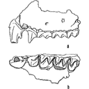
This paper proposes a systematic revision of the Oligocene Mongolian Ctenodactylidae, on the basis of abundant material obtained by screen/washing operations in stratified localities of the Ulantatal area (Inner Mongolia) (UTL1, 2, 3, 4, 5, 7, 6 & 8). A Chinese-German team has collected several thousands of isolated rodent teeth, and a number of fragmentary jaws. A new genus is identified (Alashania nov. gen. tengkoliensis nov. sp.), and eight former species are reevaluated, Karakoromys decessus, Tataromys sigmodon, T. minor, T. plicidens, Yindirtemys ulantatalensis, Y. bohlini, Y. deflexus, with several synonymies. A new Yindirtemys species is described: Y. shevyrevae nov. sp. and another one close to that: Y. aff. shevyrevae nov. sp. Four new species, which are rare in the localities, remain in open nomenclature because they are not well-represented. Yindirtemys differs from the other genera by the permanence of crescentic structures, while the other genera show a general reduction of the trigonoid area (= anterior valley). We define a range of size variation for each well documented population. Although the dental morphology shows a wide range of variation, given that transitional morphologies occur in a single locality, it is possible to provide a clear definition for most species. We show that dental patterns of the different genera can be derived from the pattern of Karakoromys. As a number of Tataromyinae have been determined in several localities from China, Kazakhstan and Mongolia, usually on the basis of scarce material, or surface collections, the present study would be used to re-evaluate their attribution inasmuch as the taxa are now placed in the Oligocene stratigraphy. The diversity of sizes and forms reflects the adaptive radiation of the family during the Oligocene, within a forested environment where the vegetation was probably abundant.
PV article infos
Published in Vol. 34, Fasc. 3-4 (2006)
|
PDF
|

|
Morphological description and identification of an extraordinary new elephant cranium from the early Pliocene of Ileret, Kenya
Published online: 21/10/2021
Keywords:
Elephantidae; Loxodonta adaurora; cranium; early Pliocene; Ileret; Kenya
https://doi.org/10.18563/pv.44.2.e3
Abstract
Abstract: Paleontological exploration in the Turkana Basin near Ileret, Kenya yielded the most complete adult elephant cranium (KNM-ER 63642) known from the late Miocene to mid-Pliocene. KNM-ER 63642 derives from the lower Lonyumun Mb. of the Koobi Fora Fm. and dates to the early Pliocene, >4.3 Ma. The cranium is immense in size and preserves most of its structures including left and right M2-3, permitting its comprehensive comparative study and secure taxonomic assignment to Loxodonta adaurora. Features distinctive of the species and exhibited by KNM-ER 63642 include very elongate, divergent tusk alveoli, a short, biconvex cranial roof, anterosuperior angulation of the occipital planum, non-inflated occipital planum and absence of supralateral parietal "bossing," broad, flat premaxillary nasal processes, broad, laterally downturned nasal aperture superior to the level of the orbits, and M3s with wide, subhypsodont plates that are parallel-faced and separated by U-shaped transverse valleys. The M3s also exhibit characteristic L. adaurora traits of greatest width at their bases, rounded cross-sectional shape, thick enamel, abundant cementum, and strong anterior and posterior accessory conules. Of extant taxa, KNM-ER 63642 most closely resembles crania of African elephants. Its inclusion in the Loxodonta clade is tenuous, however, because shared features are either symplesiomorphic or are difficult to test for synapomorphy due to the poor fossil record of crania of late Miocene-early Pliocene elephants. Overall, the cranial morphology of KNM-ER 63642 is unexpectedly advanced for an elephant of its antiquity. Its anteroposterior compression and height are concordant with efficient proal masticatory action, indicating that by the early Pliocene L. adaurora evolved craniodental adaptations in phase with feeding preference for C4 grasses. The advantage of synchrony of morphology and behavior is reflected by the dominance of the species in the greater Turkana Basin during that interval.
PV article infos
Published in 44-2 (2021)
|
PDF
|

|
Observations sur l'anatomie crânienne du genre Palaeotherium (Perissodactyla, Mammalia): mise en évidence d'un nouveau sous-genre, Franzenitherium
Published online: 01/12/1992
Keywords:
Palaeotherium; Paléogène; Perissodactyla; skull anatomy; Systematics
https://doi.org/10.18563/pv.21.3-4.203-224
Abstract
The skull remains referred to the genus Palaeotherium are recorded and described. Biometrical tests are made to elucidate intrageneric allometric relationships and to allow comparisons with various other perissodactyls. Apart from the well known shortness of post canine diastems and deepness of the narial opening, the genus is characterized by a great lengthening of the splanchnocranium, owing to a spreading of the post-orbital facial region, by a reduced area of the eye-socket and by the prevalence of the temporal muscle with regard to the masseter; this original shape of the masticatory apparatus needs to be related to the morphology of the jugal teeth and particularly to their asymmetrical semi-hypsodonty.
These animals, whose running ability was evidently poor, appear to have been adapted to rather closed environments, feeding on relatively soft vegetable matter; olfactory sense was likely to play a leading part in interindividual and environmental relationships. Such evolutionary trends might explain the disappearance of most of them, as clirnatic conditions deteriorated at the end of the Eocene, before the "Grande Coupure" which affected mammalian faunas at that time.
Although the present paper is not directly concemed with phylogenetics, it invalidates the supposed ancestor-descendant relationship between P. castrense and P. magnum, and it suggests a possible emergence of the P. medium lineage from a P. siderolithicum stock. Moreover, the structure of the post-orbital facial area allows the erection of a new sub-genus, Franzenitherium, for the species lautricense and duvali.
PV article infos
Published in Vol. 21, Fasc. 3-4 (1992)
|
PDF
|
|
Book of Abstracts of the XXII Annual Meeting of the European Association of Vertebrate Palaeontologists, 30 June–5 July 2025, Kraków, Poland
Published online: 20/06/2025
Keywords:
Abstracts; EAVP
https://doi.org/10.18563/pv.eavp2025
Abstract
Book of Abstracts of the XXII Annual Meeting of the European Association of Vertebrate Palaeontologists, 30 June–5 July 2025, Kraków, Poland.
PV article infos
Published in 48-1 (2025)
|
PDF
|
|
Preliminary report on the fishes (Chondrichthyes & Teleostei) from the lower Oligocene (Rupelian) Red Bluff Clay at site AMo-9, Monroe County, Alabama, USA
Published online: 26/06/2024
Keywords:
Batomorphii; Elasmobranchii; Galeomorphi; Gulf Coastal Plain; Vicksburg Group
https://doi.org/10.18563/pv.47.2.e2
Abstract
Herein we describe a small but relatively diverse assemblage of fossil fishes derived from the lower Oligocene (Rupelian) Red Bluff Clay at site AMo-9 in Monroe County, Alabama, USA. Identified amongst the remains are 15 unequivocal taxa representing 11 families within five orders, and one additional taxon represents an unknown order and family. Taxa identified include Eostegostoma sp., Otodus (Carcharocles) sp., Mitsukurinidae/Carchariidae indet., Macrorhizodus praecursor, Galeorhinus sp., Negaprion gilmorei, Physogaleus sp., “Sphyrna” sp., Galeocerdo sp., cf. “Aetobatus” sp., Sphyraena sp., Xiphiorhynchus kimblalocki, Xiphiorhynchus sp., Cylindracanthus ornatus, and C. rectus. Several additional fossils could not be identified beyond Lamniformes, Carcharhiniformes, and Teleostei, but they likely belong to one of the identified taxa within this paleofauna. All of the fishes previously reported from the Red Bluff Clay within the entirety of the Gulf Coastal Plain of the USA are otolith-based, and each of the 15 unequivocal taxa reported herein are important new records for this lithostratigraphic unit. In particular, the Eostegostoma sp. and Xiphiorhynchus spp. specimens represent the first occurrences of these taxa in Alabama. The specimens of C. ornatus, Eostegostoma sp., and X. kimblalocki are stratigraphic and temporal range extensions from the middle and late Eocene into the Rupelian Stage of the Oligocene. Other described taxa may represent transitional forms between those described from the late Eocene and late Oligocene within the region. This study provides a tantalizing preliminary view into faunal transitions that occurred amongst marine fishes across the Eocene/Oligocene boundary within the Gulf Coastal Plain of the USA.
PV article infos
Published in 47-2 (2024)
|
PDF
|
|
The beginning of the adaptive radiation of Theridomorpha (Rodentia) in Western Europe: morphological and phylogenetic analyses of early and middle Eocene taxa; implications for systematics
Published online: 20/09/2021
Keywords:
characters analyses; Dental morphology; Eocene; Rodentia; variability
https://doi.org/10.18563/pv.44.2.e2
Abstract
This paper provides a revision of the early and middle Eocene European rodents previously referred to as Ischyromyoidea, including taxa considered to be at the origin of the Theridomorpha. The use of an accurate dental terminology and a better understanding of the size and shape of their infra-orbital foramen (i.o.f.) led us to a substantial revision of this group, which allowed to better characterize them and to appreciate their variability. On these bases, phylogenetic analyses (cladistic and standard Bayesian
approaches) of early Ypresian to late Priabonian European rodent species were undertaken in order to highlight the root of the early Theridomorpha and its content. In this paper, the phylogeny was established based on 343 characters (338 dental) through 45 early Paleogene taxa using both cladistic and bayesian analyses. The ingroup included on one hand a few North American genera (Reithroparamys, Microparamys, and Acritoparamys) and European ones (Eogliravus, Ailuravus, Corbarimys, Meldimys, Euromys, Plesiarctomys, and Pseudoparamys) considered until now as being related with the North American superfamily Ischyromyoidea. On the other hand, it included genera close to the root of the Theridomorpha (Sparnacomys, Pantrogna, and Hartenbergeromys) and early Theridomyoidea (Masillamys, Protadelomys, and some Pseudosciuridae). The phylogenetic results obtained via the two
distinct reconstruction approaches are consistent in virtually all relationships. The proposed systematics here derives from these phylogenetic results. This phylogenetic context led us to change the suprafamilial, familial, subfamilial or generic attribution of several species. Characters of Theridomorpha, like the obliquely developed postprotocristid allied with the occurrence of a metalophulid I, have been found in genera previously considered as Ischyromyidae (Pseudoparamys, Euromys, Sparnacomys, Meldimys, Pantrogna, and Hartenbergeromys) as well as the large i.o.f., when preserved (Pseudoparamys, Hartenbergeromys, and Masillamys). Based on these morphological observations and new phylogenetic considerations, the content of the Theridomorpha clade is here enlarged, thereby extending back the first theridomorph radiations to the early Eocene. Aside, a new taxon (Reinomys rhomboides gen and sp. nov.) is described from Avenay. In addition, a new genus, Auroremys, is created for the species subita (Comte et al., 2012) from Chery-Chartreuve.
PV article infos
Published in 44-2 (2021)
|
PDF
S.I. Data
|

|
Decouverte d'un nouveau Diacodexis (Artiocactyla, Mammalia) dans l'Eocène inférieur de Silveirinha, Portugal.
Published online: 15/09/1989
Keywords:
Artiodactyla; Eocene; Migration; Portugal; Silveirinha
https://doi.org/10.18563/pv.19.1.29-44
Abstract
A new artiodactyl, Diacodexis antunesi n.sp., is described from the early Eocene of Silveirinha, Portugal. Comparisons are made with Diacodexis gazini GODINOT, 1978, D. varleti SUDRE et al., 1983, D. cf. varleti from Paris Basin sites, D. sp. from Dormaal and from localities in Spain and England, D. secans from North America and D. pakistanensis from Asia; affinities and evolutive tendencies are discussed. The presence of Diacodexis in the locality of Silveirinha confirms the very early Eocene age of the latter. As Diacodexis antunesi appears ta be more primitive than D. gazini from Rians (early Eocene of France), it lends corroboration to the interpretation (essentially based previously on condylarths) of the Silveirinha assemblage as the oldest Eocene fauna known in Europe and supports the hypothesis that early artiodactyls migrated from Europe to North America.
PV article infos
Published in Vol. 19, Fasc. 1 (1989)
|
PDF
|

|
La plus ancienne faune de mammifères du Quercy : Le Bretou
Published online: 01/12/1974
Keywords:
Le Bretou; Quercy Phosphorites
https://doi.org/10.18563/pv.6.3-4.177-196
Abstract
Redécouvertes en 1968 dans le bois du Bretou, les deux poches à phosphorites qui ont livré cette faune sont parmi les plus méridionales du plateau quercynois. De faible profondeur, huit mètres, les cavités sont de petite taille et font partie d'un complexe de sept fissures réparties dans les bois du Bretou. Les autres trous n'ont livré aucun fossile, soit parce qu'ils avaient été intégralement vidés de leur sédiment, soit que des venues d'eau en interdisent l'accès.
Dans les deux poches fossilifères, proches de quelques mètres, le sédiment que l'on rencontre en placage sur les parois présente un aspect particulier. Il s'agit en effet d'une brèche très indurée, où la calcite domine, et contenant assez peu de sédiments sidérolithiques. Les ossements contenus dans cette brèche sont assez nombreux, très souvent brisés. Les dents isolées y sont relativement rares. Exploitées séparément, les deux poches ont livré des faunes identiques, aucune différence chronologique ne pouvant être décelée entre elles. Les formes rencontrées dans une seule des cavités sont rares mais la poche la plus riche est celle dénommée Le Bretou 2.
L'intérêt de la faune récoltée réside dans son ancienneté. On sait en effet que H.G. Stehlin avait signalé dans les collections du Quercy des restes attribuables à Lophiodon lautricense, mais la provenance exacte de ces fossiles reste inconnue. Au Bretou si nous n'avons pas rencontré à ce jour le Lophiodon, il n'en demeure pas moins que l'âge de la faune récoltée doit être proche de celui de la faune de Robiac.
PV article infos
Published in Vol. 06, Fasc. 3-4 (1975)
|
PDF
|

|
Contribution à l'étude des genres Gliravus et Microparamys (Rodentia) de l'Eocène d'Europe.
Published online: 15/03/1971
Keywords:
Eocene; Gliravus; Microparamys; Rodentia
https://doi.org/10.18563/pv.4.4.97-135
Abstract
Based on material found in about 15 localities the relationships of the genera Microparamys and Glirarus have been studied. One new genus, two subgenera and three species [Microparamys (Sparnacomys) chandoni n. subgen. and n. sp., Microparamys (Pantrogna) russelli n. subgen., Eoglirarus wildi n. gen. and n. sp., Gliravus meridionalis n. sp.] as well as the publication
of numerous new facts concerning species previously reported, support the phyletic scheme proposed. The latter shows that the origin of the Gliravinae is to be sought among the very small and still rather poorly known Microparamys species of the early Eocene. Gliravus: robiacensis can be considered as the common ancestor to different lineages not only of Glirarus but also of modern genera (Peridyromys, Glirudinus and Microdyromyx), at the origin of which Peridyromys micio, although difficult to interpret, occupies a similar place.
The stratigraphic conclusions permit more detail in the chronologie succession of the localities studied. The paleoecologic and biogeographic aspects lead one to the problem of the oligocene "Grande Coupure" through the study of this group.
PV article infos
Published in Vol. 04, Fasc. 4 (1971)
|
PDF
|
|
Les Chiroptères du Miocène inférieur de Bouzigues. 1- Etude systématique.
Published online: 17/04/1968
Keywords:
bats
https://doi.org/10.18563/pv.1.3.65-133
Abstract
In recent years, the techniques of chemical preparing have permitted a rich paleontologic material to be obtained from the phosporitic sediment of Bouzigues (Hérault, France). The fauna of this locality is comprised of quite varied microvertebrates, amphibians, reptiles, birds, mammals. Twenty five species of the latter, belonging to seven orders, are today known from the site. Among them, the numerous rodents have allowed L. Thaler to chronologically situate this fauna in the Zone of Laugnac (<< late Aquitanian ›> of some authors).
The chiropterans are, with the rodents, the best represented of the locality's mammals. Three families comprise the bat fauna, with nearly complete dominance by one of them (Hippoxideridae) over the two others (Megadermatidae and Vespertílionidae)
Six forms are described, of which three are new species and one a new sub-genus.
Megaderma braillomi n. sp., an animal of rather large size, shows like the Miocene megaderms several evolved dental characters, translating the adaptation of these animals to a partially carnivorous regime. The Bouzigues species seems, however, to represent a particular lineage.
Hipposideros (Brachipposideros n. subgen.) dechaseauxi n. sp. and Hípposideros (Brachipposideros) cf. collongenris Depéret, small sized forms, belong to a group rather well represented in the late Oligocene and early Miocene of Europe, and not distinguished until now within the genus Hipposideros.
Hipposideros (Pseudorhinolophus) bouziguensis n. sp., is the most abundant mammal in the locality and, occuring at the Oligocene-Miocene limit, the last representative known of the subgenus Pseudorhínolophus, common in Europe from the middle Eocene.
However, beyond Neogene and Quaternary times, certain among the numerous living species of Hipposideros are close to Pseudorhinolophus and others to Brachipposíderos. 'This fact would in the future justify a global revision of the genus, on the basis of comparative anatomy of the squeleton and of the teeth.
The bat fauna of Bouzigues is completed by two small Vespertilionidae, rare forms, Myoris sp. I and sp. II.
PV article infos
Published in Vol. 01, Fasc. 3 (1968)
|
PDF
|

|
Field trip guides of the 20th Annual Conference of the European Association of Vertebrate Palaeontologists, 26th June – 1st July 2023, Sabadell (Barcelona), Spain
Published online: 16/06/2023
Keywords:
https://doi.org/10.18563/pv.eavp2023fieldtrip
Abstract
PV article infos
Published in special issue 1-2023 (2023)
|
PDF
|

|
L'occlusion dentaire chez Peradectes, Amphiperatherium et Peratherium, Marsupiaux du tertiaire d'Europe.
Published online: 01/10/1980
Keywords:
Didelphidae; Eocene; Mastication; Oligocene; Wear facets
https://doi.org/10.18563/pv.9.ext.79-89
Abstract
The general principles guiding the study of wear facets which develop during mastication in mammals possessing tribosphenic molars are named. The application of this method of study to the molars of European Tertiary Didelphidae shows that the lineage of this family as represented by the species Peratherium cuvieri (Upper Eocene), P. elegans (Lower-middle Oligocene) and P. antiquum (Upper Oligocene) has propessively evolved toward a more carnivorous diet.
PV article infos
Published in Vol. 9, Ext (1980)
|
PDF
|

|
Les Amphibiens et les reptiles du Pliocène supérieur de Balaruc II (Herault, France)
Published online: 15/09/1989
Keywords:
amphibians; Europe; France; Pliocene; Reptiles
https://doi.org/10.18563/pv.19.1.7-28
Abstract
The late Pliocene site (MN 16) of Balaruc II (Hérault, France) has provided remains of the following amphibians and reptiles: Chelotriton pliocenicus nov. sp. and Triturus marmoratus (Salamandridae), cf. Rana (Ranidae), cf. Blanus (Amphisbaenidae), cf. Agama (Agamidae), Gekkonidae indet., Lacerta s.l. (Lacertidae), "Ophisaurus" sp. (Anguidae), Michauxophis occitanus (Aniliidae), Erycinae indet. (Boidae), Elaphe cf. E. longissima and Malpolon sp. (Colubridae), cf. Naja (Elapidae) and Vipera sp. (Viperidae). The salamandrid Chelotriton pliocenicus and the aniliid Michauxophis occitanus constitute, up to now, the only records of these groups in the European Pliocene. The fauna is indicative of a warm, dry
subtropical climate.
PV article infos
Published in Vol. 19, Fasc. 1 (1989)
|
PDF
|

|
Contribution à la classification des Pistes de Vertébrés du Trias : les types du Stormberg d'Afrique du Sud (2 ème Partie: le Stormberg supérieur - 1. Le biome de la zone B/1 ou niveau de Moyeni: ses biocénoses).
Published online: 01/12/1974
Keywords:
biocenosis; Footprints; South Africa; Stormberg; Trias
https://doi.org/10.18563/pv.6.ext
Abstract
Les Pistes de Vertébrés du Stormberg Supérieur ("Trias terminal à Rhétien"), ou Quthingien
Si les zones du Stormberg inférieur se sont révélées contenir de nombreuses traces, surtout dans les faciès dits "Molteno moyen et supérieur", représentant apparemment la base du Keuper, il est frappant de voir pratiquement l'ensemble de cette grosse faune "Molteno" disparaître avec la fin de cette période, que nous avons appelée le "Maphutsengien".
Dès les premières zones du Stormberg supérieur, que nous nommons le"Quthingien" la zoocénose et la phytocénose, en même temps que les données d'ensemble manifestées par l'environnement, sont modifiées. Nous ne verrons plus guère de dépôts marécageux à flore riche et variée, parfois même luxuriante. Les fougères elles-mêmes ont disparu. Elles sont remplacées par de maigres plantes, aux feuilles très souvent filiformes qui paraissent témoigner d'un climat continental. Le sol est devenu de plus en plus rouge, avec des variations latérales beaucoup plus accusées. Les fleuves amenant des galets des monts du "Grand Sud" ont tari. La faune va en subir les conséquences. Certaines des espèces se révèleront sautillantes ou coureuses, pour un grand nombre plus légères et pour la quasi-totalité d'apparence carnivore ou entomophages, les phytophages devant se contenter d'un régime ingrat,difficile ou à tout le moins irrégulier,les dépôts le montrent.
C'est dans ces conditions que s'inaugure notre Etage nouveau,quelque peu discordant sur les zones A/5, A/6 ou A/7 du Stormberg inférieur (Maphutsengien). Le Stormberg supérieur (ou Quthingien) commence avec le paléopaysage remarquable dit de Moyeni, que nous allons maintenant étudier, typologiquement, avec ses homologues du même âge. Quelques 38 types d'animaux tous nouveaux vont défiler à nos yeux lors de la zone de base de cet Etage, ou zone B/1.
L'on nous avait proposé d'intituler ce Ile Tome de la série : "La grande Dalle de Moyeni et ses homologues. Paléo-spectacles, scènes et paysages animaux au Lesotho à l'approche du Trias finissant". Nous avons préféré garder le sous-titre plus haut, peut-être plus prosaïque.
Un llle Tome est en préparation : "Les développements ultérieurs et terminaux de la faune du Gondwana".
PV article infos
Published in Vol. 6, Ext (1974)
|
PDF
|
|
A partial skeleton of Metaxytherium medium from the middle Miocene of La Morfassière quarry (Indre-et-Loire, France)
Published online: 16/01/2025
Keywords:
Faluns; France; Metaxytherium; Miocene; Sirenia
https://doi.org/10.18563/pv.48.1.e1
Abstract
Sirenians are among the common marine fossil remains found in the Faluns deposits of western France. We describe new material of a Dugongidae sirenian from the middle Miocene Savignean facies of La Morfassière quarry (Indre-et-Loire, northwestern France) that includes a well preserved and almost complete skull associated with its mandible, several vertebrae and ribs. The cranial remains exhibit features that allow to attribute them to Metaxytherium medium, a species recorded from the middle and early late Miocene of European and Mediterranean coasts. The discovery of an associated skull and mandible of this species is unusual in this area and deserves to be reported, mostly because its preservation contributes to a better knowledge of the variable structure of its skull and teeth anatomy. For the first time the body size and weight of M. medium can be assessed through regression equations based on skull measurements. The particularly good condition of the material can be explained by the calm and deep marine environment in which it was deposited.
PV article infos
Published in 48-1 (2025)
|
PDF
|

|
La poche à Phosphate de Ste-Néboule (Lot) et sa faune de vertébres du Ludien supérieur. 5-Squamates
Published online: 25/09/1978
Keywords:
Eocene; Quercy Phosphorites
https://doi.org/10.18563/pv.8.2-4.201-215
Abstract
The Squamates from Sainte-Néboule are charateristic of the Upper Eocene from the Phosphorites du Quercy. Most genera known from this locality do not survive the « Grande Coupure ». Cadurcoboa insolita gen. nov., sp. nov. is a small Boidae very characteristic of the Upper Eocene.
PV article infos
Published in Vol. 08, Fasc. 2-4 (1978)
|
PDF
|

|
The Quaternary avifauna of Crete, Greece.
Published online: 01/09/1988
Keywords:
Avifauna; Crete; Quaternary; Systematics
https://doi.org/10.18563/pv.18.1.1-94
Abstract
Pleistocene bird fossils have been studied from nine localities on Crete. Part of this material was described earlier by the author (Weesie, 1982) and will not be treated here in extenso, the results will be incorporated. More than one third of the over 10,000 fossil bird bones available could be identified ; they were found to represent at least 65 bird species. The following species of the Pleistocene Cretan avifauna are new to the fauna of Crete : Branta ruficollis, Haliaeetus albicilla, Gyps melitensis, Aquila chrysaetos simurgh n. ssp., Ketupa zeylomensis, Aegolius funereus, Dendrocopos leucotos, Zoothera dauma, Turdus iliacus and Pyrrhula pyrrhula. The Pleistocene Cretan avifauna differs less from comparable mainland avifaunas than (fossil) avifaunas from oceanic islands do. Still, the Pleistocene Cretan avifauna has two qualities that are characteristic of island avifaunas : the almost complete absence of a group of birds (the Galliformes) and the presence of two endemic (sub)species : the giant eagle Aquila chrysaetos simurgh n. ssp. and the long-legged owl Athene cretensis (Weesie, 1982). The new subspecies is described in the present study.
These endemic birds of prey were found in association with their supposedly principal prey species (now extinct as well) : endemic mice for the owl and endemic deer for the eagle. Endemic mammals have been found in association with endemic birds of prey on many islands, not only in the Mediterranean. There is evidence that the size of endemic birds of prey becomes optimally adapted to their feeding on certain endemic mammals, especially rodents. Another characteristic of the Pleistocene Cretan avifauna is the great number of species of birds of prey. This appears to be a common characteristic of fossil avifaunas from caves on Mediterranean islands as well as from caves on the European mainland. However, we think that ecological conditions on Pleistocene Crete (especially the abundant presence of mice) helped to account for the high representation of birds of prey. Furthemore, the fossil avifauna enables us to draw some conclusions about the climate and vegetation on Pleistocene Crete : it is concluded that the climate was cooler than today and that Crete was largely covered with forests. Finally, the reasons for the extinction or disappearance from Crete of some bird species of the Pleistocene Cretan avifauna are discussed.
PV article infos
Published in Vol. 18, Fasc. 1 (1988)
|
PDF
|

|
Book of Abstracts of the 20th Annual Conference of the European Association of Vertebrate Palaeontologists, 26th June – 1st July 2023, Sabadell (Barcelona), Spain
Published online: 15/06/2023
Keywords:
Abstracts; EAVP
https://doi.org/10.18563/pv.eavp2023
Abstract
PV article infos
Published in special issue 1-2023 (2023)
|
PDF
|
|
First report of Cylindracanthus (Osteichthyes) from the Eocene of India
Published online: 25/03/2024
Keywords:
Cylindracanthus; Eocene; histology; rostrum; Umarsar mine.
https://doi.org/10.18563/pv.47.1.e2
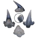
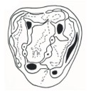
Abstract
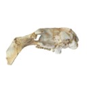
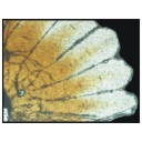
Fossils of the endangered sturgeons and peddlefishes are widely distributed. We here report for the first time the presence of one of the extinct osteichthyes genus Cylindracanthus (Liedy 1856a) from the Early Eocene lignite-bearing successions of the Kutch Basin, India. The present well preserved rostrum is characterised by numerous wedge-shaped components encircling the central canal that runs along its length, paired at the base and each wedge contributing to the formation of a ridge. The rostrum lacks teeth. The present find extends the palaeobiogeographical distribution of Cylindracanthus considerably and supports its Eocene age as dental remnants preserved in Cylindracanthus sp. shows a decrease in remanent dentition and tooth bases from the Cretaceous to the Eocene. Cylindracanthus is an useful palaeoenvironmental indicator as it has been found associated typically with deposits of nearshore marine environments.
PV article infos
Published in 47-1 (2024)
|
PDF
|

|
A New caseid Synapsid from the Permian (Guadalupian) of the Lodève basin (Occitanie, France)
Published online: 18/07/2022
Keywords:
; Caseidae; France; Guadalupian; semi-aquatic lifestyle
https://doi.org/10.18563/pv.45.2.e2
Abstract
Lalieudorhynchus gandi gen. nov. and sp. nov. is a new caseid synapsid from the Permian of the Lodève Basin, Occitanie, France. This new taxon is represented by a partial but well-preserved postcranial skeleton, and is characterized by the following apomorphies: a transverse section of the sacral and anterior caudal neural spines with a very thin keel-like process anteriorly, a slender dorsal tip of the dorsal and caudal spines, a narrow distal end of the first sacral rib, a fossa on triceps process of metacoracoid, and a very large distal tarsal 1 of same width than the astragalus, with nearly all sides being shallowly concave.
The skeleton corresponds to a sub-adult individual that was excavated from the La Lieude Formation dated as Roadian-Capitanian (Guadalupian). A sedimentological and taphonomical analysis of the type locality, together with preliminary osteohistological observations, suggest that this new French caseid was rather aquatic, as already hypothesised for other large forms.
A phylogenetic analysis of caseids is performed to test the position of this new taxon and to better understand the evolution of the clade: interestingly, Lalieudorhynchus gandi gen. nov. et sp. nov. is closer to the NorthAmerican “Cotylorhynchus” hancocki than to the other French caseids Ruthenosaurus and Euromycter from the Artinskian of the geographically closer Rodez Basin. These two last caseids document the Artinskian radiation of the clade, which remained diverse until Olson’s extinction. Caseids survived, as Lalieudorhynchus is one of the youngest representatives of the clade, and may have used novel ecological strategies to access their vegetarian food sources.
PV article infos
Published in 45-2 (2022)
|
PDF
|
