
Abstract book of the 18th Conference of the EAVP
Pterosaurs from Coahuila
Pliocene-Pleistocene large mammals from Le Riège and Saint-Palais
Les sélaciens du Miocène de la région de Montpellier
Muridae du Pliocène supérieur d'Espagne et du midi de la France.
Contribution à l'étude des genres Gliravus et Microparamys.
Eocene (57) , Quercy Phosphorites (38) , Systematics (32) , Rodents (29) , Mammalia (27) , Rodentia (25) , Miocene (24)
|
Batoids (Rajiformes, Torpediniformes, Myliobatiformes) from the Sülstorf Beds (Chattian, Late Oligocene) of Mecklenburg, northeastern Germany: a revision and description of three new speciesThomas ReineckePublished online: 24/06/2015Keywords: Batoids; Chattian; Elasmobranchii; North Sea Basin; Oligocene https://doi.org/10.18563/pv.39.2.e2 Abstract Bulk-sampling of fossil-rich tempestites from the Chattian Sülstorf Beds of PV article infos Published in Vol.39-2 (2015) |
|
|
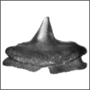
New Squalicorax species (Neoselachii: Lamniformes) from the Lower Maastrichtian of Ganntour phosphate deposit, MoroccoHenri Cappetta
Published online: 05/12/2014 |
|

|
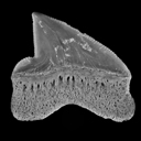
Additions to the elasmobranch fauna from the upper Cretaceous of New Jersey (middle Maastrichtian, Navesink Formation)Gerard R. Case and Henri Cappetta
Published online: 15/12/2004 |
|
|
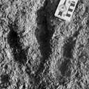
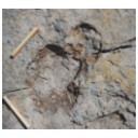
Nouvelles données sur les Ichnites de dinosaures d'El Bayadh (Crétacé Inférieur, Algérie)Mostefa Bessedik
Published online: 16/12/2008 |
|
|
Small sauropod tracks in the Hettangian of Southern France – A case of ichnite fossilization in an intertidal zonePierre Demathieu, Alain Izart, André Charrière
Published online: 28/06/2022 |
|
|
Latest Early-early Middle Eocene deposits of Algeria (Glib Zegdou, HGL50), yield the richest and most diverse fauna of amphibians and squamate reptiles from the Palaeogene of AfricaJean-Claude Rage
Published online: 08/02/2021 |
|
|
|
|

|
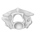
Die Ohr-Region der Paulchoffatiidae (Multituberculata, Ober-Jura).Gerhard HahnPublished online: 15/11/1988Keywords: Multituberculata; Ober-Jura; Paulchoffatiidae; Petrosum; Portugal https://doi.org/10.18563/pv.18.3.155-185 Abstract The petrosal of the Paulchoffatiidae HAHN, 1969 is described and compared with that of younger multituberculates and of other Mesozoic mammals. The "Morrison petrosal", described by Prothero (1983), is also discussed; it probably belongs to the multituberculates. The reconstruction of the ventral side of the Paulchoffatiinae-skull, given by Hahn in 1987, is completed by addition of the otic and the occipital region. PV article infos Published in Vol. 18, Fasc. 3 (1988) |
|

|
Rongeurs caviomorphes de l'Oligocène de Bolivie. 1 Introduction au deseadien de BolivieRobert HoffstetterPublished online: 01/08/1976Keywords: Rodentia; South America https://doi.org/10.18563/pv.7.3.1-16 Abstract The Tertiary of the Salla-Luribay basin consists of red beds affected by the second period of the andine compression, of Miocene ending age. The Tertiary layers are exposed at an approximate elevation of 3.500 to 4.000 meters. Two stratigraphic units can be distinguished in them: the Luribay conglomerates, in which vertical clifts result from erosion, and the Salla layers consisting mostly of consolidated clays. These clays are very fossiliferous and have furnished a rich vertebrate fauna which gave to R. Hoffstetter the possibility to establish the Oligocene age of these beds. Sediments of same age has been reported to be present in several other places of Bolivia, particularly near Lacayani, where have been collected highly hypsodont Rodents, different from those found in Salla-Luribay basin. PV article infos Published in Vol. 07, Fasc. 3 (1976) |
|

|
An unusual cranial fossil of the giant lower Pliocene shrew (Paranourosorex gigas Rzebik-Kowalska, 1975) from Podlesice, PolandDavid L. Harrison and Barbara Rzebik-Kowalska
Published online: 29/10/1991 |
|

|
Préface au mémoire jubilaire en hommage à René LavocatJacques MichauxPublished online: 01/10/1980Keywords: Editorial https://doi.org/10.18563/pv.9.ext.1-13 Abstract Monsieur René Lavocat, Directeur du Laboratoire de Paléontologie des Vertébrés de la troisième section de l'Ecole Pratique des Hautes Etudes, quittait le service actif en l'année 1979. View editorial Published in Vol. 9, Ext (1980) |
|

|
Neue Beobachtungen zum Schädel-und Gebiss-Bau der Paulchoffatiidae (Multituberculata,Ober-Jura).Gerhard HahnPublished online: 15/12/1987Keywords: Dentition; Paulchoffatiidae; Portugal; Skull structure; Upper Jurassic https://doi.org/10.18563/pv.17.4.155-196 Abstract The ventral face of the Paulchoffatiinae skull (Multituberculata, Lower Kimmeridgian, Portugal) is new reconstructed. Some details hitherto unknown are added, as the presence of jugals, the structure of the palatine and the extension of the pterygoids. The situation of the m2/ is discussed. Kielanodon hopsoni n. g., n. sp. is erected, known by its upper p3-5/. From Guimarotodon leiriensis the mandible with its dentition is made known. New informations concerning the milk-dentition and the replacement of teeth are also added. PV article infos Published in Vol. 17, Fasc. 4 (1987) |
|

|
Insectivores pliocènes du Sud de la France (Languedoc-Roussillon) et du Nord-Est de l'Espagne.Jean-Yves CrochetPublished online: 31/10/1986Keywords: Biostratigraphy; Insectivora; Languedoc; Pliocene; Spain; Systematics https://doi.org/10.18563/pv.16.3.145-171 Abstract The first lists of Insectivores (Erinaceidae, Talpidae and Soricidae) from the Pliocene beds of Southern France and North-East Spain are given in this paper. The material from twelve localities is studied. These localities are geographically situated in Languedoc (Celleneuve, Vendargues, Nîmes, Sète, Balaruc 2 and Seynes), in Roussillon (Terrats, Serrats-d'en-Vacquer, Château d'eau and Mont-Hélène) and in North-East Spain (Layna, Medas Islands and Puebla de Valverde). These faunas correspond to the Early, Middle and Late Pliocene. 1 to 8 taxa are identified in these localities and 14 specific taxa are presently listed for this period in this area. Two new specific taxa are described as Galerix depereti nov. sp. from all the Early Pliocene localities in the North-Pyrenean area and as Desmanella gardiolensis nov. sp. from Balaruc 2. For this small mammals, two faunal assemblages are recognized. The first one is dated from the Early Pliocene (F 1, 2 and 3 zones in Aguilar et Michaux) and is characterized by Galerix depereti and rare and little diversified Soricids. The second one is Late Pliocene in age (zones G 2 and G 3). The fossils of the genus Talpa are relatively abundant and the Soricids are diversified and very abundant. The Middle Pliocene (zone G 1) is a transitional period. ln these faunas, most of the insectivore genera are known from the European Late Miocene beds (8 on 10). This fact demonstrates a relative continuity between the invectivore faunas from the Late Miocene to the Early Pliocene. In conclusion, somme paleoecological considerations are suggested. PV article infos Published in Vol. 16, Fasc. 3 (1986) |
|

|
A new Ardynomys (Rodentia,Cylindrodontidae) from the Eocene of the eastern Gobi Desert, Mongolia.Demberelyin DashzevegPublished online: 16/12/1996Keywords: Ardynomys; Eocene; Mongolia; Rodentia; Systematics Abstract A partial skull of Ardynomys russelli sp. nov. (Rodentia, Cylindrodontidae) is described. This was collected in the late Eocene of Alag Tsab locality in the eastem Gobi Desert, Mongolia. Ardynomys russelli sp. nov. is characterized by small size, brachyodont molars, and retention of P3. It represents the earliest record of the genus Ardynomys MATTHEW & GRANGER, 1925, in Asia. PV article infos Published in Vol. 25, Fasc. 2-4 (1996) |
|

|
Revision des faunes de vertébrés du site de Provenchères-sur-Meuse (Trias terminal, Nord-Est de la France)Gilles Cuny
Published online: 14/06/1995 |
|

|
The microfauna of the Djebel Qafze CaveG. HaasPublished online: 15/09/1972Keywords: Micromammals; Rodents https://doi.org/10.18563/pv.5.5.261-270 Abstract This study examines the microfauna from Djebel Qafze Cave, focusing on mammalian remains, with Microtus guentheri as the dominant species and a notable absence of Cricetinae (hamsters). The richest layers (XVIa, XVII, XVIII) suggest accumulation by raptors, particularly owls, given the high presence of nocturnal species like voles and shrews. Two significant species, Arvicanthis cf. ectos and the newly described Rattus (? Mastomys) nazarensis, are highlighted. Reptiles (gekkos, Ophisaurus) and birds are also present, while hedgehogs and lagomorphs are absent. The findings underscore the role of avian predators in fossil deposition and refine the chronology of regional Pleistocene fauna. PV article infos Published in Vol. 05, Fasc. 5 (1972) |
|

|
Contribution à l'étude des Cricétidés oligocènes d'Europe occidentaleMonique Vianey-Liaud
Published online: 20/01/1972 |
|
|
Contributions à l'étude de l'anatomie crânienne des rongeurs. 1- Principaux types de cricétodontinésJean-Louis HartenbergerPublished online: 25/09/1967Keywords: Cricetodon; Cricetodontinae; Miocene https://doi.org/10.18563/pv.1.2.47-64 Abstract Description, for the first time, of the skull of Ruscinomys Depéret on the basis of a nearly complete specimen, and description of a new facial part of a Megacricetodon Fahlbusch skull (material from upper Miocene, Spain). New description of the skull (facial part) of " Cricetodon" incertum Schlosser on the basis of the specimen from the Oligocene of Quercy phosphorites already published by S. Schaub. PV article infos Published in Vol. 01, Fasc. 2 (1967) |
|
|
Terrestrial vertebrate paleocommunities from the Cerro del Pueblo Formation (Late Cretaceous; Late Campanian) at Las Aguilas, Coahuila, MexicoHéctor E. Rivera-Sylva
Published online: 16/07/2019 |
|
|
Late Campanian theropod trackways from Porvenir de Jalpa, Coahuila, MexicoHéctor E. Rivera-Sylva
Published online: 29/11/2017 |
|