|
The geologically youngest remains of an ornithocheirid pterosaur from the late Cenomanian (Late Cretaceous) of northeastern Mexico with implications on the paleogeography and extinction of Late Cretaceous ornithocheirids
Published online: 21/07/2020
Keywords:
Coahuila; Late Cenomanian; north-eastern Mexico; Ornithocheiridae; Pterosauria
https://doi.org/10.18563/pv.43.1.e4
Abstract
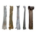
Ornithocheirid pterosaurs were the largest of the toothed pterodactyloids and had a worldwide distribution, although their fossil record is fragmentary, with the exception of the north-eastern Brazilian Crato and Santana Formations (Aptian, ?Albian, Early Cretaceous). With Istiodactylidae, they were also the only toothed pterosaurs that survived into the Cenomanian (Late Cretaceous), becoming extinct at the end of this period. Here we report on an ornithocheirid metacapus from the Late Cenomanian laminated limestone of north-eastern Mexico discovered about 120 km north-west of Ciudad Acuña, northern Coahuila at the south banks of Rio Bravo. The specimen comprises a fragmentary distal syncarpal, a crushed but complete metacarpal IV, two fragmentary preaxial metacarpals and a possible fragmentary terminal left wing finger phalanx. It represents the geologically youngest known ornithocheirid worldwide. We suggest that ornithocheirid pterosaurs may have become extinct because of massive sea level fluctuations during the mid to late Cretaceous that may have obliterated their breeding sites on coastal plains and low lying islands.
PV article infos
Published in Vol 43-1 (2020)
|
PDF
|

|
Pantolestidae nouveaux (Mammalia, Insectivora) de l'Eocène moyen de Bouxwiller (Alsace).
Published online: 31/03/1970
Keywords:
Bouxwiller; Insectivora; Mammalia; Middle Eocene; Pantolestidae
https://doi.org/10.18563/pv.3.3.63-82
Abstract
The Pantolestidae from the middle eocene of Bouxwiller are the subject of a detailed study. Buxolestes hammeli (n. g., n. sp.) is not closely related to any other European or North American form described until now; it presents, however, some characters in common with Pantolestes, a form of the same age from North America. A parallel evolution from a common ancestral form could explain this ressemblance.
Another form (gen. and sp. indet.) accompanies Buxolertes hammeli in the Bouxwiller fauna.
PV article infos
Published in Vol. 03, Fasc. 3 (1970)
|
PDF
|

|
La poche à phosphate de Sainte-Néboule (Lot) et sa faune de vertébrés du Ludien supérieur. 13-Rongeurs
Published online: 25/09/1978
Keywords:
Eocene; Quercy Phosphorites
https://doi.org/10.18563/pv.8.2-4.313-318
Abstract
Sainte-Néboule has yielded only 4 species of Rodents. But the Theridomyids (Blainvillimys rotundidens and Patriotheridomys altus) are very significative of the age of the locality: Ste-Néboule is lower than the marker level of Escamps
PV article infos
Published in Vol. 08, Fasc. 2-4 (1978)
|
PDF
|

|
Compléments sur les Chiroptères de l'Eocène moyen d'Europe. Les genres Palaeochiropteryx et Cecilionycteris.
Published online: 01/10/1980
Keywords:
Chiroptera; Geiseltal; Messel; Middle Eocene
https://doi.org/10.18563/pv.9.ext.91-126
Abstract
New dental and skeletal material referable to Palaeochiropteryx tupaiodon from the Middle Eocene locality of
Messel (G.F.R.) is studied, which provides additions to the previously gained knowledge of this european genus. Dental specimens from Geiseltal (G.D.R.), also of Middle Eocene age, allow us to analyze Cecilionycteria prisca. Some of these are separated to establish a new genus, Matthesia, and two new species, M. germanica and M. ? insolita.
PV article infos
Published in Vol. 9, Ext (1980)
|
PDF
|

|
Observations sur des remaniements structuraux post-mortem dans des dents de mammifères fossiles provenant des phosphorites du Quercy
Published online: 01/12/1974
Keywords:
Quercy Phosphorites; rearrangements; Teeth
https://doi.org/10.18563/pv.6.3-4.163-176
Abstract
Deux types de remaniements post mortem me paraissent caractéristiques de l'état de conservation des dents de mammifères fossiles dans les Phosphorites du Quercy :
1) Des destructions localisées d'origine biologique, sous forme de galeries de morphologie très variable creusées dans la dentine et le cément, et impliquant sans doute la participation de différents types de micro-organismes. Ces altérations se sont développées peu de temps après la mort, avant la fossilísation proprement dite et se sont rapidement arrêtées après l'enfouissement dans le sédiment phosphaté.
2) Des perturbations dans les structures de la dentine liées aux variations locales de minéralisation, provoquées par une imprégnation diffuse des zones les moins calcifiées par divers minéraux et probablement surtout de l`apatite.
PV article infos
Published in Vol. 06, Fasc. 3-4 (1975)
|
PDF
|

|
Les traces de pas de Dinosaures et autres Archosaures du Lias inférieur des grands Causses, Sud de la France
Published online: 15/12/2002
Keywords:
Dinosauroid footprints; France; Grands-Causses; Hettangian; ichnostratigraphy; paleoenvironments; Sinemurian; statistical results
https://doi.org/10.18563/pv.31.1-4.1-143
Abstract
The Causses" is a near 3400 km2 large plateau located in the south of France. Here the first dinosaur footprints where found in 1935. After this, this area has yielded an ever-increasing number of ichnites now in excess of 500 specimens. These latter, 15 to 50 cm long, tridactyl or tetradactyl footprints of generally biped animals, were discovered at the surface of Hettangian to lower Sinemurian dolomite layers within 4 distinct stratigraphic units. The 35 sites bearing ichnites are located on the plateau margin. For the first time, morphologic characters studied through descriptive statistic methods with the usual parameters and classical Student and Snédecor tests, allowed us, to divide the whole set of biped traces into 6 ichnospecies. Their definitions are further constrained by multivariate statistical results using Principal Component Analysis (PCA), Factor Analysis of correspondances (FAC) and Discriminant Analysis (DA). All have confirmed the morphologic observations. So that now, the following taxa are identified : Grallator variabilis, G. lescurei, G. sauclierensis, G. minusculus, Eubrontes giganteus, Dilophosauripus williamsi, cf. Moraesichnium, Orníthopus fabrei nov ichnosp. The more immediately visible differences relate to the interdigital II-IV divarication and the digit length ratio. To this panel, we must add Batrachopus deweyi and shapes suggesting Trisauropodichnus and/or Anomoepus. Among all ichnite associations described in the lower Liasic, the New England assemblage presents the most affinities with ours. It shows the ichnotaxa Grallator, Dilophosauripus, Eubrontes, Batrachopus without forgetting Ornithopus fabrei nov. ichnosp. which is close to Ornithopus gallinaceus from the Massachusetts and Connecticut basins. On comparing the present early Jurassic ichnofauna of the Causses with the ones of the Middle and Upper Triassic formations of the eastem border of the Massif Central (France), it appears that tridactyl footprints become more and more numerous and large from Triassic to Early Jurassic. In the Causses, these latest are prevalent but in Quercy (France), Poland, Italy, USA, they are also associated with Omithopoda, Thyreophora and Sauropoda ichnites. Footprint areas considered here were generaly under an arid climate. Animals that passed by were heavy and bulky possible Megalosaur trackmakers, and lighter and slender Coelophysids or Ceratosaurs. For all, these areas were pathways as the orientations of the trackways seem point out. The directions followed by these reptiles were without any important variation during the Hettango-Sinemurian stages. These areas were also used from time to time by Crocodilomorpha and may be tetradactyl (I-IV) bipedal avian Theropods. However, the number of such trackways in sites, sometimes substantial, should not lead us to overestimate the trackmakers populations. These last were probably relatively moderately abondant in this inter-supratidal swamp environment. In the Causses, ichnites are connected with former algo-laminated deposits (Algal mats) which were rapidly hardened by means of calcitisation of cyanobacteria. The result has been a moderate depth of footprints; autopodia disturbing only a few cm of the carbonate substrate. Other fossils have been discovered : invertebrates with thin bivalve and gastropod shells, crustaceans tests and plants. These latter suggest the existence of paleomangroves like environments but also continental vegetation periodically overruning the swamp environment during regression/transgression cycles. At these times, wooded parts of it, could become protecting, feeding, resting and nesting places.
PV article infos
Published in Vol. 31, Fasc. 1-4 (2002)
|
PDF
|

|
The skull of Arsinoitherium (Mammalia, Embrithopoda) and the higher order interrelationships of ungulates
Published online: 17/12/1992
Keywords:
Arsinoitherium; PHYLOGENY; Skull; Ungulate
https://doi.org/10.18563/pv.22.1.1-43
Abstract
Detailed anatomical description of arsinoithere cranial remains from the Lower Oligocene, Fayum Depression, Egypt, provides the basic data for a systematic investigation. All cranial and some postcranial features are assessed from a phylogenetic standpoint. Several soft tissue characters are then added to a cladistic analysis based on 54 derived ungulate morphological characters. The resulting phylogenetic hypothesis implies that perissodactyls, sirenians, proboscideans and arsinoitheres constitute a monophyletic unit (5 synapomorphies). However, increasing the tree length by 3 steps reveals a closer association between hyraxes and perissodactyls. Nevertheless, 13 synapomorphies link proboscideans, sirenians and arsinoitheres to the exclusion of all other ungulates. Form of the sphenopalatine and ethmoid foramina, recurved posttympanic process, absence of a fenestra rotundum in the petrosal, vestigial paroccipital process of the exoccipital and the highly unusual absence of a hypoglossal foramen in the skull, imply a robust sister-group relationship between arsinoitheres and proboscideans. In this analysis artiodactyls share only one derived character with all other ungulates studied. Monophyly of Ungulata, including Artiodactyla, is therefore only weakly supported. It is argued that pedal anatomy of hyraxes is non-homologous with that of Tethytheria. Arsinoitherium should now be classified within Tethytheria, sharing a sister-group relationship with Proboscidea. Hyraxes are excluded, thus refuting the concept of Paenungulata. However, monophyly of the wider concept, Pantomesaxonia, containing hyraxes, perissodactyls, sirenians, proboscideans and now, arsinoitheres, is supported by this study.
PV article infos
Published in Vol. 22, Fasc. 1 (1992)
|
PDF
|

|
Agriotherium intermedium (Stach 1957) from a Pliocene fissure filling of Xiaoxian County (Anhuei Province, China) and the phylogenetic position of the genus.
Published online: 30/09/1983
Keywords:
Carnivora; China; PHYLOGENY; Pliocene; skull anatomy; Ursidae
https://doi.org/10.18563/pv.13.3.65-81
Abstract
A fragmentary mandible and maxilla of a small sized Agriotherium of a young individual discovered from a Pliocene fissure filling in Xiaoxian county (Anhuei Province, China) are described. Judging from the morphology of the dentition and its dimensions the new material can be identified as Agriotherium inlermedium (STACH l957). Hendey's proposition (1980) that the Agriotherium species are derived from Indarctos is reconsídered on the basis of the new documents. As a result of a more general phylogenetic discussion it can be stated, that: 1. the supposed size increase as well as other trends, leading from Indarctos to Agriotherium are untenable ; 2. there are no positive indications to assume a phylogenetic transition of these two genera. 3. there are no real arguments in favor of an adaptational reversal in the evolution of Agriotherium. Hence, many features of that genus supposed by Hendey to be derived are plesiomorphic ; 4. regardless of the previous points it is methodologícally impossible to establish direct ancestor - descendant relationships between Indarctos and Agriotherium species, as Hendey did. Based on the data available and especially on the characters of the new material from China it is more likely that Agriotherium and Indarctos are two genera which developed independently. While advanced Agriotherium species, e.g. A. africanum, resemble in some respects Indarctos by adaptational analogies, more primitive species, e.g. Agriotherium intermedium, are quite dissimilar to lndarctos. While Indarctos might be derived from an Ursavus like forerunner, Agriotherium has its roots more likely somewhere in between Ursavus and the Hemicyon-group.
PV article infos
Published in Vol. 13, Fasc. 3 (1983)
|
PDF
|
|
A new and primitive species of Protophiomys (Rodentia, Hystricognathi) from the late middle Eocene of Djebel el Kébar, Central Tunisia
Published online: 02/06/2014
Keywords:
Adaptive radiation; Bartonian; Dental morphology; North Africa; Paleobiogeography
https://doi.org/10.18563/pv.38.1.e2
Abstract
Based on fossil discoveries and phylogenetic studies, an Eocene Asian origin for hystricognathous rodents and anthropoid primates has gained strong support in recent years. The two groups then invaded both Africa and South America, which promoted their evolutionary success. However, the fossil record has so far failed to constrain the nature and precise timing of these pivotal dispersal events. In Africa, given the apparent absence of hystricognaths and anthropoids in early to early middle Eocene localities, it is suggested that these mammal groups dispersed from Asia to Africa sometime during the middle Eocene. In this paper, we report the discovery of several isolated teeth of a rodent from a new vertebrate locality situated in central Tunisia (Djebel el Kébar, KEB-1), dating from the late middle Eocene (Bartonian, ~39.5 Myr). These fossils document a diminutive new species of Protophiomys (P. tunisiensis nov. sp.), a basal genus of hystricognathous rodents which is well known from several North African mammalian-bearing localities of the end of the Eocene. The teeth of P. tunisiensis display a suite of anatomical details comparable with those observed in the other species of the genus, but with a lesser degree of development. Such an apparent primitive evolutionary stage is corroborated by the greater antiquity of this Tunisian species. P. tunisiensis nov. sp. is so far the most ancient representative of hystricognaths in Africa. However, it can be expected that hystricognaths were already present on that landmass given the new data on early caviomorphs recently reported from South America (at ~41 Myr). The arrival of hystricognaths in Africa from South Asia certainly predates the depositional period of the Kébar sediments, but perhaps not by much time.
PV article infos
Published in Vol.38-1 (2014)
|
PDF
|

|
Le genre Plagiolophus (Palaeotheriidae, Perissodactyla, Mammalia): révision systématique, morphologie et histologie dentaires, anatomie crânienne, essai d'interprétation fonctionnelle
Published online: 15/12/2004
Keywords:
New taxa; Paléogène; perissodactyls; skull anatomy; tooth histology
Abstract
The genus Plagiolophus is documented, almost solely in Western Europe, from the middle Eocene up to the mid Oligocene (MP 12 to MP 25), i.e. more than for 15 MY. Seventeen species are now recorded whose two of them are new, P. ringeadei nov. sp. and P. mamertensis nov. sp. Some anatomical variations and the deflection of certain evolutionary trends justify the distinction of three subgenera, Paloplotherium, Fraasiolophus nov. and Plagiolophus s.s. The genus displays a wide range in size and weight (between 10 and 150 kg). The detailed description of the skull of several species is here given for the first time.
Despite important evolutionary drifts during this long time span, the dentition shows a great structural homogeneity, which renders difficult the determination of fragmentary specimens or isolated teeth. It is characterized by a great heterodonty; premolars are little molarized and present a certain regression through time with paradoxically some progress in the molarization. The hypsodonty increases: the first Plagiolophus are hardly less brachyodont than Propalaeotherium, and the last ones are nearly as hypsodont as Merychippus from the early Miocene. The upper molars change from a wide crown pattern, with an open occlusal surface, lightly oblique transverse lophs and rounded internal cusps, to a narrower pattern, with a frontally constricted occlusal surface and internal lophs aligned parallel to the ectoloph. The M3/3 become always longer.
The dental enamel displays horizontal Schreger-bands with imprecise limits occupying only the middle part of the enamel layer. The dentine is remarkable by its high rate of pericanalicular dentine. The crown cementum, lacking in earlier forms, increases to the point where it fills the occlusal valleys of the
teeth.
The masticatory musculature shows a increasing prominence of the temporal, with probably an important role devoted to the pterygoid muscles in lateral movements related to a two-phase type of chewing.
The evolution of the dentition, of the masticatory musculature and of the repartition of masticatory forces indicate that the Plagiolophus have known different diets through their long evolutionary history; at first browsers they became mixed feeders and finally grazers. Their relatively long neck allowed these animals to reach different vegetal layers. The strength of the nuchal crests also suggests that they were able to have strong backwards movements of the head to pull up their food.
This evolution of diet seems related to the slow degradation of environmental conditions attested during this period in western Europe, with the generalization of more open landscapes, increasing aridity and more marked seasons.
Besides, a remodeling of the face is ontogenetically and along time observed, in relation with the evolution of the masticatory apparatus and especially with that of the mandibular lever arm. The postcanine diastemata become longer in the course of evolution; the free extremities of the nasals are always relatively long which contradicts the hypothesis according to which Paloplotherium may have had a trunk. At last the lineage Fraasiolophus can be distinguished by the presence of a deep malar fossa, probably related to a strong development of the maxillo-labialis superior muscle.
The orbit is always large and tends to increase in size, which indicates a good development of the vision and its increasing role in the life relations. A peculiar type of epitympanic sinus could have been used as a resonance chamber insuring a certain amplification of sounds before their transmission to the eardrum. The endocranial cast reveals a relatively large brain with an advanced degree of gyrencephaly. Beside the role eventually played in food research and social relations, these neurophysiological abilities, also related to an advance in cursorial fitness, could have contributed to the survival of these animals facing the predation pressure of the first fissipede carnivores and the competition with new immigrant herbivores after the "Grande Coupure".
On the basis of some shared apomorphies with the Pachynolophinae, which prevent from considering the latter as Equidae (molarization of the premolars, reduction of the premaxilla dorsal apophysis, peculiar epitympanic sinus, splitting of the jugular process), the hypothesis of an autochthonous origin of Plagiolophus issued from a form near Propalaeotherium, is once again proposed and discussed. Finally, intra-generic relationships are taken into consideration.
PV article infos
Published in Vol. 33, Fasc. 1-4 (2004)
|
PDF
|
|
Lissamphibians from Dams (Quercy, SW France): Taxonomic identification and evolution across the Eocene-Oligocene transition
Published online: 25/06/2025
Keywords:
Eocene-Oligocene; Grande Coupure; Lissamphibia; Quercy Phosphorites
https://doi.org/10.18563/pv.48.1.e3
Abstract
The locality of Dams (Quercy, southwestern France) has yielded two fossil assemblages, one from the late Eocene and another from the early Oligocene, making it one of the few localities with infillings across the Eocene-Oligocene transition. At least 24 taxa (13 mammals, 11 snakes) have been identified in this locality. Study of the lissamphibian remains from Dams yields an Eocene and an Oligocene assemblage, with a total of eight taxa. The Eocene assemblage includes two unnamed salamandrine species, one unnamed pelobatid species and one pyxicephalid species (Thaumastosaurus). The Oligocene assemblage includes two unnamed pleurodeline species, one salamandrine species (Salamandra sansaniensis) and an unnamed pelobatid species. Among the eight taxa from Dams, one Eocene salamandrine and one Oligocene pleurodeline are identified for the first time in the Quercy. A review of the lissamphibians from the Quercy area identifies eleven taxa for the Late Eocene (MP19) and eight taxa for Early Oligocene (MP22), with a major turnover at the Eocene-Oligocene transition. This turnover occurs in a time of major climatic changes, with a significant decrease in temperature and precipitation and concurrent increase in seasonality in Europe, likely affecting specialized taxa.
PV article infos
Published in 48-1 (2025)
|
PDF
|
|
Eocene Teleostean Otoliths, Including a New Taxon, from the Clinchfield Formation (Bartonian) in Georgia, USA, with Biostratigraphic, Biogeographic,
and Paleoecologic Implications
Published online: 03/01/2022
Keywords:
climate; Congridae; Ophidiidae; Sciaenidae; tectonics
https://doi.org/10.18563/pv.45.1.e1
Abstract
Investigations of the Clinchfield Formation (middle Eocene, upper Bartonian) exposed at the Hardie Mine (Wilkinson County, Georgia, USA), produced 4,768 actinopterygian otoliths representing 14 taxa and increased the number of bony fishes threefold from the site. The somewhat limited richness was characterized by bonefishes, mud eels, conger eels, sea catfishes, cusk-eels, snooks, grunts, drums and croakers, and porgies. The assemblage had a relatively even distribution with Ophidiidae, Congridae, and Sciaenidae most common. Included in the otolith taxa was a new sciaenid genus and species, Eosciaena ebersolei, with unknown relationships to other Sciaenidae. The Clinchfield otoliths were compared to other middle and late Eocene in age otolith assemblages in Alabama, Mississippi, and Louisiana utilizing percentage similarity measurements. Analysis indicated that the Clinchfield otoliths were not greatly similar or greatly unlike the Moodys Branch and Yazoo Clay otolith assemblages. However, the Clinchfield showed little relationship to the slightly older Lisbon Formation in adjacent Alabama and is postulated to be related to global climatic and plate tectonic events. Biostratigraphically, the Clinchfield otolith taxa are essentially the same as the other formations except for the Lisbon, which has at least ten unique species. Abundances of Clinchfield otolith taxa indicate a possible sub-bioprovince in the eastern Gulf Coastal Plain. The Clinchfield otoliths indicate a tropical to perhaps subtropical, soft substrate, mainly normal marine to slightly reduced salinities, inner shelf (0–20 m) paleoenvironment with indications of proximal continental coastlines. This investigation represents an initial step in addressing the immensely understudied Paleogene otolith assemblages in Georgia.
PV article infos
Published in 45-1 (2022)
|
PDF
|

|
Study of the Turolian hipparions of the lower Axios valley (Macedonia, Greece). 4. Localities of Dytiko.
Published online: 15/12/1988
Keywords:
Equidae; Greece; Hipparion; Lower Axios Valley; Macedonia; Mammalia; Turolian
https://doi.org/10.18563/pv.18.4.187-239
Abstract
The hipparions from the Dytiko localities of the lower Axios valley (Macedonia, Greece) are studied. The material comes from three localities Dytiko-l, 2, 3 (DTK, DIT, DKO), which are situated near the village of Dytiko, about 60 km northwest to Thessaloniki. Three species have been determined, the medium-sized H. mediterraneum, the small-sized H. matthewi and the very small-sized H. periafricanum. The determined Hipparion species, their morphological characters and their comparison with the other Axios valley material indicate a Late Turolian age for the Dytiko localities.
PV article infos
Published in Vol. 18, Fasc. 4 (1988)
|
PDF
|

|
Sur les empreintes de pas des gros mammifères de l'Eocène supérieur de Garrigues-ste-Eulalie (Gard)
Published online: 01/10/1980
Keywords:
Eocene; Euzet; Footprints; Ichnofauna
https://doi.org/10.18563/pv.9.ext.37-78
Abstract
Is hereby described an impressive lchnoiauna belonging to the Lower to Middle Ludian of the Gard (S. France). The slab, already cleaned over a length of 18 m, is located near the top of the Potamides aporoschema lacustrine limestone (Lower Ludian, Euzet zone). It is therefore older than the Célas sandstone deposit, and still more than the Melanoides albigensis and M. acutus marly limestone corresponding to the Upper Levels of the Ludian stage. Although biostratigraphically older than the La Débruge and Montmartre zone, the biotope shows already a sampling of very tall Artiodactyles, Perissodactyles and Carnivorous. One of the most « majestic ›› Artiodactyles, Anopolotheriipus lavocati, nov., points out a huge size type. To mention also among the Ichnotypes described, 10, the big Perissodactyle Palaeotheriipus similimedius, nov., and the big Carnivorous Hyaenodontipus praedator, nov.
PV article infos
Published in Vol. 9, Ext (1980)
|
PDF
|

|
Nouvelle quantification de l'Hypsodontie chez les Theridomyidae : l'exemple de Theridomys ludensis nov. sp.
Published online: 30/12/1985
Keywords:
Dental morphology; evolution; Hypsodonty; Oligocene; Theridomyidae
https://doi.org/10.18563/pv.15.3.159-172
Abstract
A new example of parallelism in the dental pattern ofthe Theridomyidae is illustrated by the description ofa new species: Theridomys Iudensis from the standard level of Antoingt (middle Oligocene). Considering the occurence ofthis parallelism phenomenon. the use of numerous qualitative and quantitative criteria is essential to characterize the different stages ofthe different evolutive lineages. Thus, a new simple parameter is proposed (CHY = H+l/0,5 L) to estimate hypsodonty of the medium hypsodont Rodentia.
PV article infos
Published in Vol. 15, Fasc. 3 (1985)
|
PDF
|

|
An Australian Miocene Brachipposideros (Mammalia, Chiroptera) related to Miocene representatives from France
Published online: 15/12/1982
Keywords:
Australia; bats; Chiroptera; Miocene
https://doi.org/10.18563/pv.12.5.149-172
Abstract
A new middle Miocene hipposiderid bat is described from a limestone deposit on Riversleigh Station in north-western Queensland. Hipposideros (Brachipposideros) nooraleebus n. sp. is the first record of this subgenus from anywhere in the world outside of France. The palaeoecological setting of the fossil bats appears to have been a relatively quiet, sunny lime-enriched tropical pool that contained tortoises, crocodiles and fish. It is possible that the bats were washed into the pool from an adjacent cave.
The Riversleigh bat most closely resembles the French Burdigalian (early middle Miocene) bat H. (B.) aguilari. It is also possible that it may have been closely related to the original Australian hipposiderid stock that ultimately gave rise to the endemic monotÿpic Rhinonycteris aurantius. The disjunct distribution of species of H. (Brachipposideros) suggests that representatives of this subgenus will be found in at least tropical southern Asia.
PV article infos
Published in Vol. 12, Fasc. 5 (1982)
|
PDF
|

|
Arvicolinae (Rodentia) du Pliocène terminal et du quaternaire ancien de France et d'Espagne.
Published online: 30/10/1971
Keywords:
Arvicolinae; France; Pleistocene; Pliocene; Spain
https://doi.org/10.18563/pv.4.5.137-214
Abstract
Two steps can be distinguished in the history of the first invasion of western and south western Europe by the arvicolines. The first step corresponds to the installation of these rodents with the immigration of Promimonys inxuliferus Kowalski, then of Mimomys stehlini Kormos and of Mimomys gracilis (Kretzoi). The second is characterized by the establishment of a geographic differentiation in the arvicoline fauna between the south of France and Spain, from where are described new species of Mimomys (Mimamys cappettai, Mimomys septimanus, Mimonys medasensis), and the rest of France, where are found only elements already known from central Europe or England (Mimomys polonicus Kowalski, Mimomys pliocaenicus F. Major, Mimomys reidi Hinton, or forms very close to the latter). This geographic differentiation, which is very certainly the consequence of the division of Europe into distinct climatic provinces, one of them being the southern province comprising at least Spain and southern France, could result from a cladogenetic evolution of Mimomys stehlini and Mimomys gracilis after their immigration. The present work is also a contribution to the search for correlations between the diverse micromammal localities of the latest Pliocene (or early Villafranchian) and of the early Quaternary of Europe.
PV article infos
Published in Vol. 04, Fasc. 5 (1971)
|
PDF
|
|
Autopsie d’une radiation adaptative : Phylogénie des Theridomorpha, rongeurs endémiques du Paléogène d’Europe - histoire, dynamique évolutive et intérêt biochronologique
Published online: 15/12/2016
Keywords:
Diversification; Extinction; Paléoenvironnements; Rodentia; Theridomyoidea
https://doi.org/10.18563/pv.40.3.e1
Abstract
Résumé :
L’étude des rongeurs Theridomorpha permet de suivre le déroulement d’une radiation adaptative pendant toute sa durée (Eocène moyen-Oligocène terminal), sur un territoire restreint à l’extrémité ouest de l’Europe Occidentale. Dans ce papier, la phylogénie de ce groupe est établie à partir d’une analyse cladistique reposant sur l’examen de 315 caractères (310 dentaires). Le groupe d’intérêt comprend 110 des 132 espèces (24 genres) de Theridomyoidea et deux genres encore inclus jusqu’ici dans les Reithroparamyinae qui rejoignent les Theridomorpha. Les groupes externes comprennent des Glires basaux, Cocomys, Tanquammys et 16 Ischyromyiformes. Un cadre phylogénétique robuste est produit, qui permet de clarifier la systématique des Theridomorpha. La position des Remyoidea (nov. sup.fam.) au sein des Ischyromyiformes, extérieure aux Theridomorpha, est confortée. Les Protadelomys et Tardenomys sont à la base des Theridomyoidea, avant la séparation en deux clades correspondant aux familles Pseudosciuridae et Theridomyidae. Les sous-familles sont consolidées : Pseudosciurinae et Sciuroidinae pour les Pseudosciuridae ; Issiodoromyinae, Oltinomyinae, Columbomyinae, Theridomyinae, auxquelles s’ajoute au moins une nouvelle sous-famille (Patriotheridomyinae), pour les Theridomyidae. La topologie des chrono-espèces (sensu Simpson), traitées antérieurement comme lignées évolutives, apparaît dans la plupart des cas sous forme de clades successifs dans lesquels les espèces sont le plus souvent arrangées de manière pectinée, émergeant dans l’ordre stratigraphique. L’analyse des caractères aux principaux nœuds permet de consolider les caractères diagnostiques des taxons et les tendances évolutives, ainsi que de discuter des divers parallélismes et convergences dans l’évolution des structures et patrons dentaires (e.g., émail des incisives unisérié chez les Issiodoromyinae et les Patriotheridomyinae, ou pseudo-multisérié chez les Blainvillimys les plus hypsodontes, les Protechimys et Archaeomys ; patrons dentaires téniodontes ; allongement des dents déciduales chez les Patriotheridomyinae, Issiodoromyinae et Theridomyidae ; sélénodontie ou lophodontie). Les dynamiques évolutives traduites par les changements morphologiques sont mises en relation avec les variations environnementales. Enfin, les implications biochronologiques de l’évolution des Theridomyoidea sont discutées.
Abstract:
The adaptive radiation of the rodents Theridomorpha occurred during a limited time window (middle Eocene to late Oligocene), on an area restricted to Western Europe. In this paper, the phylogeny of this group is established via a cladistic assessment of 315 morphological characters (310 dental). The group of interest encompasses 110 upon 132 species (24 genera) of Theridomyoidea, and two genera formerly included within the Reithroparamyinae, and which are included here within the Theridomorpha. The outgroups include basal Glires, Cocomys, Tanquammys and 16 Ischyromyiformes. A robust phylogenetic frame is produced, which allows clarifying the systematics of the Theridomorpha. Within the Ischyromyiformes, the Remyoidea (nov. supfam.) are set apart from the Theridomorpha. Protadelomys and Tardenomys represent the earliest offshoots of the Theridomyoidea, before the dichotomy between Pseudosciuridae and Theridomyidae. It supports the former subfamilies Pseudosciurinae and Sciuroidinae within the Pseudosciuridae; and for the Theridomyidae: the Issiodoromyinae, Oltinomyinae, Columbomyinae, Theridomyinae, with at least one new subfamily (Patriotheridomyinae). The topologies of the chronospecies (sensu Simpson), formerly considered as evolutionary lineages, appear in most cases as successive clades, in which the species are generally pectinately arranged and emerging in the stratigraphic order. The analysis of characters supporting the main nodes allow consolidating the diagnosic characters of the taxa and their evolutionary trends, as well as discussing the various cases of parallelism and convergence in the evolution of structures and dental patterns (e.g., uniserial incisor enamel for Issiodoromyinae and Patriotheridomyinae, or pseudo-multiserial for the most hypsodont Blainvillimys, Protechimys and Archaeomys; taeniodont dental patterns; lengthening of deciduous premolars for Patriotheridomyinae, Issiodoromyinae and Theridomyidae; selenodonty or lophodonty).
Evolutionary dynamics are analysed with respect to environmental changes. Finally, biochronological implications of the evolution of Theridomyoidea are discussed.
PV article infos
Published in Vol 40-3 (2016)
|
PDF
S.I. Data
|

|
Rongeurs (Mammalia, Rodentia) du Miocène de Beni-Mellal
Published online: 15/02/1977
Keywords:
Morocco; Neogene
https://doi.org/10.18563/pv.7.4.91-125
Abstract
The rodent fauna of Beni-Mellal is characterized by the abundance of ctenodactylids and cricetids. The latter are represented by four distinct species, among which a new form. Dakkamys zaiani nov. gen., nov. sp. is described. A detailed morphological analysis shows that, contrary to that which had been established before, « Cricetodon ›› atlasi Lavocat, 1961, is not closely related to any European form known; this species is attributed, in consequence, to the new genus Mellalomys. A simple biometric analysis has shown that the genus Myocricetodon Lavocat, 1952, is represented in this locality by two distinct species. The systematic homogeneity of the Beni-Mellal cricetids is also demonstrated: they can, as a matter of fact, all be referred to the subfamily Myocricetodontinae. The definition of this subfamily is completed. The sciurids and glirids are also reviewed in the light of new systematic and biogeographic information established ln Europe. A new species of Atlantoxevus from the early Pleistocene of Morocco, A. huvelini nov. sp., is described. It is probably the descendant of A. tadlae from Beni-Mellal. Biogeographic analysis leads one to consider this fauna as the result of geographic isolation in the Maghreb since the late Oligocene or the early Miocene. In particular no direct European influence can be discerned. Stratigraphic considerations resulting from the discovery of new localities in North Africa lead to the confirmation of the ante-Vallesian age of this fauna and to its parallelism with the faunas of La Grive in Western Europe and Fort Ternan in East Africa. The peculiar geologic nature of this locality is discussed.
PV article infos
Published in Vol. 07, Fasc. 4 (1977)
|
PDF
|

|
Prospection paléontologique de la région de Torralba de Ribota (Burdigalien du bassin de Calatayud, prov. de Zaragoza, Espagne)
Published online: 01/10/1980
Keywords:
Faunal assemblage; Macromammals; Spain; Zaragoza prov.
https://doi.org/10.18563/pv.9.ext.233-247
Abstract
The study of another faunal assemblage (mostly macromammals) from Torralba de Ribota (Calatayud, Zara-
goza Prov.) demonstrates the Middle "Burdigalian" age of the deposit, MEIN zone 4a. Some ten genera have been recognized. a.o. Anchitherium, Aceratherium and Lagopsis.
PV article infos
Published in Vol. 9, Ext (1980)
|
PDF
|
