
Abstract book of the 18th Conference of the EAVP
Pterosaurs from Coahuila
Pliocene-Pleistocene large mammals from Le Riège and Saint-Palais
Les sélaciens du Miocène de la région de Montpellier
Muridae du Pliocène supérieur d'Espagne et du midi de la France.
Contribution à l'étude des genres Gliravus et Microparamys.
Eocene (57) , Quercy Phosphorites (38) , Systematics (32) , Rodents (29) , Mammalia (27) , Rodentia (25) , Miocene (24)

|
La poche à phosphate de Ste-Néboule (Lot) et sa faune de vertebres du Ludien supérieur. 9- Primates et ArtiodactylesJean SudrePublished online: 25/09/1978Keywords: Eocene; Quercy Phosphorites https://doi.org/10.18563/pv.8.2-4.269-290 Abstract La faune d'artiodactyles de Ste-Néboule, qui comprend neuf espèces, présente de nombreux PV article infos Published in Vol. 08, Fasc. 2-4 (1978) |
|

|
Rongeurs Caviomorphes de l'Oligocène de Bolivie. 2 Rongeurs du Bassin Deseadien de Salla-Luribat.René LavocatPublished online: 01/08/1976Keywords: cranium; Paleobiogeography; Rodentia https://doi.org/10.18563/pv.7.3.15-71 Abstract The fauna studied in the following work involves the dentitions and skulls more or less complete of 5 genera, among which only Cephalomys was previously known by its skull. One must notice that the Salla's species of this genus is a new one. Sallamys, rather small, shows a dentition rather similar to that of Platypittamys Wood from Patagonia. The upper molars, more primitive than those of this last genus, according to the smaller dimensions of the hypocone, retain a distinct metaloph. This metaloph tends to be reduced in a way which may give us a possibility to understand how it disappeared in Platypittamys. The upper P4 can be compared as well to that of Platypittamys as to that of Gaudeamus from the African Oligocene. The lower P4, more molarized than that of Platypittamys, is already moving towards the miocene type of structure. The infraorbital foramen is wide and the insertion of the masseter on the muzzle is spacious. Branisamys, genus of a great size, shows an auditory region partly preserved, peculiarly the promontorium with the fenestra rotunda, entirely of the Hystricognathi type. Upper molars are very clearly pentalophodont. A new reconstruction is proposed for the tooth called Villarroelomys by Hartenberger. This tooth is shown to be a lower D4, perhaps of Branisamys , certainly of a rather nearly allied form, and Hartenberger does agree with the essential part of this new conclusion. Of Incamys, two incomplete skulls are known, each one being admitted to be the type of a distinct species, the first one being I. bolivianus, I. pretiosus the second. The infraorbital foramen is of a great size and the impression of the masseter on the muzzle is spacious. The sphenopalatine foramen is widely developed and of a really very uncommon great size. Only Thryonomys from Africa shows a similar tendency to the enlargement of this foramen, but not so extreme. The main basicranial foramina can be observed. The upper teeth, hemi-hypsodont, show, either a vestigial metaloph, similar to that of recent Thryonomys from Africa, associated with a well developed mesoloph, either a well developed metaloph, while the mesoloph is reduced or absent. Cephalomys was previously known by anterior parts of the skull showing a wide infraorbital foramen and a spacious facial insertion of the masseter. Its lacrymal is of the phiomorph type and the spheno-palatine foramen is seemingly of great size, like in Incamys. The species is new. The varied peculiarities of the upper teeth of these genera can be easily understood if we refer to the plan of the teeth of Phiomys andrewsi from the Oligocene and Miocene of Africa. The structure of this genus, clearly more primitive, still typically brachyodont, shows and clearly explains the fundamental coherence of the varied realisations arised from such a structure. Luribayomys n.g. is known only by an anterior half of a skull without teeth. It is remarquable by the great development of the masseter's insertions on the muzzle and by the lacrymal region, well preserved, typically phiomorphid. The classification previously published by A.E. Wood and B. Patterson is granted in its essential parts, provisionally, but not as a definitive solution. Nevertheless the Dasyproctidae are integrated within the Cavioidea, following the conclusions of Bugge and of Vucetich, reached independently. The conclusion emphasizes the exceptional meaning of the fauna of Salla-Luribay. This shows that Platypittamys, while interesting, can no more be supposed certainly representative of the normal structure of the Oligocene Caviomorph, and not even of their ancestors. The anatomical peculiarities exhibited in these new samples, auditory region, lacrymal, spheno-palatine foramen, reinforce the primitive structural identity with the Phiomorpha. Similarly, the new lower D4 favour very close relationships, ever if the affinities of the D4 has been questioned or minimized by Wood and Patterson. It is certainly possible to admit that parallelism could explain limited similarities, like the presence in North America of Rodents with an hystricomorph type of infraorbital foramen and an hystricognath mandible. But if the parallelism could be a sufficient explanation of the identical association of multiple and complete structures observed in the Caviomorpha and Phiomorpha, all the Zoological systematic would have to be questioned. The last positions of A.E. Wood on the subject (1975) are revised and criticised, and the recent publications studying the problems of distance between Africa and South America in Eocene time, as a consequence of the drift, are quoted; the possibility of transportation by rafts is shown. A new hypothesis is proposed about the interrelationships of Pentalophodont Rodents, with interesting paleobiogeographic implications. PV article infos Published in Vol. 07, Fasc. 3 (1976) |
|

|
Essai de filiation des campagnols et des lemmings (Arvicolidae, Rodentia) en zone holartique d'après la morphologie dentaire.Jean ChalinePublished online: 01/10/1980Keywords: Arvicolidae; Dental morphology; Paleogeography; phyletic relationships https://doi.org/10.18563/pv.9.ext.375-382 Abstract The Arvicolid evolution results in an increase of the dental structure complexity. The M3/ differenciation seems to characterise the tribe subdivisions, that of M/1 being variable from one to another lineage. The phyletic relationships of fossil lineages are discussed from a paleogeographic point of view. PV article infos Published in Vol. 9, Ext (1980) |
|

|
On the genus Dikkomys (Geomyoidea, Mammalia)Morton Green and Philip R. BjorkPublished online: 01/10/1980Keywords: Dikkomys; Geomyoidae; North America https://doi.org/10.18563/pv.9.ext.343-353 Abstract The geomyoid genus Dikkomys is well represented in a sample from the Black Bear Quarry Il local fauna of Early Hemingfordian age in Bennett County, South Dakota. Isolated unworn P/4's of Dikkomys matthewi WOOD have a prominent median cristid (sagicristid) with a connection to the metaconid and the hypolophid. With wear, P/4 does not become as molariform as P/4 because of this cristid. PV article infos Published in Vol. 9, Ext (1980) |
|

|
Analysis of changing diversity patterns in Cenozoic land mammal age faunas, South AmericaLarry G. Marshall and Richard L. CifelliPublished online: 30/03/1990Keywords: Cenozoic; Chronofaunas; diversity; Equilibrium theory; Extinction; Land mammal faunas; Origination; South America https://doi.org/10.18563/pv.19.4.169-210 Abstract Comparison of various measurements of taxonomic evolution using stratigraphic range data for orders, families and genera of land mammals indicates several means by which deficiencies of the South American fossil record (e.g., presence of hiatuses, unequal temporal and geographic representation of ages, unequal systematic treatment) may be normalized, thus permitting a less distorted appreciation of diversity pattern and trend. Initial radiation of native taxa resulted in a relative equilibrium by early Eocene time. Subsequent increases in absolute diversity were apparently induced by immigration at the family level and by environmental factors at the generic level. Miocene through Pleistocene phases of faunal stability, herein characterized as chronofaunas, are punctuated by rapid turnover events resulting from a complex of factors, including adaptive radiation of immigrant taxa into unoccupied eco-space; environmental and concomitant habitat change induced by orogenic events of the Andes; and biotic interactions between native and immigrant taxa, including competition and prey naivete. The first two factors account for major faunal transitions in the South American middle and late Tertiary; immigration-induced turnover may have been of greater importance in shaping the character of the fauna upon the Great American Interchange and the arrival of man in the Neotropics PV article infos Published in Vol. 19, Fasc. 4 (1990) |
|

|
Pronycticebus neglectus - an almost complete adapid primate specimen from the Geiseltal (GDR)Urs Thalmann, Hartmut Haubold and Robert D. MartinPublished online: 04/12/1989Keywords: Adapiformes; Eocene; Paleoecology; PHYLOGENY; Pronycticebus neglectus https://doi.org/10.18563/pv.19.3.115-130 Abstract In the course of the current revision of adapid primates from the Eocene Geiseltal, an almost complete specimen was found in the Geiseltal Museum collections. The fossil, the most complete adapid specimen so far discovered in Europe, has been determined as Pronycticebus neglectus n. sp. PV article infos Published in Vol. 19, Fasc. 3 (1989) |
|

|
Mammals and stratigraphy : the Paleocene of EuropeDonald E. Russell, Jean-Louis Hartenberger, Charles Pomerol, Sevket Sen
Published online: 01/12/1982 |
|

|
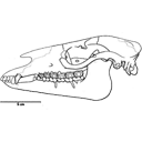
Un nouveau genre de ?Palaeotheriidae (Perissodactyla, Mammalia) décelé dans les phosphorites du Quercy (Eocène supérieur ou Oligocène) d'après un arrière crâne sans dents.Jean-Albert RemyPublished online: 15/06/1999Keywords: endocranial cast; Epitympanic sinus; Palaeotheriidae; Paléogène; Quercy Phosphorites; skull anatomy Abstract A rear skull from the Quercy Phosphorites is described. It documents a new perissodactyl genus, likely assignable to the family Palaeotheriidae and probably paleogene of age. Owing to the lack of any tooth, this family assignment remains however somewhat hypothetical. The specimen is firstly characterised by the presence of a wide epitympanic sinus swelling and hollowing the squamosal shell and the post-glenoid process. This cavity might make up a peculiar pattern of improvement for the hearing apparatus by carrying out a kind of drum near the middle ear, whereas the bony tympanic remains barely bulged and forms but a few developed auditory bulla. This pattern appears an outcome of a trend observed with many palaeotheriids, such as Plagiolophus. Furthermore, the endocranial cast shows a rather high degree of gyrencephaly for a paleogene mammal. The prominent lambdoidal crest points out a powerful nape musculature and a lowered head bearing. Consequently, it is assumed that such an animal was probably living in somewhat open places, at the expense of rather tough vegetables. It might have been accordingly provided with a semi-hypsodont, cement covered, "plagiolophoid" dentition. PV article infos Published in Vol. 28, Fasc. 1 (1999) |
|

|
Stratigraphy and Oligocene-Miocene mammalian biochronology of the Aktau Moutains, Dzhungarian Alatau Range, KazakhstanElena G. Kordikova
Published online: 16/12/1996 |
|

|
Contributions à l'étude des micromammifères du gisement Miocène supérieur de Montredon (Hérault). 3- Les insectivoresJean-Yves Crochet and Morton GreenPublished online: 30/06/1982Keywords: Hérault; Insectivora; Late Miocene; Micromammals; Montredon https://doi.org/10.18563/pv.12.3.119-131 Abstract This paper presents a preliminary list of insectivores from the Vallesian beds at Montredon (France). The associated rodent fauna has established a Vallesian age for the fauna. Eleven species belonging to the Soricidae, Talpidae, Erinaceidae, and Dimylidae are identified of which four only are referred with certainty to forms already named. PV article infos Published in Vol. 12, Fasc. 3 (1982) |
|
|
Révision systématique des Anchilophini (Palaeotheriidae, Perissodactyla, Mammalia).Jean-Albert RemyPublished online: 15/10/2012Keywords: Anchilophus; Eocene; new genus; new species; Palaeotheriidae; Paranchilophus; Perissodactyla; Systematics https://doi.org/10.18563/pv.37.1-3.1-165 Abstract The knowledge of the Anchilophini has been lately renewed by the discovery of a rather large amount of new material still largely unpublished. This new material offers the opportunity of a systematic revision of this tribe gathering those of European Eocene Equoidea which bear no mesostyle on upper check teeth and display a heavy trend to the molarization of premolars. PV article infos Published in Vol. 37, Fasc. 1-3 (2012) |
|
|
Discovery of the most ancient Notidanodon tooth (Neoselachii: Hexanchiformes) in the Late Jurassic of New Zealand. New considerations on the systematics and range of the genus
|
|

|
A new rodent from Quaternary deposits of the Canary Islands and its relationships with Neogène and recent murids of Europe and Africa.Rainer Hutterer
Published online: 15/12/1988 |
|

|
Un gisement à mammifères dans la formation lacustre d'âge Miocène moyen du Collet Redon près de St-Cannat (Bouches-du-Rhone). Implications stratigaphiquesJean-Pierre Aguilar and G. ClauzonPublished online: 10/04/1979Keywords: France; Neogene; Rodentia https://doi.org/10.18563/pv.8.5.327-341 Abstract The new fauna of Collet Redon (Bouches-du-Rhône, France) is dated by three rodents: Megacricetodon aff. bavaricus, Democricetodon affinis mutilus and Peridyromys cf. hamadryas. They correlate this locality with Oggenhof and Ohningen in Bavaria (Western Germany). As the radiometric age of Ohningen is estimated between 14 and 13 M.Y., these three localities are of Serravallian age. This datation brings a complete readjusment of the stratigraphy of the section of Collet Redon formerly described by Collot and Combaluzier. The marine deposits with underly the continental formation with the mammal fauna, are Burdigalian. The angular unconformity between the marine and the continental deposits gives evidence of an episode of emersion on the margin of a sedimentary basin, with deformation and erosion. Owing to the newly discovered fauna, this geodynamical event is clearly settled within the regional geographical and chronological context. Lacustrine and continental deposits of such an age were up to now unsuspected in this area. PV article infos Published in Vol. 08, Fasc. 5 (1979) |
|

|
La poche à phosphate de Ste-Néboule (Lot) et sa faune de vertébres du Ludien Supérieur. 2- Amphibiens. Etude PreliminaireJean-Claude Rage
Published online: 25/09/1978 |
|

|
Hexanchiforme nouveau (Neoselachii) du Crétacé inférieur du Sud de la FranceHenri Cappetta
Published online: 18/12/1990 |
|

|
Révision des Rhombodontidae (Neoselachii Batomorphii) des bassins à phosphate du MarocAbdelmajid Noubhani and Henri Cappetta
Published online: 20/05/1994 |
|

|
Contributions à l'étude du gisement Miocène supérieur de Montredon (Hérault). Les grands mammifères. 7 - Les proboscidiens DeinotheriidaeHeinz TobienPublished online: 15/11/1988Keywords: allometry; Astaracian; Deinotherium; Montredon; Systematics; taphonomy; Vallesian https://doi.org/10.18563/pv.18.ext.135-175 Abstract Some complete tooth rows and about one hundred isolated teeth enabled the identification of the deinothere of the Vallesian site Montredon (Hérault) as Deinotherium giganteum KAUP 1829, mainly by comparisons with the likewise Vallesian sample of the type locality Eppelsheim (Rheinhessen, F.R.G.). PV article infos Published in Vol. 18, Ext (1988) |
|

|
Middle Eocene rodents from the Subathu group, Northwest Himalya.Kishor Kumar
Published online: 15/12/1997 |
|

|
Long-term fidelity of megaoolithid dinosaurs to a large breeding-ground in the Upper Cretaceous of Aix-en-Provence (southern France).Géraldine Garcia
Published online: 15/12/2003 |
|