|
Critical comments on the genus Propachynolophus Lemoine, 1891 (Mammalia, Perissodactyla, Equoidea)
Published online: 27/09/2017
Keywords:
Eocene; Eurohippus; Pachynolophus; Propalaeotherium; tooth morphology
https://doi.org/10.18563/pv.41.1.e3
Abstract
Abstract
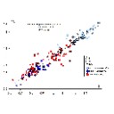
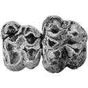
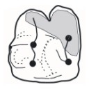
The validity of Propachynolophus Lemoine, 1891, supposedly an intermediate between Hyracotherium Owen, 1841 and Pachynolophus Pomel, 1847, has been questioned for a long time. A detailed analysis of features on which this genus is based further supported by a formal cladistic analysis demonstrates that Propachynolophus is not a valid taxon. The type species, “Propachynolophus gaudryi Lemoine, 1891” shall be assigned to Propalaeotherium Gervais, 1849, under the new combination Propalaeotherium gaudryi (Lemoine, 1891). “Pachynolophus maldani Lemoine, 1878”, later assigned to Propachynolophus, typifies the new genus Orolophus, under the binomen Orolophus maldani (Lemoine, 1878). The other referred species, “Propachynolophus levei Hooker, 1994” and “P. remyi Checa-Soler, 1997” are poorly documented, and both species shall be provisionally referred to as “Hyracotherium” levei (Hooker, 1994) and “Hyracotherium” remyi (Checa-Soler, 1997), pending new discoveries.
PV article infos
Published in Vol 41-1 (2018)
|
PDF
S.I. Data
|

|
La poche à phosphate de Ste-Néboule (Lot) et sa faune de vertebres du Ludien supérieur. 7- Didelphides (Marsupiaux)
Published online: 25/09/1978
Keywords:
Eocene; Quercy Phosphorites
https://doi.org/10.18563/pv.8.2-4.231-242
Abstract
The family Didelphidae is represented by three species in the Sainte-Néboule site, phosphorites of Quercy (lower Oligocene, San Cugat's nivel): Amphiperatherium minutum (Aymard), Amphiperatherium sp. and Peratherium cuvieri (Fischer). Only the first and third species are abundant (88 and 97 pieces). This two populations are described. The marsupial fauna of the european lower Oligocene is not recognized in its entirety in this site.
PV article infos
Published in Vol. 08, Fasc. 2-4 (1978)
|
PDF
|

|
Les Issiodoromyinae (Rodentia, Theridomyidae) de l'Eocène supérieur à l'Oligocène supérieur en Europe occidentale
Published online: 15/05/1976
Keywords:
climate; Faunal turnover; Paléogène
https://doi.org/10.18563/pv.7.1-2.1-115
Abstract
Based on material from 30 localities, morphologic dental, cranial and biometric analyses have permitted the characterization of two parallel Issiodoromyine lineages, and also the definition of diverse species representing several evolutive stages. Thus it is that new lineages complete the contribution made by the Theridomyinae and Cricetidae and permit, for the Quercy in particular, additional precision in the biochronologic succession of the localities. One of the lineages is limited to the genus Pseudoltinomys LAVOCAT; the other evolves from the genus Elfomys HARTENBERGER to the genus Issiodoromys BRAVARD in GERVAIS. The latter is affected by profound anatomical changes due to a functional modification of the mastication apparatus. These changes seem to be able to be put in relation with the aridification and cooling of the climate at the end of the Eocene. At the end of the middle Oligocene, a new chewing structure is achieved. It is found in diverse living rodents that inhabit a rather arid steppe environment (Cavia, Pedetes, Ctenodactylus). To these supposed nearby ecologic conditions, these rodents have responded in a convergent fashion. It is possible to attribute to the extreme specialization of Issiodoromys its incapacity to adapt to the new climatic crisis of the end of the Oligocene. The arrival of immigrants may be considered as another cause of its disappearance at this time, complementary or not with the first.
PV article infos
Published in Vol. 07, Fasc. 1-2 (1976)
|
PDF
|

|
Sur les Condylarthres Cernaysiens Tricuspiodon et Landenodon (Paléocène supérieur de France)
Published online: 01/10/1980
Keywords:
Arctocyonidae; Condylarths; Late Paleocene; Tricuspiodontidae
https://doi.org/10.18563/pv.9.ext.127-166
Abstract
The numerical importance of the Condylarths in the Cernaysian fauna is discussed. The Condylarth family, Tricuspiodontidae, is reviewed in the light of new material and its close relationships to the Phenacodontidae is suggested ; one new species is recognized : Tricuspiodon sobrinus. European Arctocyonidae are reviewed and the recentclassification of Van Valen is briefly commented on. Also, the arctocyonine Landenodon is described for the first time in Thanetian (Late Paleocene) sediments ; two new species are proposed : T. lavocati and T. phelizoni.
PV article infos
Published in Vol. 9, Ext (1980)
|
PDF
|

|
Contributions à l'étude du gisement Miocène supérieur de Montredon (Hérault). Les grands mammifères. 8 - Analyse paléoécologique de la faune mammalienne
Published online: 15/11/1988
Keywords:
France; Mammalia; Montredon; Paleoecology; Upper Miocene
https://doi.org/10.18563/pv.18.ext.177-186
Abstract
The species diversity of the mammalian fauna from Montredon (Hérault, France, late Miocene) is examined in terms of richness and abundance. A cenogramic analysis of the fossil mammalian community suggests the prevalence of open habitats, with the presence of marshes and of a poorly developed galery forest, and a climate rather warm and dry.
PV article infos
Published in Vol. 18, Ext (1988)
|
PDF
|

|
Contributions à l'étude du gisement Miocène supérieur de Montredon (Hérault). Les grands mammifères. 3 - Les artiodactyles ruminants
Published online: 15/11/1988
Keywords:
Artiodactyla; France; Montredon; Ruminentia; Upper Miocene
https://doi.org/10.18563/pv.18.ext.43-56
Abstract
The remains of Ruminantia are very rare at Montredon. No specific determination was possible. We have only one Micromeryx, one small cervid, one tragocere and one (?) gazella. The faunal spectrum is in good correlation with the general retreat of the cervids in the European upper Miocene, but in contrast it is not convenient for the bovids, which are in expansion at the same time. For them, we have to invoke the local ecological conditions.
PV article infos
Published in Vol. 18, Ext (1988)
|
PDF
|

|
Une nouvelle espèce de Steneosaurus (Thalattosuchia, Teleosauridae) dans le Callovien du Poitou (France) et la systématique des Steneosaurus longirostres du Jurassique moyen d'Europe Occidentale.
Published online: 15/09/1998
Keywords:
middle Jurassic; nov. sp.; phylogenetic relationships; skulls; Steneosaurus pictaviensis; Systematics; thalattosuchian crocodile
Abstract
The study of all the available skulls allows us to review the systematic relationships of the longirostrine Steneosaurus from the Middle Jurassic of western Europe. Up to now, Aalenian and Bajocian deposits have not yielded any significant Steneosaurus remain. In the Bathonian, the only valid longirostrine species, S. megistorhynchus, is known in the Britain-Normandy Basin, the Poitou and the Lorraine. In the Callovian, most of the longirostrine Steneosaurus remains can be attributed to the species S. leedsi. Nevertheless, some remains from the Middle Callovian of Poitou (France) show important differences with S. leedsi. A new Steneosaurus species, only known in Poitou, is created and named S. pictaviensis. The specific characters are carried by the skull (preorbital pit well marked, orbit and ptetygoid fossae shapes), by the mandible (symphysis shape) and by the teeth (ornamentation). S. megistorhynchus is probably situated near the stem of the Callovian species but remains from the Bathonian and Lower Callovian are very scarce and it is very difficult to precise the phylogenetic relationships between the longirostrine species of the Middle Jurassic.
PV article infos
Published in Vol. 27, Fasc. 1-2 (1998)
|
PDF
|

|
Pseudorhyncocyon cayluxi Filhol, 1892 insectivore géant des phosphorites du Quercy
Published online: 15/11/1974
Keywords:
Insectivores; Leptictidae; Quercy Phosphorites
https://doi.org/10.18563/pv.6.1-2.33-46
Abstract
Une hémimandibule et une molaire supérieure recueillies dans le gisement oligocène inférieur d'Escamps (phosphorites du Quercy) fournissent de nouvelles informations sur le genre Pseudorhyncocyon FllHOL, grand insectivore longirostre du Paléogène d'Europe, fossile très mal connu jusqu'ici. Des comparaisons avec les macroscélididés africains, géolabididés nord-américains, et leptictidés euraméricains permettent de rattacher cet amimal aux leptictidés, et de le rapprocher du genre Lepticidium TOBIEN, au sein de la sous-famille européenne nouvelle des pseudorhyncocyoninés.
PV article infos
Published in Vol. 06, Fasc. 1-2 (1974)
|
PDF
|

|
Lower Paleogene crocodilians from Silveirinha, Portugal.
Published online: 15/10/2003
Keywords:
?Upper Paleocene / Lowermost Eocene; Crocodilians; Ecology; Portugal
Abstract
The presence at Silveirinha of one of the earliest, ? Late Paleocene or Lowermost Eocene, european representatives of the genus Diplocynodon is based mostly on isolated bones and teeth (often from juveniles). This small-sized form is the only crocodilian so far recognized in this site. The longevity of Diplocynodon in Portugal becomes much extended; the genus survived there until the Middle Miocene at least. A discussion on the possible affinities with other eocene Díplocynodon and especially those from Cubillos-Valdegallina (Zamora, Spain) is presented. On the other hand, differences have been detected in comparison with: Díplocynodon tormis, from the middle Eocene of the Douro basin in Spain, which may belong to another phyletic line; and the aff. Diplocynodon from Dormaal (Belgium) and Le Quesnoy (France), nearly contemporaneous of Silveirinha. The Silveirinha Diplocynodon and many other data strongly suggest moist, subtropical, quite limited in space environments related to an alluvial plain crossed by small, meandering channels.
PV article infos
Published in Vol. 32, Fasc. 1 (2003)
|
PDF
|

|
Les gangas (Aves, Columbiformes, Pteroclidae) du Paléocène et du Miocène inférieur de France.
Published online: 11/02/1993
Keywords:
Birds; evolution; Lower Miocene; New taxa; Oligocene; Paleoecology; Paulhiac; Quercy Phosphorites; Saint-Gérand-Ie-Puy; Sandgrouse; Upper Eocene
https://doi.org/10.18563/pv.22.2-3.73-98
Abstract
The two species of Sandgrouse from Quercy, Pterocles validus MILNE-EDWARDS and P. larvatus MILNE-EDWARDS, are ascribed to the genus Archaeoganga MOURER-CHAUVIRÉ which includes a third, very large species, A. pinguis. The Sandgrouse of Saint-Gérand-le-Puy, Pterocles sepultus MILNE-EDWARDS, is ascribed to a new genus, Leptoganga. This form appears in the Upper Oligocene of Quercy, in Pech Desse and Pech du Fraysse localities, and is still present in the Lower Miocene of Saint-Gérand-le-Puy and Paulhiac. Recent Sandgrouse live in semidesert or desert areas. The indications provided by mammal and bird faunas in the localities where sandgrouse were found, confirm that the paleoenvironment was open and arid. The morphological study of these fossils indicates that, in the Upper Eocene, the Pteroclidae were already completely individualized with respect to Charadriiformes.
PV article infos
Published in Vol. 22, Fasc. 2-3 (1993)
|
PDF
|

|
The evolution of the molar pattern of the Erethizontidae (Rodentia,Hystricognathi) and the validity of Parasteiromys Ameghino, 1904.
Published online: 15/06/1999
Keywords:
Argentina; Erethizontidae; Hystricognathi; Miocene; Molar evolution; Porcupines; Rodentia; Systematics
Abstract
The genus Parasteiromys AMEGHINO, 1904 is revalidated, and P. friantae sp. nov. (Hystricognathi, Erethizontidae) from Colhuehuapian (early Miocene) sediments of the southern cliff of Colhue-Huapi Lake (Province of Chubut, Argentina), is described. The molar morphology of these taxa and of living porcupines adds new elements to understand the dental evolution of the Erethizontidae, and to propose the hypothetical ancestral molar pattern for this family. This pattern does not correspond to any of the morphologies traditionally proposed as ancestral for South American hystricognathous rodents. The proposed pattern is characterized by a metaloph disconnected from the posteroloph and oriented towards the hypocone, and the third loph incompletely developed with the lingual portion homologous to the mesolophule of Baluchimyinae (Chapattimyidae) from the Miocene of Pakistan. The inferred steps of the molar evolution of erethizontids towards the pentalophodont condition, considered derived for the family, are illustrated. This study strengthens the hypothesis placing erethizontids in a basal position among rodents of the suborder Hystricognathi.
PV article infos
Published in Vol. 28, Fasc. 1 (1999)
|
PDF
|

|
Evolution of the Rhizomyine zygoma
Published online: 30/12/1985
Keywords:
parallel evolution; Rhizomyidae; Rodentia; Siwalik; zygoma
https://doi.org/10.18563/pv.15.3.129-138
Abstract
Cranial anatomy of a late Miocene rhizomyid, Brafhyrhizomys cf. B. pilgrimi, provides new evidence on the origin of the dorsal, round infraorbital foramen of living rhizomyines. Primitive rhizomyids retain a myomorphous keyhole foramen with a long ventral slit that retracts upward during the evolutionary history of the Rhizomyidae. The primitive condition of the elongated ventral slit is represented by Kanisamys sivalensis. Among later burrowers the foramen shows progressive dorsal migration, the ventral slit terminating midway up the snout in B.tertracharax and B. choristos ; well above the midline of the snout in Brachyrhizamys cf. B. pilgrimi. Apparently this transformation began earlier among Rhizomyinae than among Tachyoryctinae and continued to a more derived stage in rhizomyines. ln living Rhizomyx the ventral slit is absent and only a high round hole remains at the anterior end of the zygomatic arch.
PV article infos
Published in Vol. 15, Fasc. 3 (1985)
|
PDF
|

|
Nouveaux Mammifères Eocènes du Sahara Occidental
Published online: 01/11/1979
Keywords:
Eocene; Mammals; Occidental Sahara
https://doi.org/10.18563/pv.9.3.83-115
Abstract
The fossil mammals collected from the Eocene of Hammada du Dra (northwest Sahara. Algeria) and two fragmentary teeth from the Lutetian of M'Bodione Dadere (Senegal) are described.
The fossils from the northwest Sahara come from a lacustrian deposit dated by charophytes (Raskyella aff. pecki, Raskyella n. sp.. Maedleriella lavocati, Maedleriella sp. et ? Peckichara sp.) as Middle Eocene or perhaps Lower Eocene (Gevin, Feist and Mongereau, 1974). Several hyracoids (3 or 4) identified from this formation extends the age of the family Pliohyracidae Osborn in Africa. Three forms appear to belong in the genera Megalohyrax, Titanohyrax and perhaps Bunohyrax which have been know until now only from the lower Oligocene of the Fayum (M. gevini n. sp. ; T. mongereaui n. sp.. ? Bunohyrax or Megalohyrax indet.). Another hyracoidof small size is referred to a new genus, Microhyrax (M. lavocati n. sp.).
Helioseus insolitus n. g. n. sp. is described without ordinal assignment. Azibius (Sudre, 1975) which has been the subject of questions and interpretations is reviewed.
Only one tooth from the Lutetian of M'Bodione Dadere is complete enough to interpret. lt probably belongs to a condylarth and demonstrates for the first time, the presence of the order in Africa. The second tooth is too fragmentary for comment.
In conclusion., the paleobiogeographic role of Africa at the end of the cretaceous and the beginning of the Cenozoic is discussed.
PV article infos
Published in Vol. 09, Fasc. 3 (1979)
|
PDF
|

|
Evolution des Aplodontidae Oligocènes Européens
Published online: 01/10/1979
Keywords:
Aplodontidae; Europe; Oligocene
https://doi.org/10.18563/pv.9.2.33-82
Abstract
Until now Aplodontidae of the European Oligocene have been documented by four species only. The phylogenetic relations remained obscure. as the distribution of only one species has been known in some detail. New material made it possible to define the stratigraphic range of two of the already existing species (Plesispermophilus angustidens, Sciurodon cadurcense) and to follow their development during the Oligocene beginning with the event of the « Grande Coupure ››. Sciurodon remained nearly without change until the end of the Middle Oligocene. Plesispermophilus angustidens split into two distinct phyletic lines, one of which (P. macrodon n. sp.) reaching considerable size, is represented till the beginning of the Upper Oligocene (Pech de Fraysse, Gaimersheim). The other line leads to Plesispermophilus ernii (basal Upper Oligocene of Burgmagerbein 1. terminal Upper Oligocene of Coderet). Besides the already known forms a new small-sized species (P. atavus n. sp.) is described, which by its primitive features closely resembles the genus Plesispermophilus. Two other small-sized species already known from the Upper Oligocene (? P. argoviensis) and Lower Miocene (? P. descedens) seem to be closely related to the new species. It cannot be decided whether they are descendents of this line or have developed independently, because of their poor fossil record.
Comparison of the evolutionary modalities in the different phylogenetic lines reveals general trends. the most striking of which is the complication of the tooth pattern by the development of additional crests. In the lower molars the cusps diminuate in size and are more and more transformed into ridges. ln addition new connection between the crests appear. in the upper molars, the « selenodont » shape of the teeth becomes more and more dominant, and in the two main evolutionary lines of Plesispermophilus the metaconulus becomes duplicated. A further evolutionary trend is the size increase of the premolars compared to the molars, which is even more pronounced in the Miocene Aplodontidae.
Phylogenetic relations between the primitive Plesispermophilus and certain « prociurines ›› of Northern America as well as between Plesispermophilus (P. angustidens) and more progressive forms of the Upper Oligocene (P. ernii, P. macrodon n. sp.) can be documented. In this light, the taxonomic distinction between Prosciurinae (bunodont) and Allomyinae (selenodont) sensu Rensberger 1976 can be shown to be artificial, because it separates forms from each other, which are evidently closely related. Consequently the separation into two subfamilies has been abolished.
PV article infos
Published in Vol. 09, Fasc. 2 (1979)
|
PDF
|

|
Les Pseudosciuridae (Mammalia, Rodentia) de l'Eocène moyen de Bouxwiller, Egerkingen et Lissieu.
Published online: 30/10/1969
Keywords:
Bouxwiller; cranium; Egerkingen; Middle Eocene; Rodents
https://doi.org/10.18563/pv.3.2.27-64
Abstract
The description of new material from three classic middle Eocene localities of western Europe permits the addition of details to the systematics of primitive Pseudosciurids. The cranial anatomy of Protadelomys cartieri (STEHLIN and SCHAUB) from Egerkingen is described here and compared to that of the Adelomyines from the late Eocene, until now the only ones known. The morphologic and biometric study of the dentition of P. cartieri compared to that of P. alsaticus n. sp. from Bouxwiller and P. Iugdunensis n. sp. from Lissieu, forms respectively older and younger than P. cartieri, permits the evolutionary tendencies of the group to be demonstrated and shows that notable differences in age exist between these localities. This ensemble of forms can constitute a valuable guide lineage in the establishment of a fine stratigraphy of the period. Other less well known lineages are present at Egerkingen along with P. cartieri. They can be related to genera that have been noted int he late Eocene. In conclusion, a criticism of recent zonation proposals, made by divers authors, completes this article.
PV article infos
Published in Vol. 03, Fasc. 2 (1969)
|
PDF
|
|
Les rongeurs de Chéry-Chartreuve et Rocourt-Saint-Martin (est du bassin de Paris; Aisne, France). Leur place parmi les faunes de l'Eocène Moyen d'Europe
Published online: 15/12/2012
Keywords:
Biochronology; evolution; Middle Eocene; Paris basin; Rodents; Systematics
https://doi.org/10.18563/pv.37.4-5.167-271
Abstract
This paper is mainly devoted to the systematics of rodents from two middle Eocene (Bartonian) localities: Chéry-Chartreuve and Rocourt-Saint-Martin (Aisne, Eastern Paris Basin). These two localities are stratigraphically located slightly above the Auversian sands. The two faunas, which comprise 11 and 8 taxa, respectively, are very different in their composition. That of Rocourt-Saint-Martin shows strong similarities with that of the geographically very close locality of Grisolles, referred to the MP16 mammalian Reference level. The very distinct fauna of Chéry-Chartreuve includes a new species of Ailuravinae, Ailuravus nov.sp, and some teeth of the theridomyid Protadelomys, which represent archaic elements in the fauna. The most abundant species of the locality represents a new genus of primitive Theridomyidae. The presence of some teeth belonging to a new species of large Remyinae, Remys nov. sp., of Elfomys engesseri HOOKER & WEIDMANN, and a population of small dimensions referred to the genus Estellomys allow a correlation with Les Alleveys (Switzerland), with however some differences that would indicate an older age for Chéry-Chartreuve. Situated at the base of the "Marinesian" from the Bassin de Paris, this fauna is unquestionably different from those referred to the MP16 reference level and could represent an older level for which the macrofauna remains very poorly known. Conversely, the comparison of rodents from La Livinière II with those present in MP16 faunas, especially those of Robiac (Gard), shows a great similarity between both localities. This casts doubts on whether to keep this La Livinière II faunule to define the current MP15 reference level, while the biostratigraphical position of Pontils 26 (Spain), previously referred to this level, is reconsidered. Chery Chartreuse could be a good candidate for a new definition of the MP15 reference level.
PV article infos
Published in Vol. 37, Fasc. 4-5 (2012)
|
PDF
|
|
A new study of the anthracotheres (Mammalia, Artiodactyla) from pondaung formation, Myanmar: systematics implications
Published online: 16/12/2008
Keywords:
Anthracohyus; Anthracokeryx; Anthracotherium; Pondaung Formation; sexual dimorphism; Siamotherium; South East Asia; taxonomy
https://doi.org/10.18563/pv.36.1-4.89-157
Abstract
Anthracotheres from the Pondaung Formation, Myanmar, are considered as one of the most primitive artiodactyl groups and they represent the oldest known record in the world. Thus, the understanding of this group has numerous implications for evolutionary biology and biochronological correlations. However, the systematlcs of these mammals has been interpreted in different ways, and the main debate focuses on the number of taxa represented in the Pondaung Formation. The revised taxonomy proposed here is mainly based on the relative development of the upper molar W-shaped ectoloph, system of crests and stylar cusps, and on body size. On the basis of these characters, they are classified into four genera including six different species. Two well-known genera, Anthracotherium and Anthracokeryx, are validated and more precisely diagnosed. Anthracokeryx possesses a better developed W-shaped ectoloph, system of crests and stylar cusps than Anthracotherium, which displays notable differences with the more derived representatives of this genus. Both of these Pondaung genera show evidence for sexual dimorphism. However, the incompleteness of fossil material fueled a debate concerning the status of two additional Pondaung anthracotheres, Siamotherium and Anthracohyus. The latter genus is of uncertain affinities, but it has been considered as a hippopotamid ancestor. Despite new material attributed to these two forms, additional discoveries are still required to establish their taxonomic status. The hypothesis that Southeast Asia was the centre of origin of Anthracotheriidae is supported by the retention of numerous primitive dental characters in these taxa and by the antiquity of the Pondaung Formation, to which an age of 37 My is now generally accepted.
PV article infos
Published in Vol. 36, Fasc. 1-4 (2008)
|
PDF
|
|
A late Eocene palaeoamasiine embrithopod (Mammalia, Afrotheria) from the Adriatic realm (Island of Rab, Croatia)
Published online: 14/12/2023
Keywords:
Balkanatolia; Grande Coupure; Great Adria; Paleobiogeography; Systematics
https://doi.org/10.18563/pv.47.1.e1
Abstract
A cheek tooth recently unearthed in the Lopar Sandstone unit, of late Eocene age, in the northern part of Rab Island, Croatia, is one of the very few Eocene mammalian remains found in the Adriatic area. Thorough comparison of this tooth with those of Old-World Palaeogene mammalian orders suggests that it is a M3 belonging to an embrithopod afrothere. The specimen is referred to as Palaeoamasia sp. This genus was formerly known only in Eocene deposits of Anatolia but with close relatives in Romania among Palaeoamasiinae. The geographical distribution of this subfamily perfectly matches the recently-named Balkanatolian landmass, which experienced in-situ evolution of endemic mammals prior to the Grande Coupure event that occurred around the Eocene–Oligocene transition. This last event is characterised by massive Asian immigration in Western Europe and the supposed extinction of many endemic Central and Western European mammals, including Palaeoamasiinae.
PV article infos
Published in 47-1 (2024)
|
PDF
|
|
First early Eocene tapiroid from India and its implication for the paleobiogeographic origin of perissodactyls
Published online: 08/09/2015
Keywords:
Ceratomorpha; Helaletidae; Paléogène; Tapiromorpha; Vastan
https://doi.org/10.18563/pv.39.2.e5
Abstract
The presence of cambaytheres, the sister group of perissodactyls, in western India near or before the time of collision with Asia suggests that Perissodactyla may have originated on the Indian Plate during its final drift towards Asia. Herein we reinforce this hypothesis by reporting two teeth of the first early Eocene tapiromorph Perissodactyla from the Cambay Shale Formation of Vastan Lignite Mine (c. 54.5 Ma), Gujarat, western India, which we allocate to a new genus and species, Vastanolophus holbrooki. It presents plesiomorphic characters typical of the paraphyletic “Isectolophidae,” such as small size and weak lophodonty. However, the weaker hypoconulid and low paralophid, higher cusps, lower cristid obliqua, and the lingual opening of the talonid are found in Helaletidae, the most primitive tapiroid family. V. holbrooki, gen. et sp. nov., may be the oldest and the most primitive tapiroid, suggesting that at least tapiroid perissodactyls originated on India.
PV article infos
Published in Vol.39-2 (2015)
|
PDF
|

|
La poche à phosphate de Ste-Néboule (Lot) et sa faune de vertebres du Ludien supérieur. 12- Fissipèdes (Carnivores)
Published online: 25/09/1978
Keywords:
Carnivora; Eocene; Quercy Phosphorites
https://doi.org/10.18563/pv.8.2-4.301-311
Abstract
Les Carnivores Fissipèdes de Sainte-Néboule appartiennent tous au genre Cynodictis et semblent constituer une population homogène. Celle-ci se distingue suffisamment des espèces déjà décrites pour constituer un taxon particulier : Cynodictis lacustris neboulensis n. s. sp. . L'étude des variations à l'intérieur de cette population nous a conduit à reconsidérer les critères utilisés pour définir les espèces existantes et à regrouper certaines d'entre elles. Il semble qu'il demeure cependant trois lignées distinctes dans le genre Cynodictis mais le matériel nous paraît encore insuffisant pour traduire cette remarque en termes de systématique.
PV article infos
Published in Vol. 08, Fasc. 2-4 (1978)
|
PDF
|
