
Abstract book of the 18th Conference of the EAVP
Pterosaurs from Coahuila
Pliocene-Pleistocene large mammals from Le Riège and Saint-Palais
Les sélaciens du Miocène de la région de Montpellier
Muridae du Pliocène supérieur d'Espagne et du midi de la France.
Contribution à l'étude des genres Gliravus et Microparamys.
Eocene (57) , Quercy Phosphorites (38) , Systematics (32) , Rodents (29) , Mammalia (27) , Rodentia (25) , Miocene (24)

|
Die Referenzfauna des Geiseltalium, MP levels 11 bis 13 (Mitteleozan, Lutetium)Hartmut HauboldPublished online: 04/12/1989Keywords: Eocene; Geiseltal; Land mammal ages; Mammalian reference levels https://doi.org/10.18563/pv.19.3.81-93 Abstract The Middle Eocene Fossillägerstätte of the Geiseltal lignite beds near Halle/S. (German Democratic Republic) is the reference locality of the European land mammal age Geiseltalian and of the Mammalian Paleogene reference levels MP 11 - MP 13. Due to this importance a reinvestigation is given of the lithostratigraphical development of the Geiseltal beds and of their vertebrate sites. The last are genetically related to the southwest border of the Geiseltal depression and the influx of carbonate-rich waters. The geographical distribution and stratigraphical position of the fossiliferous sites depends on subrosive and tectonically controlled distribution of coal seams. The geological factors and the known stratigraphical guide of some mammalian species suggest corrections of the age of some sites. Four of the alltogether five coal bearing phases contain the 35 sites with mammalian remains. By the distribution of the around 69 mammal species are characterized, with 5 faunal steps ranging from MP 11 to MP 14 or over the Geiseltalian up to the Lower Robiacian. Well distant are the faunas of MP 11 and MP 12. Beginning with MP 12 up to MP 13/14, the fossil record is very frequent by 27 sites. This evidence coincides somewhat more with the concept of land mammal ages compared to that of the punctual mammalian reference levels. PV article infos Published in Vol. 19, Fasc. 3 (1989) |
|

|
Le genre Leptolophus (Perissodactyla, Mammalia): morphologie et histologie dentaires, anatomie cranienne, implications fonctionnelles.Jean-Albert RemyPublished online: 15/09/1998Keywords: dental histology; Eocene; functional anatomy; Palaeotheriidae; skull anatomy; Southern France; Systematics Abstract A strong lophodonty, an extreme heterodonty, some hypsodonty and regular overlayings of coronal cement are prominent features of the genus Leptolophus (Palaeotheriinae = Palaeotheriidae s.s.). The histological pattern of the teeth unusually joins type II enamel prisms, characteristic of advanced ungulates, together with archaic features, such as an almost complete lack of Hunter-Schreger zonation and a weak expanse of peritubular dentine. The skull is narrow and slender, with an elongated ante-orbital facial region, a moderately notched nasal aperture, a rather elongated post-canine diastem, parallel zygornatic arches and a fairly dorsally located squamoso-mandibular joint.The functional analysis brings to light "ectolophodont" masticatory cycles with two phases, in which maximum power was applied, contrary to equíds, on hindmost teeth; likewise, skull accomodations to increasing height of the teeth are quite different. This study leads to the assumption that Leptolophus may have been light mammals, living in rather open surroundings, browsing on herbaceous plants or leaves cropped close to the ground. Moreover, it appears that it could have been some inadequacy of dental structures to the dietary, which leaded to quick wear of the teeth and to many enamel notches, but had been somewhat balanced by the early increase of hypsodonty, not induced in such a case by a biotop deterioration (as it will happen at the end of the Eocene). This ínadaptation might account for the short duration of the genus Leptolophus, whose the 3 species, L. stehlini, L. nouletí and L. magnus n. sp. are indeed confined in the level MP 16. Its geographical spreading (as far as known, South of western Europe) and the morphological pattern of its dentition suggest that this genus would have been related to early upper Eocene endemic spanish forms. PV article infos Published in Vol. 27, Fasc. 1-2 (1998) |
|

|
Neolicaphrium recens Frenguelli,1921,the only surviving proterotheriidae (Litopterna, Mammalia) into the south american Pleistocene.Mariano Bond
Published online: 30/07/2001 |
|

|
Rythme et modalités de l'évolution chez les rongeurs à la fin de l'Oligocène-leurs relations avec les changements de l'environnement.Bernard ComtePublished online: 15/12/2000Keywords: Environment; evolution; Oligocene; Rodents; Systematics Abstract The analysis of oxygene isotope variations as well as paleobotanical data suggest that the Oligocene/Miocene boundary corresponds to a transitional period marked by floristical and climatic variations. During this period, the pyreneo-alpine tectonics has contribued to modify the geography and western Europe landscapes. Faunal changes (appearances, extinctions, migrations) are observed in different mammalian groups, notably in the rodents. A study of the evolutionary trends and patterns in paleogene rodents is involved for the period ranging from level MP 28 of the Late Oligocene to the Early Miocene, including the Oligo-Miocene boundary. PV article infos Published in Vol. 29, Fasc. 2-4 (2000) |
|

|
Perutherium altiplanense, un Notongulé du Cretacé Supérieur du PérouLarry G. Marshall, Christian de Muizon
Published online: 30/11/1983 |
|

|
Les mammifères Montiens de Hainin (Paléocène moyen de Belgique) Part III : MarsupiauxJean-Yves Crochet and Bernard SigéPublished online: 30/09/1983Keywords: Belgium; Marsupials; Paleobiogeography; Paleocene https://doi.org/10.18563/pv.13.3.51-64 Abstract The oldest european marsupials are described from some specimens (isolated upper molars) recently found from the Hainin sediment (Middle Paleocene of Belgium). These fossils document a new species of the Peradectes genus. They give evidence of a much older occurrence of the marsupials in Europe than it was assumed. They allow us to postulate a didelphid dispersal from South America towards the western-holarctic area operating in two phases : the first one of the Peradectes genus at the end of the Cretaceous; the second one of the Didelphíni tribe at the end of the Paleocene. A central american crossing is likely for the first one, whereas a transafrican way is tentatively argued for the second one. PV article infos Published in Vol. 13, Fasc. 3 (1983) |
|

|
Les Affinités de Nyctereutes megamastoides (Pomel), canidé du gisement Villafranchien de Saint-Vallier (Drôme, France).R. MartinPublished online: 31/01/1971Keywords: Canidae; Nyctereutes; Villafranchian https://doi.org/10.18563/pv.4.2.39-58 Abstract Nyctereutes megamastoides (Pomel) from the Villafranchian of the Auvergne and from Saint-Vallier presents cranial and dental characters sufficiently close to those of the late Pliocene canid from Perpignan (Roussillon), described by Depéret under the specific name of Canis donnezani belonging to the same genus Nyctereutes. The extinction of the European "Nyctereutes" group seems due to the too great alimentary specialization of this canid, whereas the Asiatic lineage represented in the Villafranchian by Nyctereutes sinensis Schlosser and at present by Nyctereutes procyonider Gray was able to maintain itself probably by means of a profound change in its alimentary regime. PV article infos Published in Vol. 04, Fasc. 2 (1971) |
|
|
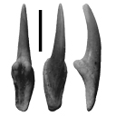
Les Paramyidae (Rodentia) de l'Eocène inférieur du bassin de Paris.Jacques MichauxPublished online: 15/07/1968Keywords: Ailuraviinae; Eocene; Paramyinae; Rodents https://doi.org/10.18563/pv.1.4.135-193 Abstract The exploitation of new early Eocene localities in the Paris Basin has resulted in the collecting of numerous mammalian remains, among which are about 300 isolated teeth representing the rodents. They belong, for the most part, to the paramyid group. Only the latest level of the early Eocene has yielded rodents belonging to the pseudosciurid group. The paramyids, the object of this study, are represented by at least 5 genera and 10 species; they are distributed among 4 clearly dilferentiated subfamilies : Paramyinae Simpson 1945, Pseudoparamyinae Michaux 1964, Ailuraviínae n. subf., Microparamyinae Wood1962. PV article infos Published in Vol. 01, Fasc. 4 (1968) |
|

|
Les traces de pas d'amphibiens, de dinosaures et autres reptiles du Mesozoïque Français : inventaire et interprétations.Georges Gand, Georges Demathieu and Christian MontenatPublished online: 15/12/2007Keywords: Footprints; France; Inventory; Mesozoic; palaeontology; palaeovenvironments; Stratigraphy https://doi.org/10.18563/pv.35.1-4.1-149 Abstract Since the 19th century, thousands of footprints were observed in the geological series of the French Mesozoic. All are located in the Triassic and Jurassic. After a promising beginning, in France, it is only a few papers which will be published in the first half 20th century, unlike the USA and of others countries of Western Europe. One ought to wait about 1950 for a revival and now they are nearly 200 papers which were devoted to the ichnofossils. The literature abundance and the renewed interest of the naturalists for the palichnologic studies decided to us to write a synthesis work. This one begins with a stratigraphic inventory in which, localisation, age and paleontological contents of about 180 fossiliferous sites are specified. After having pointed out the followed methods, the footprints paleontological interpretation is then approached in detail and the results obtained are replaced in stratigraphy to deduce the fauna evolution during the Mesozoic. So, it appears that Ichnologic data, more varied and rich in the Triassic and Liassic than those relating to the bones, very rare for the considered periods, are very informative. The middle Triassic (Anisian-Ladinian), thus reveals Cotylosauria, Lepidosauria, Crurotarsi with Rauisuchia, Ornithosuchidae, Crocodylia and Dinosauromorpha more the "Prodinosauria": Dinosamiforme whose skeletons are known in Argentina but only in Ladinian. The rather fast domination of Dinosaurs during Norian is also as well shown. The almost exclusive presence of their footprints, up to fifty cm long, in the Lower Hettangian indicates their supremacy in the environments. Footprints characterise not very deep life places located between inter-supratidal limits and often out of water. Sedimentologic and Palaeontologic studies showed that they were great coastal spaces during Middle Triassic, flood-plain with sebkhas while Upper Triassic, and a large !!coastal marsh!! in Grands-Causses during Liassic in which, mainly, fine stromatolithic layers were deposited. During the same periad, bay beaches spread in Vendée. During the Middle Jurassic, they are also brackish to lacustrine environments and recifallagoons in- the Upper Jurassic. Numerous measurements of the footprints and trackways directions showed that the animaIs moved there in weil defined directions, for long periods. They seem due to the palaeotopography of the life environments relatively stable. Also, the discovery of vegetal radicular networks and small footprints far away from the continental borderlands has suggested that the animals continuously lived in these palaeoenvironnements, belonging to large ecosystems, where the sedimentation rate was weak. This explains that thebadies could not fossilize there but only their footprints through the cyanobacterian action in main cases. From the vertical distribution of different ichnospecies, defined with adapted statistical methods, explained in this work, a palichnostratigraphy was established for the Middle Triassic. Although the footprints are also abundant in Hettango-Sinemurian of "Grands-Causses" and the Vendée, it was not possible, up to now, to establish any zonation in this series; Probably because the palichnofauna is too little diversified there, currently reduced to a majority of Theropods II-IV tridactyl traces. PV article infos Published in Vol. 35, Fasc. 1-4 (2007) |
|
|
Additions to the elasmobranch assemblage from the Bandah Formation (middle Eocene, Bartonian), Jaisalmer District, Rajasthan, India, and the palaeobiogeographic implications of the faunaRajendra S. Rana
Published online: 23/06/2021 |
|
|
The digital endocast of Necrolemur antiquusArianna Harrington
Published online: 24/07/2020 |
|
|
Difficulties with the origin of dinosaurs: a comment on the current debateMatthew G. Baron
Published online: 01/07/2020 |
|

|
The stratigraphic sequence of North American rodent faunasRobert W. WilsonPublished online: 01/10/1980Keywords: North America; Rodents; Stratigraphic sequence https://doi.org/10.18563/pv.9.ext.273-283 Abstract Rodents first appear in the latest Paleocene or earliest Eocene as very fragmentary specimens (Family Paramyidae) known largely from a single locality. After this sparse beginning, rodents are usually abundant in the North American record if proper recovery methods are used. Utilization of rodents for biostratigraphic purposes depends on 1/ extinction, and 2/ replacement by evolution of endemic groups and/or incursions of Old World rodents, and rarely and late by South American kinds. These incursions are separated by relatively long periods of isolation in the Paleogene, but more episodic in the Neogene. At least 10 rodent zones can be characterized by major distinctions, and these zones can be amplified into as many as 16 with little trouble. In general, rodent genera permit as refined a zonation as do genera of large mammals. Distinction at a specific level has not been attempted herein except in the Blancan and Post-Blancan. PV article infos Published in Vol. 9, Ext (1980) |
|

|
Les nouvelles faunes de rongeurs proches de la limite mio-pliocène en Roussillon. Implications biostratigraphiques et biogéographiquesJean-Pierre Aguilar, Jacques Michaux, Bernadette Bachelet, Marc Calvet
Published online: 29/04/1991 |
|

|
Les Otolithes de téléostéens du Miocène de Montpeyroux (Herault),France).Dirk Nolf and Henri Cappetta
Published online: 15/12/1980 |
|

|
Rongeurs nouveaux de l'Oligocène Moyen d'Espagne.Louis ThalerPublished online: 15/09/1969Keywords: Cricetidae; Oligocene; Pseudocricetodon; Rodents; Theridomys https://doi.org/10.18563/pv.2.5.191-207 Abstract Description of four new rodents from a recently discovered locality at Montalban. Theridomys crusafonti nov. sp. is considered as the ancestry of T. Iembronicus. Theridomys varian: nov. sp. includes «Theridomys» morphotypes and «Blainvilllimys» morphotypes; it could be ancestral to B. blainvillei. Pseudoltinomys nanus nov. sp. represents a new lineage paralleling in evolution that of P. gaillardi (which is equally found at Montalban). Pseudocricetodon montalbanensis nov. gen., nov. sp. designates a lineage of very small Cricetidae accompanying Eucricetodon. With these well defined new species and six others present in the locality, Montalban appears as the best faunal reference point within the biochronologic zone of La Sauvetat. PV article infos Published in Vol. 02, Fasc. 5 (1969) |
|
|
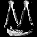
Les Palaeotheridae (Perissodactyla) de la faune de Mammifères de Fons 1 (Eocène supérieur).Jean-Albert RemyPublished online: 15/06/1967Keywords: Anchilophus; Eocene; Pachynolophus; Palaeotheriidae; Perissodactyla https://doi.org/10.18563/pv.1.1.1-46 Abstract The locality of Fons 1, one of the fossiliferous outcrops in the late Eocene limestones of Fons-outre-Gardon (Gard), has yielded varied remains of mammals. The specimens were prepared by dilute acetic acid attack on the rock and by impregnation with an acrylic resin. PV article infos Published in Vol. 01, Fasc. 1 (1967) |
|
|
Enigmatic rodents from Lavergne, a late middle Eocene (MP 16) fissure-filling of the Quercy Phosphorites (Southwest France)
|
|

|
La poche à phosphate de Ste-Néboule (Lot) et sa faune de vertébres du Ludien Supérieur. Résumé.Bernard Gèze, Jean-Claude Rage
Published online: 15/09/1978 |
|

|
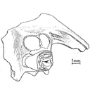
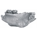
Ostéologie de la tête de Richardus excavans Lavocat,1988René LavocatPublished online: 30/10/1989Keywords: Africa; anatomy; Bathyergidae; Miocene; Rodents https://doi.org/10.18563/pv.19.2.73-80 Abstract Remarkable association of a small infraorbital foramen, similar to that in recent Heterocephalus, and of a strong muscular print on the dorsal anterior part of the zygomatic plate and on the premaxillary. Several anatomical structures to be compared with those of Heterocephalus suggest relationships with this genus. Richardus supports the ancestrality of the hystricomorph character in the bathyergids PV article infos Published in Vol. 19, Fasc. 2 (1989) |
|