|
A late Eocene palaeoamasiine embrithopod (Mammalia, Afrotheria) from the Adriatic realm (Island of Rab, Croatia)
Published online: 14/12/2023
Keywords:
Balkanatolia; Grande Coupure; Great Adria; Paleobiogeography; Systematics
https://doi.org/10.18563/pv.47.1.e1
Abstract
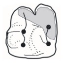
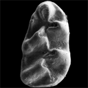
A cheek tooth recently unearthed in the Lopar Sandstone unit, of late Eocene age, in the northern part of Rab Island, Croatia, is one of the very few Eocene mammalian remains found in the Adriatic area. Thorough comparison of this tooth with those of Old-World Palaeogene mammalian orders suggests that it is a M3 belonging to an embrithopod afrothere. The specimen is referred to as Palaeoamasia sp. This genus was formerly known only in Eocene deposits of Anatolia but with close relatives in Romania among Palaeoamasiinae. The geographical distribution of this subfamily perfectly matches the recently-named Balkanatolian landmass, which experienced in-situ evolution of endemic mammals prior to the Grande Coupure event that occurred around the Eocene–Oligocene transition. This last event is characterised by massive Asian immigration in Western Europe and the supposed extinction of many endemic Central and Western European mammals, including Palaeoamasiinae.
PV article infos
Published in 47-1 (2024)
|
PDF
|
|
Nouvelles données sur les Ichnites de dinosaures d'El Bayadh (Crétacé Inférieur, Algérie)
Published online: 16/12/2008
Keywords:
Algeria; Brezina; El Bayadh; Ichnites; Lower Cretaceous; Sauropoids; Theropoids
https://doi.org/10.18563/pv.36.1-4.7-35
Abstract
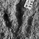
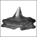
Evidence of 350 Lower Cretaceous Dinosaur footprints is pointed out in El Bayadh area. Their preliminary study allow to distinguish four trackway assemblages which reveal vertebrate bipedal presence forms of tri-and tetradactylous Dinosauroïds (Assemblages 1-3) and quadrupidal Sauropoïd (Assemblage 4).
The analysis of their footprint biometric features will attribute the quadrupidal Sauropoïd form to Brontopodus ichnogenus which is weIl known in the Jurassic and Cretaceous periods. In retum and despite their age, the dinosauroïd forms were approached, temporarily, to Grallator and Eubrontes types.
The occurrence of the dinosaur traces (Theropoïd and Sauropoïd) constitutes, in the Lower Cretaceous, an important first step of the knowlege of the marshy Reptilian fauna which takes over, from the begining of the Secondary Era, a wide paleogeographie area on the Southem Tethyan margin.
PV article infos
Published in Vol. 36, Fasc. 1-4 (2008)
|
PDF
|
|
Fossil snakes from the Palaeocene of São José de Itaboraí, Brazil Part III. Ungaliophiinae, Booids incertae sedis, and Caenophidia. Summary, update and discussion of the snake fauna from the locality
Published online: 16/12/2008
Keywords:
booid-grade incertae sedis; Brazil; Caenophidia; New taxa; Palaeocene; Russellophiidae; Snakes; tropidophiids; Ungaliophiinae
https://doi.org/10.18563/pv.36.1-4.37-73
Abstract
Aside from Madtsoiidae, anilioids, and Boidae that were studied previously, the middle Palaeocene of ltaborai (BraziI) has produced Ungaliophiinae ("tropidophiids"), booid-grade snakes incertae sedis, and a possible Russellophiidae (Caenophidia) that are described in the present article. This article is the third and final report on the snakes from the locality. The Ungaliophiinae (Paraungaliophis pricei gen. et sp. nov.) are rare whereas the booid-grade snakes incertae sedis (ltaboraiophis depressus gen. et sp. nov., Paulacoutophis perplexus gen. et sp. nov.) are more frequent. A single vertebra is referred to the Russellophiidae (Caenophidia) with reservation. An update of the whole fauna of snakes from ltaborai is provided. Hechtophis austrinus that was tentatively referred to the erycine Boidae is now regarded as a Boidae incertae sedis. Most snakes from Itaborai are known only from the locality. Astonishingly, only the ailioids Coniophis cf. C. precedens gives possible evidence of interchanges between South and North America. The fauna of snakes from Itaborai, as well as the other Palaeocene faunas of snakes from South America are distinct from those of the Cretaceous and the Eocene of South America; they appear to be more different from the Cretaceous faunas than from those of the Eocene. The fauna from Itaborai is the richest and most diverse assemblage of snakes from the Palaeocene worldwide; it shares only a few taxa with other Palaeocene localities.
PV article infos
Published in Vol. 36, Fasc. 1-4 (2008)
|
PDF
|
|
Angolabatis nom. nov.,a replacement name for the Cretaceous genus Angolaia Antunes & Cappetta, 2002 (Chondrichthyes: Rajiformes), a preoccupied name.
Published online: 15/10/2006
Keywords:
Angola; Campanian/Maastrichtian; homonymy; Hypsobatidae; nomen novum; Rajiformes
https://doi.org/10.18563/pv.34.e13
Abstract
In 2002, Antunes & Cappetta published several new taxa (genera and species) in a paper on Cretaceous elasmobranch faunas from Angola. One of the new genera was named Angolaia (Rajiformes, Hypsobatidae; type-species: Angolaia benguelaensis Antunes & Cappetta, 2002, Late Campanian/Early Maastrichtian of Angola). Recently, Dr. Christian Kammerer kindly informed us that the genus Angolaia was preoccupied by a cicadellid homoptere (Insecta), published by Linnavuori & Al-Ne'amy, 1983. So, according to mIes of the International Code of Zoological Nomenclature (ICZN 1999, articles 52, 60) this name became unavailable. Consequently, the Rajiformes genus Angolaia is a junior homonym, invalid and must be rejected. In replacement, we propose the new name Angolabatis.
PV article infos
Published in Vol. 34, Fasc. 1-2 (2006)
|
PDF
|

|
Le genre Plagiolophus (Palaeotheriidae, Perissodactyla, Mammalia): révision systématique, morphologie et histologie dentaires, anatomie crânienne, essai d'interprétation fonctionnelle
Published online: 15/12/2004
Keywords:
New taxa; Paléogène; perissodactyls; skull anatomy; tooth histology
Abstract
The genus Plagiolophus is documented, almost solely in Western Europe, from the middle Eocene up to the mid Oligocene (MP 12 to MP 25), i.e. more than for 15 MY. Seventeen species are now recorded whose two of them are new, P. ringeadei nov. sp. and P. mamertensis nov. sp. Some anatomical variations and the deflection of certain evolutionary trends justify the distinction of three subgenera, Paloplotherium, Fraasiolophus nov. and Plagiolophus s.s. The genus displays a wide range in size and weight (between 10 and 150 kg). The detailed description of the skull of several species is here given for the first time.
Despite important evolutionary drifts during this long time span, the dentition shows a great structural homogeneity, which renders difficult the determination of fragmentary specimens or isolated teeth. It is characterized by a great heterodonty; premolars are little molarized and present a certain regression through time with paradoxically some progress in the molarization. The hypsodonty increases: the first Plagiolophus are hardly less brachyodont than Propalaeotherium, and the last ones are nearly as hypsodont as Merychippus from the early Miocene. The upper molars change from a wide crown pattern, with an open occlusal surface, lightly oblique transverse lophs and rounded internal cusps, to a narrower pattern, with a frontally constricted occlusal surface and internal lophs aligned parallel to the ectoloph. The M3/3 become always longer.
The dental enamel displays horizontal Schreger-bands with imprecise limits occupying only the middle part of the enamel layer. The dentine is remarkable by its high rate of pericanalicular dentine. The crown cementum, lacking in earlier forms, increases to the point where it fills the occlusal valleys of the
teeth.
The masticatory musculature shows a increasing prominence of the temporal, with probably an important role devoted to the pterygoid muscles in lateral movements related to a two-phase type of chewing.
The evolution of the dentition, of the masticatory musculature and of the repartition of masticatory forces indicate that the Plagiolophus have known different diets through their long evolutionary history; at first browsers they became mixed feeders and finally grazers. Their relatively long neck allowed these animals to reach different vegetal layers. The strength of the nuchal crests also suggests that they were able to have strong backwards movements of the head to pull up their food.
This evolution of diet seems related to the slow degradation of environmental conditions attested during this period in western Europe, with the generalization of more open landscapes, increasing aridity and more marked seasons.
Besides, a remodeling of the face is ontogenetically and along time observed, in relation with the evolution of the masticatory apparatus and especially with that of the mandibular lever arm. The postcanine diastemata become longer in the course of evolution; the free extremities of the nasals are always relatively long which contradicts the hypothesis according to which Paloplotherium may have had a trunk. At last the lineage Fraasiolophus can be distinguished by the presence of a deep malar fossa, probably related to a strong development of the maxillo-labialis superior muscle.
The orbit is always large and tends to increase in size, which indicates a good development of the vision and its increasing role in the life relations. A peculiar type of epitympanic sinus could have been used as a resonance chamber insuring a certain amplification of sounds before their transmission to the eardrum. The endocranial cast reveals a relatively large brain with an advanced degree of gyrencephaly. Beside the role eventually played in food research and social relations, these neurophysiological abilities, also related to an advance in cursorial fitness, could have contributed to the survival of these animals facing the predation pressure of the first fissipede carnivores and the competition with new immigrant herbivores after the "Grande Coupure".
On the basis of some shared apomorphies with the Pachynolophinae, which prevent from considering the latter as Equidae (molarization of the premolars, reduction of the premaxilla dorsal apophysis, peculiar epitympanic sinus, splitting of the jugular process), the hypothesis of an autochthonous origin of Plagiolophus issued from a form near Propalaeotherium, is once again proposed and discussed. Finally, intra-generic relationships are taken into consideration.
PV article infos
Published in Vol. 33, Fasc. 1-4 (2004)
|
PDF
|

|
Contribution à la classification des pistes de vertébrés du Trias: Les types du Stormberg d'Afrique du Sud (1).
Published online: 16/10/1972
Keywords:
Footprints; South Africa; Stormberg; Trias
https://doi.org/10.18563/pv.5.ext
Abstract
Cet article explore les pistes fossiles de vertébrés du Stormberg (Trias, Afrique du Sud), révélant une diversité ichnologique inédite avec 64 ichnotypes classés en tridactyles, tétradactyles et pentadactyles. Les découvertes incluent des reptiles bipèdes comme Qemetrisauropus et Prototrisauropus, dont les empreintes suggèrent des adaptations évolutives vers la bipédie, ainsi que des pentadactyles géants comme Pentasauropus, annonçant les futurs sauropodes. L’étude combine ichnologie et restes osseux pour reconstituer des écosystèmes triasiques riches, où flore (fougères, conifères) et faune témoignent d’une transition écologique majeure avant l’essor des dinosaures.
PV article infos
Published in Vol. 5, Ext (1972)
|
PDF
|
|
New datation of the Tafna Basin (Algeria): A combination between biochronological and magnetostratigraphical data
Published online: 11/03/2015
Keywords:
correlations; Late Miocene; North Africa; Rodentia
https://doi.org/10.18563/pv.39.1.e1
Abstract
The Tafna Basin corresponds to the lowlands, which are located in front of Tessala and Traras ranges, below the Tlemcen mountains, Algeria. This basin displays a complete sedimentary cycle dominated by lagoonal-fluvial and marine deposits. The continental formations located at the base of these deposits are mainly composed of alternating sandstones and clays. An early late Miocene age has been previously attributed to them, based on direct correlations with marine deposits. Search for micromammal fossils led to the discovery of three different rodent species from a single level of the Djebel Guetaf section, located at the bottom of these deposits. The rodent assemblage indicates a late Miocene age. Combined magnetostratigraphical and biostratigraphical investigations were carried out to provide a more accurate age control of these continental deposits. Sixty-four oriented rock samples were collected for a magnetostratigraphic study along a 92 meters thick section including the fossiliferous layer. Rock magnetic investigations indicate the presence of both high and low coercivity minerals. Specimens subjected to progressive thermal demagnetization procedures show that the samples exhibit a high temperature magnetization component and display a normal polarity. Based on biostratigraphic constraints, the Guetaf section is correlated with Chron C4An, indicating an age ranging from
9.1 to 8.7 Myr.
PV article infos
Published in Vol.39-1 (2015)
|
PDF
|

|
Les sélaciens du Miocène de la région de Montpellier
Published online: 15/12/1970
Keywords:
Ichtyofauna; Miocene; Montpellier
https://doi.org/10.18563/pv.3.ext.1-139
Abstract
The utilization of screen-washing and attack by dilute acetic acid has permitted the collecting, in the Miocene of the department of Hérault (France), of a very rich ichthyofauna. This fauna is presently comprised of about 60 studied species, of which 11 are new, and represents, in the present state of knowledge, the most varied Miocene selachian fauna described in the world.
The abondance of material has allowed an overall revision to be made; it has thus been possible to complete the description and the figuration of species that were poorly known until now, and to synonymize species that were established on simple morphotypes. Paleo-ecologic study of the ichthyofauna has permitted conclusions to be drawn relative to climate and bathymetry; it was thus possible to show that the Miocene fauna of Hérault was a fauna of a subtropical sea, essentially neritic with rare pelagic contributions.
Knowing the stratigraphic position of the localities, it has been possible to distinguish three faunal assemblages based on associations of species. Some hypotheses on the evolution of certain lineages have been expressed.
The comparison of this fauna with that of other regions permitted the relationships of two diflerent faunal provinces to be specified: the first belongs to the northern domain, characterized by a fauna still subtropical but with numerous temperate water elements; the leoond belongs to the Mesogean domain characterized by warm water forms. It has also lhovm that contemporary faunas could be very different according to the bathymetric zone in which they lived, which furnishes valuable information for the paleogeographic reconstruction of sedimentary basins.
PV article infos
Published in Vol. 3, Ext (1970)
|
PDF
|
|
Batoids (Rajiformes, Torpediniformes, Myliobatiformes) from the Sülstorf Beds (Chattian, Late Oligocene) of Mecklenburg, northeastern Germany: a revision and description of three new species
Published online: 24/06/2015
Keywords:
Batoids; Chattian; Elasmobranchii; North Sea Basin; Oligocene
https://doi.org/10.18563/pv.39.2.e2
Abstract
Bulk-sampling of fossil-rich tempestites from the Chattian Sülstorf Beds of
Mecklenburg, north-eastern Germany, yielded a rich selachian fauna in which batoids
predominate by the abundance of teeth but are subordinate by the number of taxa. Thirteen
taxa are identified, among which rajiform batoids are the most diverse (six species). One
genus and three species are newly described: Raja thiedei sp. nov., Oligoraja pristina gen. et
sp. nov., and Torpedo chattica sp. nov. Two species are reallocated: Atlantoraja cecilae
(Steurbaut & Herman, 1978) new comb., and Dipturus casieri (Steurbaut & Herman, 1978)
new comb. Ontogenetic heterodonty is documented for the first time in the dental pattern of
Myliobatis sp. Stratigraphical ranges of batoid taxa in the period from Rupelian to Langhian
are presented and partly discussed in context with the palaeoclimatic evolution and
palaeogeographic situation of the North Sea Basin.
PV article infos
Published in Vol.39-2 (2015)
|
PDF
|

|
Avant-propos
Published online: 16/12/1996
Keywords:
D.E.Russell
Abstract
Le présent volume est l'aboutissement d'un projet né il y a presque cinq ans. En décembre 1991, l'un d'entre nous (MG) prenait des contacts en vue de proposer un symposium sur les mammifères fossiles, dédié à D.E. Russell, dans le programme du 4e Congrès de la European Society for Evolutionary Biology. Ce congrès, baptisé "Evolution 93", devait se tenir à Montpellier en août 1993. Son Comité d'Organisation, animé par F. Catzeflis, recherchait des organisateurs de symposiums. L'idée fut acceptée avec enthousiasme par le second d'entre nous (PDG), et le titre de notre Symposium fut précisé: " Palaeobiology and Evolution of Early Cenozoic Mammals - A Symposium in Honor of D.E. Russell". Le projet fut formellement accepté par le Comité d'Organisation en avril 1992.
PV article infos
Published in Vol. 25, Fasc. 2-4 (1996)
|
PDF
|

|
Recherches de mammifères paléogènes dans les départements de l'Aisne et de la Marne pendant la deuxième moitié du vingtième siècle
Published online: 16/12/1996
Keywords:
Biochronology; Eastern Paris Basin; Fossil localities; Mammals; paleoenvironments; Paléogène; Paleogeography
Abstract
A brief historical account of fossil vertebrate discoveries in the Eastern part of the Paris Basin between the beginning of the nineteenth century and 1950 is given. Other localities discovered since then are presented. A reconstruction of past landscapes is briefly elaborated. A biozonation based on mammals is proposed, from the Late Thanetian to the Middle Bartonian. Paleogeographical considerations are added. Suggestions regarding the search for new marnmal localities are made.
PV article infos
Published in Vol. 25, Fasc. 2-4 (1996)
|
PDF
|

|
Découverte d'un gisement de micromammifères d'âge Pliocène dans le bassin de Constantine (Algérie), présence d'un muridé nouveau : Paraethomys Athmeniae n.sp.
Published online: 16/05/1981
Keywords:
Algeria; Constantine; Micromammals; Muridae; Pliocene
https://doi.org/10.18563/pv.11.1.1-15
Abstract
The study of that locality allowed the description of a new Muridae : Paraethomys athmeniae n. sp. It reveals the existence of new rodent for Algeria : first, a Sciuridae, Atlantoxerus cf. rhodius, and second, a Gliridae, Eliomys truci. So, that work shows the presence of the genus Eliomys in North Africa before the middle of Pleistocene. Lastly, Paraethomys cf. anomalus gives an exact datation of that bed.
PV article infos
Published in Vol. 11, Fasc. 1 (1981)
|
PDF
|

|
Contributions à l'étude des micromammifères du gisement Miocène supérieur de Montredon (Hérault). 3- Les insectivores
Published online: 30/06/1982
Keywords:
Hérault; Insectivora; Late Miocene; Micromammals; Montredon
https://doi.org/10.18563/pv.12.3.119-131
Abstract
This paper presents a preliminary list of insectivores from the Vallesian beds at Montredon (France). The associated rodent fauna has established a Vallesian age for the fauna. Eleven species belonging to the Soricidae, Talpidae, Erinaceidae, and Dimylidae are identified of which four only are referred with certainty to forms already named.
PV article infos
Published in Vol. 12, Fasc. 3 (1982)
|
PDF
|

|
Crivadiatherium iliescui n. sp., nouvel Embrithopode (Mammalia) dans le Paléogène ancien de la dépression de Hateg (Roumanie).
Published online: 30/12/1985
Keywords:
Embrithopods; Late Eocene; Paleobiogeography; Romania
https://doi.org/10.18563/pv.15.3.139-157
Abstract
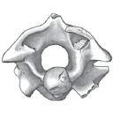
The investigations undertaken at Crivadia (Hateg Depression, Hunedoara District, Romania), the type locality of Crivadiatherium mackennai RADULESCO el al. (Radulesco, Iliesco et lliesco, 1976), led to the discovery of remains of a new Embrithopod. Close to the above mentioned species, but larger in size, this animal is here described as a new species of Crivadiatherium, C. iliescui. ln addition, the comparison made between the forms indicated above and Palaeaamasía kansui OZANSOY from the Eocene deposits of Anatolia (Ozansoy, 1966; Sen et Heintz, 1979) showed that the latter species included a heterogeneous material; this permitted us to distinguish the form in the Anatolian locality Ciçekdag-Arabin Kôyü under the name Palaeoamasia sp. The geographical distribution and diversity of the Embrithopod species under discussion (Balkan, Anatolia) support the idea of an eurasiatic origin of this group and seem to suggest the existence during the Eocene of a particular faunal province in south-eastern Europe.
PV article infos
Published in Vol. 15, Fasc. 3 (1985)
|
PDF
|

|
Systematic and evolutionary relationships of the hipparionine horses from Maragheh, Iran (Late Miocene, Turolian age)
Published online: 30/12/1985
Keywords:
evolution; Hipparionine horses; Iran; Systematics; Turolian
https://doi.org/10.18563/pv.15.4.173-269
Abstract
A systematic analysis of an hipparionine horse assemblage from Maragheh, Iran is made. A brief orientation to systematic philosophy and informal superspecific characterizations of some Old World hipparionines is given as a background to this work. A character state analysis of skulls is made, and has revealed five distinct species. A character state and stratigraphic trend analysis of isolated check tooth and postcranial remains, with known provenance, is also made. These two combined analyses reveal that the most resolute discrimination of hipparionine species and their evolutionary relationships occurs when multiple character complexes of associated skulls, maxillary and mandibular dentitions are made. When this is not possible, skulls have provided the best basis for discriminating species and their evolutionary relationships. Traditional characters of isolated cheek teeth and postcranial remains are shown here to offer limited information content for hipparionine phylogenetic systematics. The systematic portion of this study includes a comprehensive description of cranial and postcranial remains, and has further corroborated the distinction of five species which belong to at least three superspecific groups including: «Hipparion» geltyi sp. nov., Group 1; Hipparion prostylum (s. l.), and Hipparion campbelli sp. nov., Group 3; «Hipparíon» aff. moldavicum and «Hipparion» ?matthewi, Group 2. These species stratigraphic ranges and evolutionary relationships are also given here and argued to be important for establishing future hipparionine geochronologic correlations between a number of Eurasian late Miocene provinces.
PV article infos
Published in Vol. 15, Fasc. 4 (1985)
|
PDF
|

|
Rana (Amphibia : Ranidae) from the upper eocene (MP17a) Hordle Cliff locality, Hampshire, England.
Published online: 15/06/1999
Keywords:
Amphibia; England; Rana; Ranidae; Upper Eocene
Abstract
An ilium from the Upper Eocene (MP l7a) of Hordle, England, represents the first report of Rana from the Eocene of Britain. The ilium is similar to those of the water frog (Rana [ridibunda]) species group.
PV article infos
Published in Vol. 28, Fasc. 1 (1999)
|
PDF
|

|
Un nouveau genre de ?Palaeotheriidae (Perissodactyla, Mammalia) décelé dans les phosphorites du Quercy (Eocène supérieur ou Oligocène) d'après un arrière crâne sans dents.
Published online: 15/06/1999
Keywords:
endocranial cast; Epitympanic sinus; Palaeotheriidae; Paléogène; Quercy Phosphorites; skull anatomy
Abstract
A rear skull from the Quercy Phosphorites is described. It documents a new perissodactyl genus, likely assignable to the family Palaeotheriidae and probably paleogene of age. Owing to the lack of any tooth, this family assignment remains however somewhat hypothetical. The specimen is firstly characterised by the presence of a wide epitympanic sinus swelling and hollowing the squamosal shell and the post-glenoid process. This cavity might make up a peculiar pattern of improvement for the hearing apparatus by carrying out a kind of drum near the middle ear, whereas the bony tympanic remains barely bulged and forms but a few developed auditory bulla. This pattern appears an outcome of a trend observed with many palaeotheriids, such as Plagiolophus. Furthermore, the endocranial cast shows a rather high degree of gyrencephaly for a paleogene mammal. The prominent lambdoidal crest points out a powerful nape musculature and a lowered head bearing. Consequently, it is assumed that such an animal was probably living in somewhat open places, at the expense of rather tough vegetables. It might have been accordingly provided with a semi-hypsodont, cement covered, "plagiolophoid" dentition.
PV article infos
Published in Vol. 28, Fasc. 1 (1999)
|
PDF
|

|
Rythme et modalités de l'évolution chez les rongeurs à la fin de l'Oligocène-leurs relations avec les changements de l'environnement.
Published online: 15/12/2000
Keywords:
Environment; evolution; Oligocene; Rodents; Systematics
Abstract
The analysis of oxygene isotope variations as well as paleobotanical data suggest that the Oligocene/Miocene boundary corresponds to a transitional period marked by floristical and climatic variations. During this period, the pyreneo-alpine tectonics has contribued to modify the geography and western Europe landscapes. Faunal changes (appearances, extinctions, migrations) are observed in different mammalian groups, notably in the rodents. A study of the evolutionary trends and patterns in paleogene rodents is involved for the period ranging from level MP 28 of the Late Oligocene to the Early Miocene, including the Oligo-Miocene boundary.
The Rodents fauna from the sites of Venelles (Bouches-du-Rhône District, France) and Thezels (Lot, France), previously mentionned in litterature, have been studied. The first description of the Eomyidae of La Milloque (MP 29) has been completed. These faunas are compared to those from various localities dating from the considered period. In La Milloque, a new representative of the Eomys species is described next to a form close to Rhodanomys hugueneyae ENGESSER, 1987. It is the Eomys milloquensis nov. sp., the likely descendant of Eomys quercyi COMTE & VIANEY-LIAUD, 1987. Two new species are also described in Thezels: Eucricetodon thezelensis nov. sp., resulting from a likely and local evolution of Eucricetodon praecursor (SCHAUB, 1925) from La Milloque, which, in the same geographic area, could be at the origin of Eucricetodon hesperius ENGESSER, 1985 from Paulhiac. Plesiosminthus admyarion nov. sp., quite distinct from Plesiosminthus schaubi VIRET, 1926, which announces Plesiosminthus myarion SCHAUB 1930. Venelles 'Plesiosminthus schaubi population is considered as a sub-species, named Plesiosminthus schaubi meridionalis nov. subsp. New phylogenetic patterns are proposed. Among the Eomyidae, a quantification of various features of the M1-2/ crown (hypsodonty, degree of abrasion, occlusal angle, state of development of the I and V anticlines), and a comparison with the occlusal diagram of the other teeth among various other populations allows a more efficient separation of Eomys and Rhodanomys genera. In Western Europe, and within this period, it finally does not seem possible to gradually connect the genus Eomys to the genus Rhodanomys. The evolution of the Eomys quercyi - milloquensis lineage seems to underline a similar evolution to that which may have led from the Eomys to the Rhodanomys form. The latter which appears totally accomplished at level MP 29 of the Oligocene is considered as an immigrant. If we compare the most representative species of the Venelles, Thezels, and Coderet sites, (i.e. Rhodanomys, Eucricetodon, Adelomyarion, Peridyromys, Plesíosminthus), it becomes impossible to confirm their biochronological separation. The noticeable differences between the populations may be interpreted as geographical variations. An explanation to these variations, and to fauna's evolution during the Late Oligocene and Early Miocene can be found in the environmental modifications, supported by isotopic, paleobotanical and sedimentologic analysis. A tentative reconstruction of the environments is attempted by the cenogram method. The analysis of the fluctuations of fauna's diversity shows variations which may be correlated to a drop in temperature at MP 29, during the Late Oligocene, followed by an increase in temperature along with an aridity phenomenom, during the basal Miocene (MN O).The confrontation of various methods give the opportunity of reconstituting and comparing the evolution of the environment of three sequences of sites chosen from different regions. Ecological affinities of various rodents' species are being examined. It is possible to consider that the integration of all the conclusions resulting from this study should lead to an explanation to the evolution of rodents for the period around the Oligocene-Miocene boundary. The site of Coderet- level 3- would be posterior to the latter, at the beginnig of the Miocene, and would mark the level MN 0 of the Aquitanian.
PV article infos
Published in Vol. 29, Fasc. 2-4 (2000)
|
PDF
|

|
The Gliridae (Mammalia) from the oligocene (MP24) of Gröben 3 in the folded molasse of southern Germany
Published online: 28/12/2001
Keywords:
Biostratigraphy; Cyrena Beds; folded molasse; Germany; Gliridae; level MP 24; Mammals; Oligocene; Palaeoecology
Abstract
This study describes four taxa of Gliridae from the Oligocene mammal locality Gröben 3: Gliravus tenuis BAI-ILO, 1975, Bransatoglis micio (MISONNE, 1957), B. planus (BAHLO, 1975) and B. heissigi n. sp. Gliravus tenuis from Gröben 3 is somewhat more advanced than the type population found in Heimersheim. This confirms previous research suggesting that Gröben 3 should be dated earlier than Heimersheim (MP 24). The first documented occurrence of B. mício around level MP 24 was found in Gröben 3. An abundance of tooth material from B. planus in Gröben 3 makes it possible, for the first time, to observe evolutionary stages within this species from MP 21 until MP 28. B. heissigi n. sp. is restricted to level MP 24. This species is located between B. mísonnei (MP 20 - 23) and Microdyromys praemurinus (MP 25 - 28). Within the lineage Bransatoglis bahloi - B. misonnei - B. heissigi, a decrease in size is noticeable.
PV article infos
Published in Vol. 30, Fasc. 3-4 (2001)
|
PDF
|

|
Deux nouveaux primates dans l'Oligocène inférieur de Taqah (Sultanat d'Oman): premiers Adapiformes (?Anchomomyini) de la péninsule arabique?
Published online: 15/11/1993
Keywords:
Adapids; Afro-Arabian plate; Early Oligocene; New taxa; Primates; Trans-Tethyan dispersals
https://doi.org/10.18563/pv.22.4.141-196
Abstract
Two new species, Omanodon minor n. g., n. sp. and Shizarodon dhofarensis n. g., n. sp., known from fifteen isolated teeth, are described here as the first adapiform primates (?Anchomomyini) recognizable to date in the Taqah material (early Oligocene of Sultanate of Oman).
Omanodon minor n. g., n. sp. displays special morphological similarity to the adapid tribe Anchomomyini from the Eocene of Europe, and especially to the Anchomomys lineage. Resemblances with the extant lemurifonn Microcebus are also noticeable and could be regarded as supporting Schwartz & Tattersall (1983) hypothesis of special relationships between the anchomomyine adapids and the cheirogaleid lemuriformes. However, these morphological affmities can be interpreted, altematively, as the results of parallelisms: important differences in upper molars indicate that the resemblances of cheirogaleids and Omanodon minor n. g., n. sp. are indeed probably due to parallelisms. Phyletic relationship of O. minor n. g., n. sp. to Anchomomyini is finally the most likely hypothesis.
Shizarodon dhofarensis n. g., n. sp., although much more poorly known, is closely related to Omanodon minor n. g., n. sp., at least at a familial level. The general morphology of this species suggests
also a close link with adapid Anchomomyini, although precise relationships within this tribe remain obscure. Interesting resemblances of Shizarodon dhofarensis n. g., n. sp. to Djebelemur martinezi lower molars (early Eocene of Tunisia) are also noticeable. These resemblances are even stronger than those betwen Omanodon minor and Djebelemur martinezi. However the very bunodont upper molars referred to D. martinezi are unusual for adapids, and there are moreover some notable differences in their lower molars. Thus resemblances in Djebelemur and Shizarodon are probably due to paralellisms.
Because of the fragmentary nature of the material and of possible parallelisms, the systematic position of Omanodon and Shizarodon within adapiformes cannot however yet be established definitively.
PV article infos
Published in Vol. 22, Fasc. 4 (1993)
|
PDF
|
