
Abstract book of the 18th Conference of the EAVP
Pterosaurs from Coahuila
Pliocene-Pleistocene large mammals from Le Riège and Saint-Palais
Les sélaciens du Miocène de la région de Montpellier
Muridae du Pliocène supérieur d'Espagne et du midi de la France.
Contribution à l'étude des genres Gliravus et Microparamys.
Eocene (57) , Quercy Phosphorites (38) , Systematics (32) , Rodents (29) , Mammalia (27) , Rodentia (25) , Miocene (24)

|
The Pleistocene vertebrate fauna of Robinson Cave, Overton County, TennesseeJ. E. Guilday, H. W. Hamilton and A. D. Mc CradyPublished online: 20/01/1969Keywords: Fauna; Mammalia; Pleistocene; Tennessee https://doi.org/10.18563/pv.2.2.25-75 Abstract A late Pleistocene deposit of 60 species of vertebrates and 12 of invertebrates is described from Robinson Cave, Overton County, Tennessee, U.S.A. Forty-eight species of mammals are represented by at least 2,483 individuals; 10 % are extinct, 10 % occur in the state only as boreal relicts in the Great Smoky Mountains; 23 % no longer occur as far south as Tennessee; 57 % occur at or near the site today. Nínety-one percent of the Recent mammal species can be found living today in the Minnesota-Wisconsin area, approximately 10 degrees farther north. Fluorine analysis suggests a long period of accumulation. The following 10 mammalian species are recorded from Tennessee for the first time. Sorex arcticus, Microsorex hoyi, Citellus tridecemlineatus, Clethrionomys gapperi, Microtus pennsylvanicus, Synaptomys cooperi, Synaptomys borealis, Zapus nudsonius, Napaeozapus insignis, Martes americana. Six additional species are present as boreal relicts in the Great Smoky Mountains of eastern Tennessee but not at the site today : Sorex cinereus, Sorex dispar, Sorex palustris, Parascalops breweri, Glaucomys sabrinus, Mustela nivalis. Six forms are extinct: Canis dirus, Ursus americanus amplidens, Sangamona furtiva, Dasypus bellus, Mammut americanus,Megalonyx jeffersoni. Twenty-six additional species of mammals, all of the snails, birds, reptiles, and amphibians recovered from the fauna still inhabit the area today: The fauna is indicative of a cold-temperate climatic episode associated with the Wisconsin glaciation, but may be chronologically mixed. PV article infos Published in Vol. 02, Fasc. 2 (1969) |
|

|
The Quaternary avifauna of Crete, Greece.Peter D. WeesiePublished online: 01/09/1988Keywords: Avifauna; Crete; Quaternary; Systematics https://doi.org/10.18563/pv.18.1.1-94 Abstract Pleistocene bird fossils have been studied from nine localities on Crete. Part of this material was described earlier by the author (Weesie, 1982) and will not be treated here in extenso, the results will be incorporated. More than one third of the over 10,000 fossil bird bones available could be identified ; they were found to represent at least 65 bird species. The following species of the Pleistocene Cretan avifauna are new to the fauna of Crete : Branta ruficollis, Haliaeetus albicilla, Gyps melitensis, Aquila chrysaetos simurgh n. ssp., Ketupa zeylomensis, Aegolius funereus, Dendrocopos leucotos, Zoothera dauma, Turdus iliacus and Pyrrhula pyrrhula. The Pleistocene Cretan avifauna differs less from comparable mainland avifaunas than (fossil) avifaunas from oceanic islands do. Still, the Pleistocene Cretan avifauna has two qualities that are characteristic of island avifaunas : the almost complete absence of a group of birds (the Galliformes) and the presence of two endemic (sub)species : the giant eagle Aquila chrysaetos simurgh n. ssp. and the long-legged owl Athene cretensis (Weesie, 1982). The new subspecies is described in the present study. PV article infos Published in Vol. 18, Fasc. 1 (1988) |
|

|
Norselaspis glacialis n.g., n.sp, et les relations phylogénétiques entre les kiaeraspidiens (Osteostraci) du dévonien inférieur du Spitsberg.Philippe Janvier
Published online: 15/06/1981 |
|

|
Perutherium altiplanense, un Notongulé du Cretacé Supérieur du PérouLarry G. Marshall, Christian de Muizon
Published online: 30/11/1983 |
|

|
Données nouvelles sur le genre Stehlinia (Vespertilionoidea, Chiroptera) du Paléocène d'EuropeBernard SigéPublished online: 01/12/1974Keywords: Chiroptera; Palaeocene; Vespertilionoidea https://doi.org/10.18563/pv.6.3-4.253-272 Abstract Cet article présente une étude détaillée du genre Stehlinia, un chiroptère du Paléogène d'Europe, basé sur un matériel abondant issu notamment du gisement d'Escamps (Quercy). L'analyse révèle que Stehlinia possède un mélange de caractères primitifs (comme une denture tribosphénique complète et des prémaxillaires soudés) et évolués (notamment dans le squelette post-crânien), le rapprochant des vespertilionoïdes actuels, en particulier des Kerivoula. PV article infos Published in Vol. 06, Fasc. 3-4 (1975) |
|
|
Discovery of the most ancient Notidanodon tooth (Neoselachii: Hexanchiformes) in the Late Jurassic of New Zealand. New considerations on the systematics and range of the genus
|
|

|
La poche à phosphate de Ste-Néboule (Lot) et sa faune de vertébres du Ludien Supérieur. 2- Amphibiens. Etude PreliminaireJean-Claude Rage
Published online: 25/09/1978 |
|

|
Contribution à la classification des Pistes de Vertébrés du Trias : les types du Stormberg d'Afrique du Sud (2 ème Partie: le Stormberg supérieur - 1. Le biome de la zone B/1 ou niveau de Moyeni: ses biocénoses).Paul EllenbergerPublished online: 01/12/1974Keywords: biocenosis; Footprints; South Africa; Stormberg; Trias https://doi.org/10.18563/pv.6.ext Abstract Les Pistes de Vertébrés du Stormberg Supérieur ("Trias terminal à Rhétien"), ou Quthingien PV article infos Published in Vol. 6, Ext (1974) |
|
|
A new study of the anthracotheres (Mammalia, Artiodactyla) from pondaung formation, Myanmar: systematics implicationsAung N. SoePublished online: 16/12/2008Keywords: Anthracohyus; Anthracokeryx; Anthracotherium; Pondaung Formation; sexual dimorphism; Siamotherium; South East Asia; taxonomy https://doi.org/10.18563/pv.36.1-4.89-157 Abstract Anthracotheres from the Pondaung Formation, Myanmar, are considered as one of the most primitive artiodactyl groups and they represent the oldest known record in the world. Thus, the understanding of this group has numerous implications for evolutionary biology and biochronological correlations. However, the systematlcs of these mammals has been interpreted in different ways, and the main debate focuses on the number of taxa represented in the Pondaung Formation. The revised taxonomy proposed here is mainly based on the relative development of the upper molar W-shaped ectoloph, system of crests and stylar cusps, and on body size. On the basis of these characters, they are classified into four genera including six different species. Two well-known genera, Anthracotherium and Anthracokeryx, are validated and more precisely diagnosed. Anthracokeryx possesses a better developed W-shaped ectoloph, system of crests and stylar cusps than Anthracotherium, which displays notable differences with the more derived representatives of this genus. Both of these Pondaung genera show evidence for sexual dimorphism. However, the incompleteness of fossil material fueled a debate concerning the status of two additional Pondaung anthracotheres, Siamotherium and Anthracohyus. The latter genus is of uncertain affinities, but it has been considered as a hippopotamid ancestor. Despite new material attributed to these two forms, additional discoveries are still required to establish their taxonomic status. The hypothesis that Southeast Asia was the centre of origin of Anthracotheriidae is supported by the retention of numerous primitive dental characters in these taxa and by the antiquity of the Pondaung Formation, to which an age of 37 My is now generally accepted. PV article infos Published in Vol. 36, Fasc. 1-4 (2008) |
|

|
The evolution of the molar pattern of the Erethizontidae (Rodentia,Hystricognathi) and the validity of Parasteiromys Ameghino, 1904.Adriana M. Candela
Published online: 15/06/1999 |
|

|
Rongeurs du Miocène supérieur de Chorora, Ethiopie: Murinae, Dendromurinae et conclusions.Denis Geraads
Published online: 30/07/2001 |
|

|
Nouvelles espèces de Dendromus (Rongeurs,Muriodea) à Langebaanweg (Pliocène,Afrique du Sud) conséquences stratigraphiques et PaléoecologiquesChristiane Denys
Published online: 20/05/1994 |
|

|
Contribution à l'étude des genres Gliravus et Microparamys (Rodentia) de l'Eocène d'Europe.Jean-Louis HartenbergerPublished online: 15/03/1971Keywords: Eocene; Gliravus; Microparamys; Rodentia https://doi.org/10.18563/pv.4.4.97-135 Abstract Based on material found in about 15 localities the relationships of the genera Microparamys and Glirarus have been studied. One new genus, two subgenera and three species [Microparamys (Sparnacomys) chandoni n. subgen. and n. sp., Microparamys (Pantrogna) russelli n. subgen., Eoglirarus wildi n. gen. and n. sp., Gliravus meridionalis n. sp.] as well as the publication PV article infos Published in Vol. 04, Fasc. 4 (1971) |
|

|
Neue Beobachtungen zum Schädel-und Gebiss-Bau der Paulchoffatiidae (Multituberculata,Ober-Jura).Gerhard HahnPublished online: 15/12/1987Keywords: Dentition; Paulchoffatiidae; Portugal; Skull structure; Upper Jurassic https://doi.org/10.18563/pv.17.4.155-196 Abstract The ventral face of the Paulchoffatiinae skull (Multituberculata, Lower Kimmeridgian, Portugal) is new reconstructed. Some details hitherto unknown are added, as the presence of jugals, the structure of the palatine and the extension of the pterygoids. The situation of the m2/ is discussed. Kielanodon hopsoni n. g., n. sp. is erected, known by its upper p3-5/. From Guimarotodon leiriensis the mandible with its dentition is made known. New informations concerning the milk-dentition and the replacement of teeth are also added. PV article infos Published in Vol. 17, Fasc. 4 (1987) |
|

|
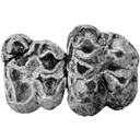
Une nouvelle espèce d'Hipparion du Miocène terminal d'Espagne.Miquel Crusafont i Pairó and P. SondaarPublished online: 31/01/1971Keywords: Hipparion; Late Miocene; Spain; Villafranchian https://doi.org/10.18563/pv.4.2.59-66 Abstract The recently discovered, very rich, Pliocene locality of Layna (Soria, Spain), has already yielded 30 species of mammals. Hipparion fissurae, described here is more dolichopodic than other Hipparion. It is related to Hipparion crusafonti from Villaroya (Villafranchian), but more slender between other characters. PV article infos Published in Vol. 04, Fasc. 2 (1971) |
|

|
Les Gliridés (Rodentia) de l'Oligocène supérieur de Saint-Victor-la-Coste (Gard).Marguerite HugueneyPublished online: 28/10/1968Keywords: Gliridae; Late Oligocene https://doi.org/10.18563/pv.2.1.1-16 Abstract The locality of St.-Victor-la-Coste (Gard) has yielded, rather abundantly, teeth of two glirids hitherto very poorly known: Glirudinus praemurinus (Freudenberg) and Glirudinus glirulus (DEHM). It has permitted, moreover, new views on the evolution of Peridyromys murinus (POMEL). Study of these forms confirms the late Oligocene age of the fauna, without allowing, however, further precision. PV article infos Published in Vol. 02, Fasc. 1 (1968) |
|

|
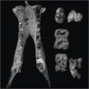
Rongeurs Caviomorphes de l'Oligocène de Bolivie. 2 Rongeurs du Bassin Deseadien de Salla-Luribat.René LavocatPublished online: 01/08/1976Keywords: cranium; Paleobiogeography; Rodentia https://doi.org/10.18563/pv.7.3.15-71 Abstract The fauna studied in the following work involves the dentitions and skulls more or less complete of 5 genera, among which only Cephalomys was previously known by its skull. One must notice that the Salla's species of this genus is a new one. Sallamys, rather small, shows a dentition rather similar to that of Platypittamys Wood from Patagonia. The upper molars, more primitive than those of this last genus, according to the smaller dimensions of the hypocone, retain a distinct metaloph. This metaloph tends to be reduced in a way which may give us a possibility to understand how it disappeared in Platypittamys. The upper P4 can be compared as well to that of Platypittamys as to that of Gaudeamus from the African Oligocene. The lower P4, more molarized than that of Platypittamys, is already moving towards the miocene type of structure. The infraorbital foramen is wide and the insertion of the masseter on the muzzle is spacious. Branisamys, genus of a great size, shows an auditory region partly preserved, peculiarly the promontorium with the fenestra rotunda, entirely of the Hystricognathi type. Upper molars are very clearly pentalophodont. A new reconstruction is proposed for the tooth called Villarroelomys by Hartenberger. This tooth is shown to be a lower D4, perhaps of Branisamys , certainly of a rather nearly allied form, and Hartenberger does agree with the essential part of this new conclusion. Of Incamys, two incomplete skulls are known, each one being admitted to be the type of a distinct species, the first one being I. bolivianus, I. pretiosus the second. The infraorbital foramen is of a great size and the impression of the masseter on the muzzle is spacious. The sphenopalatine foramen is widely developed and of a really very uncommon great size. Only Thryonomys from Africa shows a similar tendency to the enlargement of this foramen, but not so extreme. The main basicranial foramina can be observed. The upper teeth, hemi-hypsodont, show, either a vestigial metaloph, similar to that of recent Thryonomys from Africa, associated with a well developed mesoloph, either a well developed metaloph, while the mesoloph is reduced or absent. Cephalomys was previously known by anterior parts of the skull showing a wide infraorbital foramen and a spacious facial insertion of the masseter. Its lacrymal is of the phiomorph type and the spheno-palatine foramen is seemingly of great size, like in Incamys. The species is new. The varied peculiarities of the upper teeth of these genera can be easily understood if we refer to the plan of the teeth of Phiomys andrewsi from the Oligocene and Miocene of Africa. The structure of this genus, clearly more primitive, still typically brachyodont, shows and clearly explains the fundamental coherence of the varied realisations arised from such a structure. Luribayomys n.g. is known only by an anterior half of a skull without teeth. It is remarquable by the great development of the masseter's insertions on the muzzle and by the lacrymal region, well preserved, typically phiomorphid. The classification previously published by A.E. Wood and B. Patterson is granted in its essential parts, provisionally, but not as a definitive solution. Nevertheless the Dasyproctidae are integrated within the Cavioidea, following the conclusions of Bugge and of Vucetich, reached independently. The conclusion emphasizes the exceptional meaning of the fauna of Salla-Luribay. This shows that Platypittamys, while interesting, can no more be supposed certainly representative of the normal structure of the Oligocene Caviomorph, and not even of their ancestors. The anatomical peculiarities exhibited in these new samples, auditory region, lacrymal, spheno-palatine foramen, reinforce the primitive structural identity with the Phiomorpha. Similarly, the new lower D4 favour very close relationships, ever if the affinities of the D4 has been questioned or minimized by Wood and Patterson. It is certainly possible to admit that parallelism could explain limited similarities, like the presence in North America of Rodents with an hystricomorph type of infraorbital foramen and an hystricognath mandible. But if the parallelism could be a sufficient explanation of the identical association of multiple and complete structures observed in the Caviomorpha and Phiomorpha, all the Zoological systematic would have to be questioned. The last positions of A.E. Wood on the subject (1975) are revised and criticised, and the recent publications studying the problems of distance between Africa and South America in Eocene time, as a consequence of the drift, are quoted; the possibility of transportation by rafts is shown. A new hypothesis is proposed about the interrelationships of Pentalophodont Rodents, with interesting paleobiogeographic implications. PV article infos Published in Vol. 07, Fasc. 3 (1976) |
|

|
Additions to the elasmobranch fauna from the upper Cretaceous of New Jersey (middle Maastrichtian, Navesink Formation)Gerard R. Case and Henri Cappetta
Published online: 15/12/2004 |
|
|
A new species of hippopotamine (Cetartiodactyla, Hippopotamidae) from the late Miocene Baynunah Formation, Abu Dhabi, United Arab EmiratesJean-Renaud Boisserie
Published online: 07/04/2017 |
S.I. Data |

|
Relations phylétiques de Bachitherium filhol, ruminant de l'Oligocène d'Europe Occidentale.Denis Geraads
Published online: 20/06/1987 |
|