
Abstract book of the 18th Conference of the EAVP
Pterosaurs from Coahuila
Pliocene-Pleistocene large mammals from Le Riège and Saint-Palais
Les sélaciens du Miocène de la région de Montpellier
Muridae du Pliocène supérieur d'Espagne et du midi de la France.
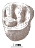
Contribution à l'étude des genres Gliravus et Microparamys.
Eocene (57) , Quercy Phosphorites (38) , Systematics (32) , Rodents (29) , Mammalia (27) , Rodentia (25) , Miocene (24)

|
La plus ancienne faune de mammifères du Quercy : Le BretouJean-Louis Hartenberger, Bernard Sigé and Jean SudrePublished online: 01/12/1974Keywords: Le Bretou; Quercy Phosphorites https://doi.org/10.18563/pv.6.3-4.177-196 Abstract Redécouvertes en 1968 dans le bois du Bretou, les deux poches à phosphorites qui ont livré cette faune sont parmi les plus méridionales du plateau quercynois. De faible profondeur, huit mètres, les cavités sont de petite taille et font partie d'un complexe de sept fissures réparties dans les bois du Bretou. Les autres trous n'ont livré aucun fossile, soit parce qu'ils avaient été intégralement vidés de leur sédiment, soit que des venues d'eau en interdisent l'accès. PV article infos Published in Vol. 06, Fasc. 3-4 (1975) |
|

|
Le genre Leptolophus (Perissodactyla, Mammalia): morphologie et histologie dentaires, anatomie cranienne, implications fonctionnelles.Jean-Albert RemyPublished online: 15/09/1998Keywords: dental histology; Eocene; functional anatomy; Palaeotheriidae; skull anatomy; Southern France; Systematics Abstract A strong lophodonty, an extreme heterodonty, some hypsodonty and regular overlayings of coronal cement are prominent features of the genus Leptolophus (Palaeotheriinae = Palaeotheriidae s.s.). The histological pattern of the teeth unusually joins type II enamel prisms, characteristic of advanced ungulates, together with archaic features, such as an almost complete lack of Hunter-Schreger zonation and a weak expanse of peritubular dentine. The skull is narrow and slender, with an elongated ante-orbital facial region, a moderately notched nasal aperture, a rather elongated post-canine diastem, parallel zygornatic arches and a fairly dorsally located squamoso-mandibular joint.The functional analysis brings to light "ectolophodont" masticatory cycles with two phases, in which maximum power was applied, contrary to equíds, on hindmost teeth; likewise, skull accomodations to increasing height of the teeth are quite different. This study leads to the assumption that Leptolophus may have been light mammals, living in rather open surroundings, browsing on herbaceous plants or leaves cropped close to the ground. Moreover, it appears that it could have been some inadequacy of dental structures to the dietary, which leaded to quick wear of the teeth and to many enamel notches, but had been somewhat balanced by the early increase of hypsodonty, not induced in such a case by a biotop deterioration (as it will happen at the end of the Eocene). This ínadaptation might account for the short duration of the genus Leptolophus, whose the 3 species, L. stehlini, L. nouletí and L. magnus n. sp. are indeed confined in the level MP 16. Its geographical spreading (as far as known, South of western Europe) and the morphological pattern of its dentition suggest that this genus would have been related to early upper Eocene endemic spanish forms. PV article infos Published in Vol. 27, Fasc. 1-2 (1998) |
|

|
Prospection paléontologique de la région de Torralba de Ribota (Burdigalien du bassin de Calatayud, prov. de Zaragoza, Espagne)Edouard Boné, Maria T. Alberdi
Published online: 01/10/1980 |
|

|
Les nouvelles faunes de rongeurs proches de la limite mio-pliocène en Roussillon. Implications biostratigraphiques et biogéographiquesJean-Pierre Aguilar, Jacques Michaux, Bernadette Bachelet, Marc Calvet
Published online: 29/04/1991 |
|
|
Rodent paleocommunities from the Oligocene of Ulantatal (Inner Mongolia, China)Helder Gomes Rodrigues
Published online: 10/06/2014 |
|

|
Les rongeurs du site Pliocène à Hominidés de Hadar (Ethiope)Maurice SabatierPublished online: 15/02/1982Keywords: Ethiopia; hominids; Muridae; Pliocene https://doi.org/10.18563/pv.12.1.1-56 Abstract The intensive exploration of the Pliocene Hadar Formation, rich in hominid remains, led us to the discovery of several micromammals levels. ln some of them, rodents are very abundant. The stratigraphic repartition of these levels do not cover the whole fossiliferous series of the formation but takes place only in the sedimentary members from Sidi Hakoma and Denen-Dora (rancing from 3.1 - 3.2 MY to 2.8 - 2.9 MY, according to the recent geochronological data). During this gap of time, the species do not show morphological changes, what allowed us to gather, in the same taxa, forms of slighty different ages. PV article infos Published in Vol. 12, Fasc. 1 (1982) |
|

|
Study of the Turolian hipparions of the lower Axios valley (Macedonia, Greece). 4. Localities of Dytiko.George D. KoufosPublished online: 15/12/1988Keywords: Equidae; Greece; Hipparion; Lower Axios Valley; Macedonia; Mammalia; Turolian https://doi.org/10.18563/pv.18.4.187-239 Abstract The hipparions from the Dytiko localities of the lower Axios valley (Macedonia, Greece) are studied. The material comes from three localities Dytiko-l, 2, 3 (DTK, DIT, DKO), which are situated near the village of Dytiko, about 60 km northwest to Thessaloniki. Three species have been determined, the medium-sized H. mediterraneum, the small-sized H. matthewi and the very small-sized H. periafricanum. The determined Hipparion species, their morphological characters and their comparison with the other Axios valley material indicate a Late Turolian age for the Dytiko localities. PV article infos Published in Vol. 18, Fasc. 4 (1988) |
|
|
A new and primitive species of Protophiomys (Rodentia, Hystricognathi) from the late middle Eocene of Djebel el Kébar, Central TunisiaLaurent Marivaux
Published online: 02/06/2014 |
|

|
Observations sur des remaniements structuraux post-mortem dans des dents de mammifères fossiles provenant des phosphorites du QuercyJean-Albert RemyPublished online: 01/12/1974Keywords: Quercy Phosphorites; rearrangements; Teeth https://doi.org/10.18563/pv.6.3-4.163-176 Abstract Deux types de remaniements post mortem me paraissent caractéristiques de l'état de conservation des dents de mammifères fossiles dans les Phosphorites du Quercy : PV article infos Published in Vol. 06, Fasc. 3-4 (1975) |
|

|
Contributions à l'étude des micromammifères du gisement Miocène supérieur de Montredon (Hérault). 1- Le gisementJean-Pierre Aguilar and Jean-Yves CrochetPublished online: 30/06/1982Keywords: Hérault; Late Miocene; Micromammals; Montredon https://doi.org/10.18563/pv.12.3.75-79 Abstract La localité fossilifère du Puech de Montredon, désignée plus communément sous le nom de Montredon, est située sur la commune de Montouliers (Hérault) à quelques 300 mètres de la limite avec le département de l'Aude. Elle a été découverte en 1845 par Narbonne, Directeur des Mines de La Caunette, et de très nombreux restes de vertébrés continentaux y ont été extraits. La plus ancienne mention de ce gisement dans la littérature semble être celle de Lartet (1859) qui signale que "M. Jourdan, de Lyon, a constaté à Montredon, près de Bize (Aude), l'association des restes de Dinotherium avec l'Hipparion". PV article infos Published in Vol. 12, Fasc. 3 (1982) |
|
|
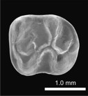
Les Paramyidae (Rodentia) de l'Eocène inférieur du bassin de Paris.Jacques MichauxPublished online: 15/07/1968Keywords: Ailuraviinae; Eocene; Paramyinae; Rodents https://doi.org/10.18563/pv.1.4.135-193 Abstract The exploitation of new early Eocene localities in the Paris Basin has resulted in the collecting of numerous mammalian remains, among which are about 300 isolated teeth representing the rodents. They belong, for the most part, to the paramyid group. Only the latest level of the early Eocene has yielded rodents belonging to the pseudosciurid group. The paramyids, the object of this study, are represented by at least 5 genera and 10 species; they are distributed among 4 clearly dilferentiated subfamilies : Paramyinae Simpson 1945, Pseudoparamyinae Michaux 1964, Ailuraviínae n. subf., Microparamyinae Wood1962. PV article infos Published in Vol. 01, Fasc. 4 (1968) |
|

|
Neolicaphrium recens Frenguelli,1921,the only surviving proterotheriidae (Litopterna, Mammalia) into the south american Pleistocene.Mariano Bond
Published online: 30/07/2001 |
|

|
New remains of the very small cuckoo, Chambicuculus pusillus (Aves, Cuculiformes, Cuculidae) from the late Early/early Middle Eocene of Djebel Chambi, TunisiaCécile Mourer-Chauviré
Published online: 15/02/2016 |
|

|
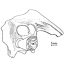
Additions of the Geiseltal mammalian faunas, Middle Eocene: Didelphidae, Nyctitheriidae, Myrmecophagidae.Gerhard Storch and Hartmut HauboldPublished online: 04/12/1989Keywords: Edentata; Geiseltalian; German Democratic Republic; Lipotyphla; Marsupialia; MP 11-13 https://doi.org/10.18563/pv.19.3.95-114 Abstract New and hitherto unpublished mammals from the stratigraphical levels Unterkohle, Untere Mittelkohle and Obere Mittelkohle of the Geiseltal near Halle, GDR, are described (= biochronological levels MP 11-13, Geiseltalian sensu Franzen & Haubold 1986a, b). The marsupial taxa Amphiperatherium aff. maximum (MP 12), A. goethei (MP 12), and Peratherium aff. monspeliense (MP 12 and 13) are recorded for the first time. A lectotype for Amphiperatherium giselense is designated, and the alleged primate Microtarsioides voigzi is assigned to Marsupialia, incertae sedis. A new insectivore species, Saturninia ceciliensis n. sp., is described (MP 13). The anteater Eurotamandua joresi is recorded for the first time outside its type locality, Grube Messel, FRG (MP 11). The present humerus and ulna display the autapomorphic features of the myrmecophagids. PV article infos Published in Vol. 19, Fasc. 3 (1989) |
|

|
The Pleistocene vertebrate fauna of Robinson Cave, Overton County, TennesseeJ. E. Guilday, H. W. Hamilton and A. D. Mc CradyPublished online: 20/01/1969Keywords: Fauna; Mammalia; Pleistocene; Tennessee https://doi.org/10.18563/pv.2.2.25-75 Abstract A late Pleistocene deposit of 60 species of vertebrates and 12 of invertebrates is described from Robinson Cave, Overton County, Tennessee, U.S.A. Forty-eight species of mammals are represented by at least 2,483 individuals; 10 % are extinct, 10 % occur in the state only as boreal relicts in the Great Smoky Mountains; 23 % no longer occur as far south as Tennessee; 57 % occur at or near the site today. Nínety-one percent of the Recent mammal species can be found living today in the Minnesota-Wisconsin area, approximately 10 degrees farther north. Fluorine analysis suggests a long period of accumulation. The following 10 mammalian species are recorded from Tennessee for the first time. Sorex arcticus, Microsorex hoyi, Citellus tridecemlineatus, Clethrionomys gapperi, Microtus pennsylvanicus, Synaptomys cooperi, Synaptomys borealis, Zapus nudsonius, Napaeozapus insignis, Martes americana. Six additional species are present as boreal relicts in the Great Smoky Mountains of eastern Tennessee but not at the site today : Sorex cinereus, Sorex dispar, Sorex palustris, Parascalops breweri, Glaucomys sabrinus, Mustela nivalis. Six forms are extinct: Canis dirus, Ursus americanus amplidens, Sangamona furtiva, Dasypus bellus, Mammut americanus,Megalonyx jeffersoni. Twenty-six additional species of mammals, all of the snails, birds, reptiles, and amphibians recovered from the fauna still inhabit the area today: The fauna is indicative of a cold-temperate climatic episode associated with the Wisconsin glaciation, but may be chronologically mixed. PV article infos Published in Vol. 02, Fasc. 2 (1969) |
|

|
A survey of Cretaceous tribosphenic mammals from middle Asia (Uzbekistan, Kazakhstan and Tajikistan), of their geological setting, age and faunal environmentLev A. Nessov, Denise Sigogneau-Russell and Donald E. RussellPublished online: 20/05/1994Keywords: Cretaceous; Environment; Middle Asia; Sharks; Tribosphenic mammals https://doi.org/10.18563/pv.23.1-4.51-92 Abstract This paper is an English concentrate of various Russian publications by the senior author presenting the mammaIian taxa from the Cretaceous (Albian through Santonian) of the region termed Middle Asia by Soviet geographers. The diagnoses are the unmodified, literal translation of the original version, but are followed with short complementary remarks; most of the species are illuslrated anew with SEM photographs, others are by normal photography. The fossiliferous formations are cited and arguments for their dating are given. Finally, the main vertebrate groups accompanying mammaIs are listed and the environment and climate at the time of deposition are suggested. In conclusion, an hypothesis on the origin and high diversity of tribosphenic mammals on the Cretaceous coastal plains of southwest Asia is proposed. In appendix the taxon Khuduklestes bohlini novo gen. novo sp. is formally defined. PV article infos Published in Vol. 23, Fasc. 1-4 (1994) |
|

|
Révision des Rhombodontidae (Neoselachii Batomorphii) des bassins à phosphate du MarocAbdelmajid Noubhani and Henri Cappetta
Published online: 20/05/1994 |
|

|
The Quaternary avifauna of Crete, Greece.Peter D. WeesiePublished online: 01/09/1988Keywords: Avifauna; Crete; Quaternary; Systematics https://doi.org/10.18563/pv.18.1.1-94 Abstract Pleistocene bird fossils have been studied from nine localities on Crete. Part of this material was described earlier by the author (Weesie, 1982) and will not be treated here in extenso, the results will be incorporated. More than one third of the over 10,000 fossil bird bones available could be identified ; they were found to represent at least 65 bird species. The following species of the Pleistocene Cretan avifauna are new to the fauna of Crete : Branta ruficollis, Haliaeetus albicilla, Gyps melitensis, Aquila chrysaetos simurgh n. ssp., Ketupa zeylomensis, Aegolius funereus, Dendrocopos leucotos, Zoothera dauma, Turdus iliacus and Pyrrhula pyrrhula. The Pleistocene Cretan avifauna differs less from comparable mainland avifaunas than (fossil) avifaunas from oceanic islands do. Still, the Pleistocene Cretan avifauna has two qualities that are characteristic of island avifaunas : the almost complete absence of a group of birds (the Galliformes) and the presence of two endemic (sub)species : the giant eagle Aquila chrysaetos simurgh n. ssp. and the long-legged owl Athene cretensis (Weesie, 1982). The new subspecies is described in the present study. PV article infos Published in Vol. 18, Fasc. 1 (1988) |
|
|
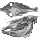
Old world hemiones and new world slender species (Mammalia, Equidae)Véra Eisenmann, John Howe and Mario PichardoPublished online: 16/12/2008Keywords: Amerhippus; biometry; Equus; Holocene; New World; Old World; Osteology; Pleistocene; Pliocene https://doi.org/10.18563/pv.36.1-4.159-233 Abstract Morphological and biometrical description of skulls, teeth, and limb bones of extant and fossil Old World herniones (including E. hydruntinus) and of New World 'stilt-Iegged' and other slender species from Blancan to Holocene. An Appendix presents ways in which the approximate size of some missing bones or dimensions may be deduced from available ones. PV article infos Published in Vol. 36, Fasc. 1-4 (2008) |
|

|
Pantolestidae nouveaux (Mammalia, Insectivora) de l'Eocène moyen de Bouxwiller (Alsace).Jean-Jacques Jaeger
Published online: 31/03/1970 |
|