
Abstract book of the 18th Conference of the EAVP
Pterosaurs from Coahuila
Pliocene-Pleistocene large mammals from Le Riège and Saint-Palais
Les sélaciens du Miocène de la région de Montpellier
Muridae du Pliocène supérieur d'Espagne et du midi de la France.
Contribution à l'étude des genres Gliravus et Microparamys.
Eocene (57) , Quercy Phosphorites (38) , Systematics (32) , Rodents (29) , Mammalia (27) , Rodentia (25) , Miocene (24)

|
Les traces de pas de Dinosaures et autres Archosaures du Lias inférieur des grands Causses, Sud de la FranceGeorges Demathieu, Georges Gand, Jacques Sciau, Pierre Freytet and Jacques GarricPublished online: 15/12/2002Keywords: Dinosauroid footprints; France; Grands-Causses; Hettangian; ichnostratigraphy; paleoenvironments; Sinemurian; statistical results https://doi.org/10.18563/pv.31.1-4.1-143 Abstract The Causses" is a near 3400 km2 large plateau located in the south of France. Here the first dinosaur footprints where found in 1935. After this, this area has yielded an ever-increasing number of ichnites now in excess of 500 specimens. These latter, 15 to 50 cm long, tridactyl or tetradactyl footprints of generally biped animals, were discovered at the surface of Hettangian to lower Sinemurian dolomite layers within 4 distinct stratigraphic units. The 35 sites bearing ichnites are located on the plateau margin. For the first time, morphologic characters studied through descriptive statistic methods with the usual parameters and classical Student and Snédecor tests, allowed us, to divide the whole set of biped traces into 6 ichnospecies. Their definitions are further constrained by multivariate statistical results using Principal Component Analysis (PCA), Factor Analysis of correspondances (FAC) and Discriminant Analysis (DA). All have confirmed the morphologic observations. So that now, the following taxa are identified : Grallator variabilis, G. lescurei, G. sauclierensis, G. minusculus, Eubrontes giganteus, Dilophosauripus williamsi, cf. Moraesichnium, Orníthopus fabrei nov ichnosp. The more immediately visible differences relate to the interdigital II-IV divarication and the digit length ratio. To this panel, we must add Batrachopus deweyi and shapes suggesting Trisauropodichnus and/or Anomoepus. Among all ichnite associations described in the lower Liasic, the New England assemblage presents the most affinities with ours. It shows the ichnotaxa Grallator, Dilophosauripus, Eubrontes, Batrachopus without forgetting Ornithopus fabrei nov. ichnosp. which is close to Ornithopus gallinaceus from the Massachusetts and Connecticut basins. On comparing the present early Jurassic ichnofauna of the Causses with the ones of the Middle and Upper Triassic formations of the eastem border of the Massif Central (France), it appears that tridactyl footprints become more and more numerous and large from Triassic to Early Jurassic. In the Causses, these latest are prevalent but in Quercy (France), Poland, Italy, USA, they are also associated with Omithopoda, Thyreophora and Sauropoda ichnites. Footprint areas considered here were generaly under an arid climate. Animals that passed by were heavy and bulky possible Megalosaur trackmakers, and lighter and slender Coelophysids or Ceratosaurs. For all, these areas were pathways as the orientations of the trackways seem point out. The directions followed by these reptiles were without any important variation during the Hettango-Sinemurian stages. These areas were also used from time to time by Crocodilomorpha and may be tetradactyl (I-IV) bipedal avian Theropods. However, the number of such trackways in sites, sometimes substantial, should not lead us to overestimate the trackmakers populations. These last were probably relatively moderately abondant in this inter-supratidal swamp environment. In the Causses, ichnites are connected with former algo-laminated deposits (Algal mats) which were rapidly hardened by means of calcitisation of cyanobacteria. The result has been a moderate depth of footprints; autopodia disturbing only a few cm of the carbonate substrate. Other fossils have been discovered : invertebrates with thin bivalve and gastropod shells, crustaceans tests and plants. These latter suggest the existence of paleomangroves like environments but also continental vegetation periodically overruning the swamp environment during regression/transgression cycles. At these times, wooded parts of it, could become protecting, feeding, resting and nesting places. PV article infos Published in Vol. 31, Fasc. 1-4 (2002) |
|

|
La poche à phosphate de Ste-Néboule (Lot) et sa faune de vertebres du Ludien superieur. 14 - Conclusions généralesJean-Louis HartenbergerPublished online: 25/09/1978Keywords: Eocene; Quercy Phosphorites https://doi.org/10.18563/pv.8.2-4.319-326 Abstract Le matériel provenant de nouvelles fouilles dans les phosphorites du Quercy, soumis aux divers spécialistes, a conduit à la soutenance de plusieurs thèses d'état: sur les rongeurs (Hartenberger, Vianey-Liaud), les Chiroptères et Insectivores (Sigé), les Artiodactyles (Sudre), les Squamates (Rage) et, en partie, les Chéloniens (De Broin). Chacun dans son domaine, à côté des conclusions d'ordre évolutif, paléogéographique ou paléoécologique, a pu établir des successions stratigraphiques des gisements du Quercy qui se sont révélées largement concordantes. Ainsi la succession des faunes du Quercy est actuellement l'une des mieux établies. Dans ce contexte, les différents spécialistes ont décidé de conjuguer leurs efforts dans l'étude monographique de plusieurs gisements repérés le long de cette échelle, afin de rassembler l'information paléontologique sur des faunes bien précises et de tenter d'obtenir des indications d'ordre taphonomique. PV article infos Published in Vol. 08, Fasc. 2-4 (1978) |
|
|
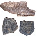
Terrestrial vertebrate paleocommunities from the Cerro del Pueblo Formation (Late Cretaceous; Late Campanian) at Las Aguilas, Coahuila, MexicoHéctor E. Rivera-Sylva
Published online: 16/07/2019 |
|
|
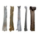
The geologically youngest remains of an ornithocheirid pterosaur from the late Cenomanian (Late Cretaceous) of northeastern Mexico with implications on the paleogeography and extinction of Late Cretaceous ornithocheiridsEberhard Frey
Published online: 21/07/2020 |
|
|
Les Palaeotheridae (Perissodactyla) de la faune de Mammifères de Fons 1 (Eocène supérieur).Jean-Albert RemyPublished online: 15/06/1967Keywords: Anchilophus; Eocene; Pachynolophus; Palaeotheriidae; Perissodactyla https://doi.org/10.18563/pv.1.1.1-46 Abstract The locality of Fons 1, one of the fossiliferous outcrops in the late Eocene limestones of Fons-outre-Gardon (Gard), has yielded varied remains of mammals. The specimens were prepared by dilute acetic acid attack on the rock and by impregnation with an acrylic resin. PV article infos Published in Vol. 01, Fasc. 1 (1967) |
|

|
Données et hypothèses sur la radiation initiale des rongeurs.Jean-Louis HartenbergerPublished online: 01/10/1980Keywords: Diversification scheme; Radiation; Rodents https://doi.org/10.18563/pv.9.ext.285-302 Abstract About the early radiation of Rodents, we have now from the early tertiary of Asia, a new fossil record, and we can do new interpretations. First the problem of the origin of Rodents is studied : considered as a sister group of Lagomorpha, it is possible to find their ancestors between the Mixodontia. Second the new facts about the origin of modern groups of Rodents are reviewed. A general scheme of this diversification can be proposed. PV article infos Published in Vol. 9, Ext (1980) |
|

|
Anatomie du membre antérieur chez un chiroptère Molossidé (Tadarida sp.) du Stampien de Cereste (Alpes-de-Haute-Provence).Bernard SigéPublished online: 01/01/1971Keywords: Chiroptera; Molossidae; Oligocene https://doi.org/10.18563/pv.4.1.1-38 Abstract The present study describes in detail the anterior limb osteology of a molossid chiropteran of the genus Tadarida, from Céreste, a Stampian locality in the Apt-Forcalquier Oligocene basin already known for its fishes, plants and insects. PV article infos Published in Vol. 04, Fasc. 1 (1971) |
|

|
Essai de filiation des campagnols et des lemmings (Arvicolidae, Rodentia) en zone holartique d'après la morphologie dentaire.Jean ChalinePublished online: 01/10/1980Keywords: Arvicolidae; Dental morphology; Paleogeography; phyletic relationships https://doi.org/10.18563/pv.9.ext.375-382 Abstract The Arvicolid evolution results in an increase of the dental structure complexity. The M3/ differenciation seems to characterise the tribe subdivisions, that of M/1 being variable from one to another lineage. The phyletic relationships of fossil lineages are discussed from a paleogeographic point of view. PV article infos Published in Vol. 9, Ext (1980) |
|

|
Evolution des Aplodontidae Oligocènes EuropéensNorbert Schmidt-Kittler and Monique Vianey-Liaud
Published online: 01/10/1979 |
|

|
Les mammifères Montiens de Hainin (Paléocène moyen de Belgique) Part1: Multituberculés.Monique Vianey-Liaud
Published online: 01/11/1979 |
|

|
Rongeurs du Miocène inférieur et moyen en Languedoc. Leur apport pour les correlations Marin-Continental et la Stratigraphie.Jean-Pierre AguilarPublished online: 31/03/1980Keywords: Languedoc; Miocene; Rodents; Southern France https://doi.org/10.18563/pv.9.6.155-203 Abstract The rodents (Cricetidae, Gliridae, Sciuridae) found in lacustrine, brackish marine and karstic sediments of Miocene age in Languedoc, assign the position of the different localities in the scale of "niveaux repères" used by mammalogists. Some detailed stratigraphical studies bring several correlations between this continental biochronological scale and the marine scale ; the most important results are the Aquitanian age of the "niveaux repères" of Coderet and Paulhiac, the Burdigalian age of Laugnac, Estrepouy, Vieux-Collonges, La Romieu and Sansan and the Langhian or Lower Serravallian age of La Grive M. The correlations between the Tethys and the Central Paratethys for the Lower Neogene profit also of these results, since the locality of Neudorf Spalte 1, 2 (Czechoslovakia) is shown to be younger than Sansan (France). The paleontological study has also several geological inferences for the Miocene of Languedoc ; with the calibration of this Miocene, we know quite precisely that the Lower Miocene is chiefly a time lacustrine sedimentation, and also that the marine Miocene sedimentation ends early in the Miocene Period, in Langhian or lower Serravallian times. PV article infos Published in Vol. 09, Fasc. 6 (1980) |
|

|
Les Otolithes de téléostéens du Miocène de Montpeyroux (Herault),France).Dirk Nolf and Henri Cappetta
Published online: 15/12/1980 |
|

|
Les mammifères de Rians (Eocène inférieur, Provence)Marc Godinot
Published online: 01/02/1981 |
|

|
The terrestrial environnement and the origin of land vertebratesJean-Louis HartenbergerPublished online: 02/12/1980Keywords: environments; Land vertebrates; Terrestrial https://doi.org/10.18563/pv.11.1.17 Abstract L'ouvrage rassemble vingt contributions présentées lors d”un colloque organisé par l'éditeur en avril 1979, à Newcastle upon Tyne. Ce sont différents aspects du problème de la << sortie des eaux ›> qui ont été abordés lors de cette réunion. Par son volume, la qualité des communications, l”abondance des illustrations, nul doute que ce livre est appelé à devenir un ouvrage de référence pour les paléontologistes qui s'intéressent de près ou de loin à ce problème : enseignants et cher cheurs y trouveront leur compte. ll est un peu regrettable toutefois qu'aucun tenant de <<l”école suédoise» n'ait eu l`occasion d`y présenter ses thèses. PV article infos Published in Vol. 11, Fasc. 1 (1981) |
|

|
Premier signalement du Monachinae (Phocidae, Mammalia) dans le Sahélien (Miocène supérieur) d'Oran (Algérie)Christian de Muizon
Published online: 15/10/1981 |
|

|
Rongeurs nouveaux de l'Oligocène Moyen d'Espagne.Louis ThalerPublished online: 15/09/1969Keywords: Cricetidae; Oligocene; Pseudocricetodon; Rodents; Theridomys https://doi.org/10.18563/pv.2.5.191-207 Abstract Description of four new rodents from a recently discovered locality at Montalban. Theridomys crusafonti nov. sp. is considered as the ancestry of T. Iembronicus. Theridomys varian: nov. sp. includes «Theridomys» morphotypes and «Blainvilllimys» morphotypes; it could be ancestral to B. blainvillei. Pseudoltinomys nanus nov. sp. represents a new lineage paralleling in evolution that of P. gaillardi (which is equally found at Montalban). Pseudocricetodon montalbanensis nov. gen., nov. sp. designates a lineage of very small Cricetidae accompanying Eucricetodon. With these well defined new species and six others present in the locality, Montalban appears as the best faunal reference point within the biochronologic zone of La Sauvetat. PV article infos Published in Vol. 02, Fasc. 5 (1969) |
|

|
Contributions à l'étude des micromammifères du gisement Miocène supérieur de Montredon (Hérault). 4 - Les chiroptèresBernard SigéPublished online: 30/06/1982Keywords: Chiroptera; Hérault; Late Miocene; Micromammals; Montredon https://doi.org/10.18563/pv.12.3.133-140 Abstract The Montredon local fauna yielded very rare bats, represented by damaged isolated teeth. Only a few documents are available for this period of the European Neogene. ln this poor state of knowledge, the material represents three undetermined species, a supposed molossid and two vespertilionids. PV article infos Published in Vol. 12, Fasc. 3 (1982) |
|

|
Les mammifères Montiens de Hainin (Paléocène moyen de Belgique) Part II : Les CondylarthresJean Sudre and Donald E. RussellPublished online: 30/12/1982Keywords: Belgium; Condylarths; Louisininae; Oxyclaeninae; Paleocene https://doi.org/10.18563/pv.12.6.173-184 Abstract The Condylarths from Hainin (Hainault, Belgium) show no affinity at the generic level to those known in other Paleocene localities of Europe and North America ; they are described as new forms : Monshyus praevius n. gen., n. sp. and Prolatidens waudruae n. gen., n. sp. Monshyus praevius, discovered in only one of the levels in the excavation at Hainin, is similar to the genera Microhyus TEILHARD and Louisina RUSSELL ; with them it is included in the subfamily Louisininae (Hyopsodontidae). With respect to Microhyus and Louisina, Monshyus is distinguished by the precociously modern aspect of its upper molars, the only teeth that are referable. Prolatidens waudruae, known only by lower molars, was found in several levels in the pit at Hainin. It is an arctocyonid presenting possible relationships to the North American form Oxyprimus galadrielae ; it therefore has been provisionally attributed to the subfamily Oxyclaeninae. If this attribution is confirmed, this species will constitute the first and only representative of the group in Europe. PV article infos Published in Vol. 12, Fasc. 6 (1982) |
|

|
Cricetid and arvicolid rodents of the California wash local fauna, late Blancan of the san Pedro Valley, Arizona.Cristiana MezzabottaPublished online: 15/12/1997Keywords: Arvicolidae; Blancan; Cenozoic; Cricetidae; Mammals Abstract An assemblage of micromammals is reported from California Wash, a fossil bearing continental deposit in the San Pedro Valley, Arizona, late Blancan in age. Cricetid and Arvicolid rodents are richly represented, including four and two species, respectively. This study mainly focuses on Sígmodon, the most abundant form. The sample of Sigmodon is compared to samples of the same genus from other localities of the San Pedro Valley of comparable age, and some inferences on the taxonomy of the genus are attempted. The specimens are referred to Sigmodon minor and Sigmodon cf. S. curtisi. Other cricetids (Onychomys pedroensis and Baiomys brachygnathus) and arvicolids (Mictomys vetus and Ondatra ídahoensis) are also recognized and described. PV article infos Published in Vol. 26, Fasc. 1-4 (1997) |
|

|
La poche à Phosphate de Ste-Néboule (Lot) et sa faune de vertébres du Ludien supérieur. 5-SquamatesJean-Claude Rage
Published online: 25/09/1978 |
|