
|
First evidence of an early Miocene marine teleostean fish fauna (otoliths) from la Paillade.(Montpellier,France)
Published online: 15/06/1999
Keywords:
Aquitanian; Biostratigraphy; La Paillade; marine deposits; Miocene; otoliths; Palaeoecology; Palaeogeography; Southern France; Teleostei
Abstract
A fossil fish fauna, based on 5533 otoliths, from the La Paillade locality at Montpellier is described and figured. The otolith-bearing marls correlate to mammal zone MN l (Aguilar, 1982), and thus represent the earliest Miocene. The fish fauna consists of 30 taxa belonging to 20 families. Two species are new: Dussumieria sittigi and Liza gaudanti. The predominant faunal element is the Lesueurigobius vicínalis-species complex, composing 73% of all investigated otoliths. The palaeoecological analysis reveals a marine to euryhaline fish fauna living under tropical to subtropical conditions in the transition zone littoral - sublittoral. Water depth probably was more than 10 m. The scarcity of pelagic físhes suggests that the habitat was either a sheltered bay and/or far away from the open sea. Furthermore, some genera represented in the La Paillade fish fauna presently live exclusively in the Indopacific realm. Their presence strongly supports a broad connection between the Indian Ocean, the Mediterranean, and the Paratethys Seas during the Early Miocene (Aquitanian). From a palaeobiogeographical point of view, faunal relationships were found between the La Paillade fish fauna and both the Paratethys fish fauna and the fish fauna from the deposits in the Upper Rhinegraben and the Mayence and Hanau Basins (Germany).
PV article infos
Published in Vol. 28, Fasc. 1 (1999)
|
PDF
|
|
First record of the family Protocetidae in the Lutetian of Senegal (West Africa)
Published online: 05/12/2014
Keywords:
innominate; Lutetian; Protocetid; Senegal
https://doi.org/10.18563/pv.38.2.e2
Abstract
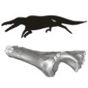
The earliest cetaceans are found in the early Eocene of Indo-Pakistan. By the late middle to late Eocene, the group colonized most oceans of the planet. This late Eocene worldwide distribution clearly indicates that their dispersal took place during the middle Eocene (Lutetian). We report here the first discovery of a protocetid fossil from middle Eocene deposits of Senegal (West Africa). The Lutetian cetacean specimen from Senegal is a partial left innominate. Its overall form and proportions, particularly the well-formed lunate surface with a deep and narrow acetabular notch, and the complete absence of pachyostosis and osteosclerosis, mark it as a probable middle Eocene protocetid cetacean. Its size corresponds to the newly described Togocetus traversei from the Lutetian deposits of Togo. However, no innominate is known for the Togolese protocetid, which precludes any direct comparison between the two West African sites. The Senegalese innominate documents a new early occurrence of this marine group in West Africa and supports an early dispersal of these aquatic mammals by the middle Eocene.
PV article infos
Published in Vol.38-2 (2014)
|
PDF
|

|
Les gangas (Aves, Columbiformes, Pteroclidae) du Paléocène et du Miocène inférieur de France.
Published online: 11/02/1993
Keywords:
Birds; evolution; Lower Miocene; New taxa; Oligocene; Paleoecology; Paulhiac; Quercy Phosphorites; Saint-Gérand-Ie-Puy; Sandgrouse; Upper Eocene
https://doi.org/10.18563/pv.22.2-3.73-98
Abstract
The two species of Sandgrouse from Quercy, Pterocles validus MILNE-EDWARDS and P. larvatus MILNE-EDWARDS, are ascribed to the genus Archaeoganga MOURER-CHAUVIRÉ which includes a third, very large species, A. pinguis. The Sandgrouse of Saint-Gérand-le-Puy, Pterocles sepultus MILNE-EDWARDS, is ascribed to a new genus, Leptoganga. This form appears in the Upper Oligocene of Quercy, in Pech Desse and Pech du Fraysse localities, and is still present in the Lower Miocene of Saint-Gérand-le-Puy and Paulhiac. Recent Sandgrouse live in semidesert or desert areas. The indications provided by mammal and bird faunas in the localities where sandgrouse were found, confirm that the paleoenvironment was open and arid. The morphological study of these fossils indicates that, in the Upper Eocene, the Pteroclidae were already completely individualized with respect to Charadriiformes.
PV article infos
Published in Vol. 22, Fasc. 2-3 (1993)
|
PDF
|
|
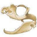
The skull of Tetraceratops insignis (Synapsida, Sphenacodontia)
Published online: 09/01/2020
Keywords:
cranium; pelycosaur; Permian; therapsid origins
https://doi.org/10.18563/pv.43.1.e1
Abstract
Tetraceratops insignis is known from a single, crushed skull from the Lower Permian of Texas. Its unique proportions and osteological details gained central meaning in the question of the origins of Therapsida since this early synapsid has been determined as the oldest and less derived therapsid. Apart from Tetraceratops, the ‘mammal-like’ Therapsida and their sister, the pelycosaur-grade Sphenacodontidae, are separated by one of the longest ghost lineages in tetrapod fossil record. However, the minor, though well justified critique faced insistent publication regarding the therapsid hypothesis. A carefull re-evaluation of the holotypic skull reveals that therapsid traits cannot be supported, including a rejection of the formerly supposed adductor shelf in the temporal fenestra. Increased understanding of ‘pelycosaur’ character variation underlines a haptodontine-grade or, less likely, sphenacodontid position for Tetraceratops.
PV article infos
Published in Vol 43-1 (2020)
|
PDF
|

|
Les rongeurs de l' Eocène inférieur et moyen d'Europe Occidentale; Systématique, phylogénie, biochronologie et paléobiogéographie des niveaux-repères MP 7 à MP 14.
Published online: 15/12/1999
Keywords:
Biochronology; Early and Middle Eocene; Gliridae; Ischyromyidae; Mammalia; MP Scale; New Genus and Species; Palaeogeography; PHYLOGENY; Rodents; Theridomyidae; Western Europe
Abstract
Fourteen distinct phyletical lineages which belong at least in three families: Ischyromyidae ALSTON, 1876, Gliridae THOMAS, 1896 and Theridomyidae ALSTON, 1876, have been identified after the study of more than 3600 rodent dental remains from about twenty Early and Middle Eocene european localities. A systematical and phylogenetical revision of these rodents has been achieved. Nearly all the specific and generic diagnosis are emended. Several new combinations and synonymies are proposed. Four new species and two new genera, Euromys nov. (Ailuravinae) and Hartenbergeromys nov. (Microparamyini), are named and described. Euromys nov. gen. is known by three distinctive ypresian (MP 7 to MP 10 european reference levels) chronospecies. This new lineage is thought to be the direct ancestor of Meldimys MICHAUX, 1968 and Ailuravus RUTIMEYER, 1891. A new species of the genus Plesiarctomys BRAVARD, 1850, Pl. lapicidinarum from Condé-en-Brie (MP 8-9 reference level), allows to relate the Plesiarctomys lineage to the Pseudoparamys MICHAUX, 1964 one. The taxa Sparnacomys HARTENBERGER, 1971, Pantrogna HARTENBERGER, 1971, and Corbarimys MARANDAT, 1989 are erected to genus rank; the last one is not thought to be an Ischyromyidae. A new chronospecies of Pantrogna, P. marandati nov. sp. from the locality of Prémontré (MP 10 reference level), is described. This lineage is at the origin of two others, namely Masillamys TOBIEN, 1954, including M. mattaueri (HARTENBERGER, 1975) nov. comb. (MP 10 reference level), and Hartenbergeromys nov. gen., known from MP 10 (H. hautefeuillei nov. sp.) and MP 11 (H. parvus TOBIEN, 1954) reference levels. The phylogenetical position of Hartenbergeromys nov. gen., at the origin of the european family Theridomyidae, is discussed. The systematical and phylogenetical status of two probable Paramyinae, "Paramys" woodi MICHAUX, 1964 and an unnamed genus and species, are discussed. New populations of the primitive Gliridae Eogliravus HARTENBERGER, 1971 and of the primitive Theridomyidae Protadelomys HARTENBERGER, 1968, are described and assigned to previously known species.
PV article infos
Published in Vol. 28, Fasc. 2-4 (1999)
|
PDF
|

|
Rongeurs du Miocène inférieur et moyen en Languedoc. Leur apport pour les correlations Marin-Continental et la Stratigraphie.
Published online: 31/03/1980
Keywords:
Languedoc; Miocene; Rodents; Southern France
https://doi.org/10.18563/pv.9.6.155-203
Abstract
The rodents (Cricetidae, Gliridae, Sciuridae) found in lacustrine, brackish marine and karstic sediments of Miocene age in Languedoc, assign the position of the different localities in the scale of "niveaux repères" used by mammalogists. Some detailed stratigraphical studies bring several correlations between this continental biochronological scale and the marine scale ; the most important results are the Aquitanian age of the "niveaux repères" of Coderet and Paulhiac, the Burdigalian age of Laugnac, Estrepouy, Vieux-Collonges, La Romieu and Sansan and the Langhian or Lower Serravallian age of La Grive M. The correlations between the Tethys and the Central Paratethys for the Lower Neogene profit also of these results, since the locality of Neudorf Spalte 1, 2 (Czechoslovakia) is shown to be younger than Sansan (France). The paleontological study has also several geological inferences for the Miocene of Languedoc ; with the calibration of this Miocene, we know quite precisely that the Lower Miocene is chiefly a time lacustrine sedimentation, and also that the marine Miocene sedimentation ends early in the Miocene Period, in Langhian or lower Serravallian times.
PV article infos
Published in Vol. 09, Fasc. 6 (1980)
|
PDF
|

|
Les mammifères de Rians (Eocène inférieur, Provence)
Published online: 01/02/1981
Keywords:
Eocene; Mammals; Provence; Rians
https://doi.org/10.18563/pv.10.2.43-126
Abstract
The fossil mammals discovered in the quarry of Rians (Sparnacian, Provence) are described. Among these forms, Hyracotherium is interesting because of the little molarization of the lower premolars and its small size, and Diacodexis by its small size and very primitive astragalus ; they may be the most primitive representatives of their respective orders. Also, Proviverra eisenmanni n. sp. is the smallest and most primitive hyaenodontid yet described. Hyopsodus itinerans is the first species of this genus described France. Among other rare fossils is a new species of bat, a small palaeoryctid, and other forms not yet identified. Marsupials are varied. Several new species are present among the rodents. The fauna is well-balanced and rich in small hyopsodontid condylarths. It is stratigraphically situated at the
Dormaal reference-level, at the base of the early Eocene, and is considered equivalent to the late Clarkforkian of North America. The hypothesis is presented that new forms appearing at the beginning of the Wasatchian in North America migrated, in fact, at that time from Europe.
PV article infos
Published in Vol. 10, Fasc. 2 (1981)
|
PDF
|
|
A late Eocene palaeoamasiine embrithopod (Mammalia, Afrotheria) from the Adriatic realm (Island of Rab, Croatia)
Published online: 14/12/2023
Keywords:
Balkanatolia; Grande Coupure; Great Adria; Paleobiogeography; Systematics
https://doi.org/10.18563/pv.47.1.e1
Abstract
A cheek tooth recently unearthed in the Lopar Sandstone unit, of late Eocene age, in the northern part of Rab Island, Croatia, is one of the very few Eocene mammalian remains found in the Adriatic area. Thorough comparison of this tooth with those of Old-World Palaeogene mammalian orders suggests that it is a M3 belonging to an embrithopod afrothere. The specimen is referred to as Palaeoamasia sp. This genus was formerly known only in Eocene deposits of Anatolia but with close relatives in Romania among Palaeoamasiinae. The geographical distribution of this subfamily perfectly matches the recently-named Balkanatolian landmass, which experienced in-situ evolution of endemic mammals prior to the Grande Coupure event that occurred around the Eocene–Oligocene transition. This last event is characterised by massive Asian immigration in Western Europe and the supposed extinction of many endemic Central and Western European mammals, including Palaeoamasiinae.
PV article infos
Published in 47-1 (2024)
|
PDF
|
|
First report of Cylindracanthus (Osteichthyes) from the Eocene of India
Published online: 25/03/2024
Keywords:
Cylindracanthus; Eocene; histology; rostrum; Umarsar mine.
https://doi.org/10.18563/pv.47.1.e2
Abstract
Fossils of the endangered sturgeons and peddlefishes are widely distributed. We here report for the first time the presence of one of the extinct osteichthyes genus Cylindracanthus (Liedy 1856a) from the Early Eocene lignite-bearing successions of the Kutch Basin, India. The present well preserved rostrum is characterised by numerous wedge-shaped components encircling the central canal that runs along its length, paired at the base and each wedge contributing to the formation of a ridge. The rostrum lacks teeth. The present find extends the palaeobiogeographical distribution of Cylindracanthus considerably and supports its Eocene age as dental remnants preserved in Cylindracanthus sp. shows a decrease in remanent dentition and tooth bases from the Cretaceous to the Eocene. Cylindracanthus is an useful palaeoenvironmental indicator as it has been found associated typically with deposits of nearshore marine environments.
PV article infos
Published in 47-1 (2024)
|
PDF
|
|
A new species of Propalaeotherium (Palaeotheriidae, Perissodactyla, Mammalia) from the Middle Eocene locality of Aumelas (Hérault, France).
Published online: 24/05/2016
Keywords:
Eocene; new species; Palaeotheriidae; Propalaeotherium
https://doi.org/10.18563/pv.40.2.e1
Abstract
A new Propalaeotherium species, clearly distinct from the genus Eurohippus, is described. It is characterized by having a similar size as P. voigti from the German Geiseltal localities (MP 11 to MP 13 reference-level), but differs in several features suggesting a slighty more derived morphology. It presents indeed less brachyodont crowns with less prominent and less elevated cingula, slightly larger relative surface of premolars, and a more marked metaconid splitting on cheek teeth. This new species is unknown from other European localities except the nearby Saint-Martin de Londres locality which has been considered older than the MP 13 level.
PV article infos
Published in Vol.40-2 (2016)
|
PDF
S.I. Data
|

|
Les vertébrés fossiles de Colombie et les problèmes posés par l'isolement du Continent sud-Américain.
Published online: 20/01/1969
Keywords:
Columbia; Cretaceous; Fauna; Quaternary; South America
https://doi.org/10.18563/pv.2.2.77-94
Abstract
A general view is given of the vertebrate faunas, Cretaceous to Quaternary of age, found in Columbia and of their principal characteristics. This view leads to the discussion of the isolation of the South American continent and of the role played by the Bolivar syncline with respect to North American immigrants during the Oligocene. The absence of marine deposits of Oligocene age in the north and northwest of Columbia suggests the possibility of a communication with Central America. This communication would have permitted the passage of hystricomorph rodents, of platyrrhine monkeys, and of colubrids. The non-occupation, until then, of the ecologie niches of these groups would have favored their installation beside the indigenous fauna. In this hypothesis it would no longer be necessary to admit that these vertebrates arrived as «island hoppers ››. The eco-biologic conditions would explain the absence of large-sized forms of North American origin.
PV article infos
Published in Vol. 02, Fasc. 2 (1969)
|
PDF
|

|
Die Referenzfauna des Geiseltalium, MP levels 11 bis 13 (Mitteleozan, Lutetium)
Published online: 04/12/1989
Keywords:
Eocene; Geiseltal; Land mammal ages; Mammalian reference levels
https://doi.org/10.18563/pv.19.3.81-93
Abstract
The Middle Eocene Fossillägerstätte of the Geiseltal lignite beds near Halle/S. (German Democratic Republic) is the reference locality of the European land mammal age Geiseltalian and of the Mammalian Paleogene reference levels MP 11 - MP 13. Due to this importance a reinvestigation is given of the lithostratigraphical development of the Geiseltal beds and of their vertebrate sites. The last are genetically related to the southwest border of the Geiseltal depression and the influx of carbonate-rich waters. The geographical distribution and stratigraphical position of the fossiliferous sites depends on subrosive and tectonically controlled distribution of coal seams. The geological factors and the known stratigraphical guide of some mammalian species suggest corrections of the age of some sites. Four of the alltogether five coal bearing phases contain the 35 sites with mammalian remains. By the distribution of the around 69 mammal species are characterized, with 5 faunal steps ranging from MP 11 to MP 14 or over the Geiseltalian up to the Lower Robiacian. Well distant are the faunas of MP 11 and MP 12. Beginning with MP 12 up to MP 13/14, the fossil record is very frequent by 27 sites. This evidence coincides somewhat more with the concept of land mammal ages compared to that of the punctual mammalian reference levels.
PV article infos
Published in Vol. 19, Fasc. 3 (1989)
|
PDF
|

|
Morphotypes dentaires actuels et fossiles des chiroptères vespertilionines. 2ème partie: implications systématique et phylogéniques.
Published online: 15/11/1987
Keywords:
Chiroptera; PHYLOGENY; Systematics; Vespertilionine
https://doi.org/10.18563/pv.17.3.77-150
Abstract
The first part of this study was devoted to a descriptive analysis of teeth morphologies among the vespertilionine bats. This leads now to a tentative synthesis, providing views on the systematics of the group. The results could be seen according to three distinct but closely related purposes : 1 - the sorting of the genera contents in order to conform the genera units to homogeneous taxa that could represent natural issues of evolutionary lineages ; 2 - the investigation of relationships between extant genera in order to infer the possibilities of common origin ; 3 - according to the preceeding items and to the observed evolutionary trends, a tentative phylogeny, modest and cautious. The contents of many genera are sorted : Leuconoe is removed from subgeneric to generic position, whereas Myotis becomes a subgenus of it ; the myotodont species are cleared away from the Pipistrellus genus ; Glischropus and Scotozous are synonymized within Pipistrellus ; Hypsugo is raised to the generic level ; some species previously ranged within Pipistrellus will form provisionally a collective group, Attalepharca nov. ; the Eptesicus genus is broken up, the excluded species being grouped within Nycterikaupius gen. nov. ; the Nycticeini tribe is defined again after exclusion of Otonycteris , Scotoecus, Scotophilus , and addition of Hesperoptenus ; the species la io and Pipistrellus tasmaniensis are removed to Eptesicus (n.s.) and Pipistrellus dormeri to Scotoecus. Groupings of genera are stated according to the main evolutionary trends of I1/. The relevance of these is often warranted by close morphologic similarities of other teeth. This leads to a recognition of the major evolutionary radiations which occurred in the group. The filiations schematized at the end of the work show the dental relationships observed between the extant genera, and could represent a phylogenic framework. Two major facts are to be underlined : 1- the early divergence of leuconoids ; 2 - the successives crossings to myotodonty from the nyctaloid flow. Fossil data from the literature are punctually and tentatively incorporated within phylogenic sketches.
PV article infos
Published in Vol. 17, Fasc. 3 (1987)
|
PDF
|

|
Le genre Plagiolophus (Palaeotheriidae, Perissodactyla, Mammalia): révision systématique, morphologie et histologie dentaires, anatomie crânienne, essai d'interprétation fonctionnelle
Published online: 15/12/2004
Keywords:
New taxa; Paléogène; perissodactyls; skull anatomy; tooth histology
Abstract
The genus Plagiolophus is documented, almost solely in Western Europe, from the middle Eocene up to the mid Oligocene (MP 12 to MP 25), i.e. more than for 15 MY. Seventeen species are now recorded whose two of them are new, P. ringeadei nov. sp. and P. mamertensis nov. sp. Some anatomical variations and the deflection of certain evolutionary trends justify the distinction of three subgenera, Paloplotherium, Fraasiolophus nov. and Plagiolophus s.s. The genus displays a wide range in size and weight (between 10 and 150 kg). The detailed description of the skull of several species is here given for the first time.
Despite important evolutionary drifts during this long time span, the dentition shows a great structural homogeneity, which renders difficult the determination of fragmentary specimens or isolated teeth. It is characterized by a great heterodonty; premolars are little molarized and present a certain regression through time with paradoxically some progress in the molarization. The hypsodonty increases: the first Plagiolophus are hardly less brachyodont than Propalaeotherium, and the last ones are nearly as hypsodont as Merychippus from the early Miocene. The upper molars change from a wide crown pattern, with an open occlusal surface, lightly oblique transverse lophs and rounded internal cusps, to a narrower pattern, with a frontally constricted occlusal surface and internal lophs aligned parallel to the ectoloph. The M3/3 become always longer.
The dental enamel displays horizontal Schreger-bands with imprecise limits occupying only the middle part of the enamel layer. The dentine is remarkable by its high rate of pericanalicular dentine. The crown cementum, lacking in earlier forms, increases to the point where it fills the occlusal valleys of the
teeth.
The masticatory musculature shows a increasing prominence of the temporal, with probably an important role devoted to the pterygoid muscles in lateral movements related to a two-phase type of chewing.
The evolution of the dentition, of the masticatory musculature and of the repartition of masticatory forces indicate that the Plagiolophus have known different diets through their long evolutionary history; at first browsers they became mixed feeders and finally grazers. Their relatively long neck allowed these animals to reach different vegetal layers. The strength of the nuchal crests also suggests that they were able to have strong backwards movements of the head to pull up their food.
This evolution of diet seems related to the slow degradation of environmental conditions attested during this period in western Europe, with the generalization of more open landscapes, increasing aridity and more marked seasons.
Besides, a remodeling of the face is ontogenetically and along time observed, in relation with the evolution of the masticatory apparatus and especially with that of the mandibular lever arm. The postcanine diastemata become longer in the course of evolution; the free extremities of the nasals are always relatively long which contradicts the hypothesis according to which Paloplotherium may have had a trunk. At last the lineage Fraasiolophus can be distinguished by the presence of a deep malar fossa, probably related to a strong development of the maxillo-labialis superior muscle.
The orbit is always large and tends to increase in size, which indicates a good development of the vision and its increasing role in the life relations. A peculiar type of epitympanic sinus could have been used as a resonance chamber insuring a certain amplification of sounds before their transmission to the eardrum. The endocranial cast reveals a relatively large brain with an advanced degree of gyrencephaly. Beside the role eventually played in food research and social relations, these neurophysiological abilities, also related to an advance in cursorial fitness, could have contributed to the survival of these animals facing the predation pressure of the first fissipede carnivores and the competition with new immigrant herbivores after the "Grande Coupure".
On the basis of some shared apomorphies with the Pachynolophinae, which prevent from considering the latter as Equidae (molarization of the premolars, reduction of the premaxilla dorsal apophysis, peculiar epitympanic sinus, splitting of the jugular process), the hypothesis of an autochthonous origin of Plagiolophus issued from a form near Propalaeotherium, is once again proposed and discussed. Finally, intra-generic relationships are taken into consideration.
PV article infos
Published in Vol. 33, Fasc. 1-4 (2004)
|
PDF
|

|
The skull of Arsinoitherium (Mammalia, Embrithopoda) and the higher order interrelationships of ungulates
Published online: 17/12/1992
Keywords:
Arsinoitherium; PHYLOGENY; Skull; Ungulate
https://doi.org/10.18563/pv.22.1.1-43
Abstract
Detailed anatomical description of arsinoithere cranial remains from the Lower Oligocene, Fayum Depression, Egypt, provides the basic data for a systematic investigation. All cranial and some postcranial features are assessed from a phylogenetic standpoint. Several soft tissue characters are then added to a cladistic analysis based on 54 derived ungulate morphological characters. The resulting phylogenetic hypothesis implies that perissodactyls, sirenians, proboscideans and arsinoitheres constitute a monophyletic unit (5 synapomorphies). However, increasing the tree length by 3 steps reveals a closer association between hyraxes and perissodactyls. Nevertheless, 13 synapomorphies link proboscideans, sirenians and arsinoitheres to the exclusion of all other ungulates. Form of the sphenopalatine and ethmoid foramina, recurved posttympanic process, absence of a fenestra rotundum in the petrosal, vestigial paroccipital process of the exoccipital and the highly unusual absence of a hypoglossal foramen in the skull, imply a robust sister-group relationship between arsinoitheres and proboscideans. In this analysis artiodactyls share only one derived character with all other ungulates studied. Monophyly of Ungulata, including Artiodactyla, is therefore only weakly supported. It is argued that pedal anatomy of hyraxes is non-homologous with that of Tethytheria. Arsinoitherium should now be classified within Tethytheria, sharing a sister-group relationship with Proboscidea. Hyraxes are excluded, thus refuting the concept of Paenungulata. However, monophyly of the wider concept, Pantomesaxonia, containing hyraxes, perissodactyls, sirenians, proboscideans and now, arsinoitheres, is supported by this study.
PV article infos
Published in Vol. 22, Fasc. 1 (1992)
|
PDF
|

|
Systematic and evolutionary relationships of the hipparionine horses from Maragheh, Iran (Late Miocene, Turolian age)
Published online: 30/12/1985
Keywords:
evolution; Hipparionine horses; Iran; Systematics; Turolian
https://doi.org/10.18563/pv.15.4.173-269
Abstract
A systematic analysis of an hipparionine horse assemblage from Maragheh, Iran is made. A brief orientation to systematic philosophy and informal superspecific characterizations of some Old World hipparionines is given as a background to this work. A character state analysis of skulls is made, and has revealed five distinct species. A character state and stratigraphic trend analysis of isolated check tooth and postcranial remains, with known provenance, is also made. These two combined analyses reveal that the most resolute discrimination of hipparionine species and their evolutionary relationships occurs when multiple character complexes of associated skulls, maxillary and mandibular dentitions are made. When this is not possible, skulls have provided the best basis for discriminating species and their evolutionary relationships. Traditional characters of isolated cheek teeth and postcranial remains are shown here to offer limited information content for hipparionine phylogenetic systematics. The systematic portion of this study includes a comprehensive description of cranial and postcranial remains, and has further corroborated the distinction of five species which belong to at least three superspecific groups including: «Hipparion» geltyi sp. nov., Group 1; Hipparion prostylum (s. l.), and Hipparion campbelli sp. nov., Group 3; «Hipparíon» aff. moldavicum and «Hipparion» ?matthewi, Group 2. These species stratigraphic ranges and evolutionary relationships are also given here and argued to be important for establishing future hipparionine geochronologic correlations between a number of Eurasian late Miocene provinces.
PV article infos
Published in Vol. 15, Fasc. 4 (1985)
|
PDF
|

|
Crivadiatherium iliescui n. sp., nouvel Embrithopode (Mammalia) dans le Paléogène ancien de la dépression de Hateg (Roumanie).
Published online: 30/12/1985
Keywords:
Embrithopods; Late Eocene; Paleobiogeography; Romania
https://doi.org/10.18563/pv.15.3.139-157
Abstract
The investigations undertaken at Crivadia (Hateg Depression, Hunedoara District, Romania), the type locality of Crivadiatherium mackennai RADULESCO el al. (Radulesco, Iliesco et lliesco, 1976), led to the discovery of remains of a new Embrithopod. Close to the above mentioned species, but larger in size, this animal is here described as a new species of Crivadiatherium, C. iliescui. ln addition, the comparison made between the forms indicated above and Palaeaamasía kansui OZANSOY from the Eocene deposits of Anatolia (Ozansoy, 1966; Sen et Heintz, 1979) showed that the latter species included a heterogeneous material; this permitted us to distinguish the form in the Anatolian locality Ciçekdag-Arabin Kôyü under the name Palaeoamasia sp. The geographical distribution and diversity of the Embrithopod species under discussion (Balkan, Anatolia) support the idea of an eurasiatic origin of this group and seem to suggest the existence during the Eocene of a particular faunal province in south-eastern Europe.
PV article infos
Published in Vol. 15, Fasc. 3 (1985)
|
PDF
|

|
Rongeurs (Mammalia, Rodentia) du Miocène de Beni-Mellal
Published online: 15/02/1977
Keywords:
Morocco; Neogene
https://doi.org/10.18563/pv.7.4.91-125
Abstract
The rodent fauna of Beni-Mellal is characterized by the abundance of ctenodactylids and cricetids. The latter are represented by four distinct species, among which a new form. Dakkamys zaiani nov. gen., nov. sp. is described. A detailed morphological analysis shows that, contrary to that which had been established before, « Cricetodon ›› atlasi Lavocat, 1961, is not closely related to any European form known; this species is attributed, in consequence, to the new genus Mellalomys. A simple biometric analysis has shown that the genus Myocricetodon Lavocat, 1952, is represented in this locality by two distinct species. The systematic homogeneity of the Beni-Mellal cricetids is also demonstrated: they can, as a matter of fact, all be referred to the subfamily Myocricetodontinae. The definition of this subfamily is completed. The sciurids and glirids are also reviewed in the light of new systematic and biogeographic information established ln Europe. A new species of Atlantoxevus from the early Pleistocene of Morocco, A. huvelini nov. sp., is described. It is probably the descendant of A. tadlae from Beni-Mellal. Biogeographic analysis leads one to consider this fauna as the result of geographic isolation in the Maghreb since the late Oligocene or the early Miocene. In particular no direct European influence can be discerned. Stratigraphic considerations resulting from the discovery of new localities in North Africa lead to the confirmation of the ante-Vallesian age of this fauna and to its parallelism with the faunas of La Grive in Western Europe and Fort Ternan in East Africa. The peculiar geologic nature of this locality is discussed.
PV article infos
Published in Vol. 07, Fasc. 4 (1977)
|
PDF
|

|
Contribution à la classification des pistes de vertébrés du Trias: Les types du Stormberg d'Afrique du Sud (1).
Published online: 16/10/1972
Keywords:
Footprints; South Africa; Stormberg; Trias
https://doi.org/10.18563/pv.5.ext
Abstract
Cet article explore les pistes fossiles de vertébrés du Stormberg (Trias, Afrique du Sud), révélant une diversité ichnologique inédite avec 64 ichnotypes classés en tridactyles, tétradactyles et pentadactyles. Les découvertes incluent des reptiles bipèdes comme Qemetrisauropus et Prototrisauropus, dont les empreintes suggèrent des adaptations évolutives vers la bipédie, ainsi que des pentadactyles géants comme Pentasauropus, annonçant les futurs sauropodes. L’étude combine ichnologie et restes osseux pour reconstituer des écosystèmes triasiques riches, où flore (fougères, conifères) et faune témoignent d’une transition écologique majeure avant l’essor des dinosaures.
PV article infos
Published in Vol. 5, Ext (1972)
|
PDF
|

|
Le genre Microstonyx en Espagne et ses relations avec les autres espèces du même genre hors d'Espagne
Published online: 01/10/1980
Keywords:
Microstonyx; Spain; Suidae
https://doi.org/10.18563/pv.9.ext.213-231
Abstract
The genus Microstonyx was found only in the north eastern part of Spain : M. antiquus, referable to the
Eppelsheim type, from the pyrenean basins ; M. antiquus, more progresive, from the Vallès Basin : M. major-erymanthius group, from the Teruel-Ademuz Area and from Pieru (Penedès Basin).
PV article infos
Published in Vol. 9, Ext (1980)
|
PDF
|
