
Abstract book of the 18th Conference of the EAVP
Pterosaurs from Coahuila
Pliocene-Pleistocene large mammals from Le Riège and Saint-Palais
Les sélaciens du Miocène de la région de Montpellier
Muridae du Pliocène supérieur d'Espagne et du midi de la France.
Contribution à l'étude des genres Gliravus et Microparamys.
Eocene (57) , Quercy Phosphorites (38) , Systematics (32) , Rodents (29) , Mammalia (27) , Rodentia (25) , Miocene (24)

|
Contributions à l'étude du gisement Miocène supérieur de Montredon (Hérault). Les grands mammifères. 7 - Les proboscidiens DeinotheriidaeHeinz TobienPublished online: 15/11/1988Keywords: allometry; Astaracian; Deinotherium; Montredon; Systematics; taphonomy; Vallesian https://doi.org/10.18563/pv.18.ext.135-175 Abstract Some complete tooth rows and about one hundred isolated teeth enabled the identification of the deinothere of the Vallesian site Montredon (Hérault) as Deinotherium giganteum KAUP 1829, mainly by comparisons with the likewise Vallesian sample of the type locality Eppelsheim (Rheinhessen, F.R.G.). PV article infos Published in Vol. 18, Ext (1988) |
|
|
Contributions à l'étude de l'anatomie crânienne des rongeurs. 1- Principaux types de cricétodontinésJean-Louis HartenbergerPublished online: 25/09/1967Keywords: Cricetodon; Cricetodontinae; Miocene https://doi.org/10.18563/pv.1.2.47-64 Abstract Description, for the first time, of the skull of Ruscinomys Depéret on the basis of a nearly complete specimen, and description of a new facial part of a Megacricetodon Fahlbusch skull (material from upper Miocene, Spain). New description of the skull (facial part) of " Cricetodon" incertum Schlosser on the basis of the specimen from the Oligocene of Quercy phosphorites already published by S. Schaub. PV article infos Published in Vol. 01, Fasc. 2 (1967) |
|

|
La poche à Phosphate de Ste-Néboule (Lot) et sa faune de vertébres du Ludien supérieur. 5-SquamatesJean-Claude Rage
Published online: 25/09/1978 |
|
|
Additions to the elasmobranch assemblage from the Bandah Formation (middle Eocene, Bartonian), Jaisalmer District, Rajasthan, India, and the palaeobiogeographic implications of the faunaRajendra S. Rana
Published online: 23/06/2021 |
|

|
Cricetid and arvicolid rodents of the California wash local fauna, late Blancan of the san Pedro Valley, Arizona.Cristiana MezzabottaPublished online: 15/12/1997Keywords: Arvicolidae; Blancan; Cenozoic; Cricetidae; Mammals Abstract An assemblage of micromammals is reported from California Wash, a fossil bearing continental deposit in the San Pedro Valley, Arizona, late Blancan in age. Cricetid and Arvicolid rodents are richly represented, including four and two species, respectively. This study mainly focuses on Sígmodon, the most abundant form. The sample of Sigmodon is compared to samples of the same genus from other localities of the San Pedro Valley of comparable age, and some inferences on the taxonomy of the genus are attempted. The specimens are referred to Sigmodon minor and Sigmodon cf. S. curtisi. Other cricetids (Onychomys pedroensis and Baiomys brachygnathus) and arvicolids (Mictomys vetus and Ondatra ídahoensis) are also recognized and described. PV article infos Published in Vol. 26, Fasc. 1-4 (1997) |
|

|
Systematic revision of Ctenodactylidae (Mammalia, Rodentia) from theMiocene of Pakistan.J.A. BaskinPublished online: 18/03/1996Keywords: Ctenodactylidae; Miocene; Prosayimys; Rodents; Sayimys; Siwalik Abstract Extensive sampling of the Siwalik deposits of the Potwar Plateau of northem Pakistan and from the Zinda Pir dome of central Pakistan has produced a fossil record of Miocene ctenodactylids that can be correlated with the paleomagnetic time scale. The early Miocene Prosayimys flynni (n. gen., n. sp.) is recognized as the first ctenodactylid in the Indian subcontinent. Prosayimys is ancestral to Sayimys. From the late early Miocene to the early late Miocene, there is an anagenetic succession of three species of Sayimys: S. cf. S. intermedius, S. sivalensis, and S. chinjiensis (n. sp.). Sayimys chinjiensis gave rise to the late late Miocene S. perplexus. A second lineage is represented by Sayimys minor, S. sp. A, and S. sp. B. PV article infos Published in Vol. 25, Fasc. 1 (1996) |
|

|
Analyse d'ouvrage: “J.J. THOMASON (Ed.): Functional morphology invertebrate paleontology (1995)”Jacques MichauxPublished online: 18/03/1996Keywords: Book review; Functional morphology Abstract Functional morphology invertebrate paleontology, édité par Jeffrey J. THOMASON, 1995. Cambridge University Press, xi + 277 p. ISBN 0-521-44095-5, f. 45,00 ($ 69,95). PV article infos Published in Vol. 25, Fasc. 1 (1996) |
|

|
Experimental taphonomy inavian eggs and eggshells: effects on early diagenesis.Ana M. Bravo, D. A. Buscalioni
Published online: 15/12/2003 |
|

|
Analysis of changing diversity patterns in Cenozoic land mammal age faunas, South AmericaLarry G. Marshall and Richard L. CifelliPublished online: 30/03/1990Keywords: Cenozoic; Chronofaunas; diversity; Equilibrium theory; Extinction; Land mammal faunas; Origination; South America https://doi.org/10.18563/pv.19.4.169-210 Abstract Comparison of various measurements of taxonomic evolution using stratigraphic range data for orders, families and genera of land mammals indicates several means by which deficiencies of the South American fossil record (e.g., presence of hiatuses, unequal temporal and geographic representation of ages, unequal systematic treatment) may be normalized, thus permitting a less distorted appreciation of diversity pattern and trend. Initial radiation of native taxa resulted in a relative equilibrium by early Eocene time. Subsequent increases in absolute diversity were apparently induced by immigration at the family level and by environmental factors at the generic level. Miocene through Pleistocene phases of faunal stability, herein characterized as chronofaunas, are punctuated by rapid turnover events resulting from a complex of factors, including adaptive radiation of immigrant taxa into unoccupied eco-space; environmental and concomitant habitat change induced by orogenic events of the Andes; and biotic interactions between native and immigrant taxa, including competition and prey naivete. The first two factors account for major faunal transitions in the South American middle and late Tertiary; immigration-induced turnover may have been of greater importance in shaping the character of the fauna upon the Great American Interchange and the arrival of man in the Neotropics PV article infos Published in Vol. 19, Fasc. 4 (1990) |
|
|
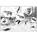
Essai de reconstitution d'un paysage du Quercy vers -35 Ma. (Esquisse de Christian Pondeville, 1977).Monique Vianey-Liaud
Published online: 15/09/1978 |
|

|
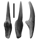
La poche à phosphate de Ste-Néboule (Lot) et sa faune de vertebres du Ludien supérieur. 11- CréodontesBrigitte Lange-BadréPublished online: 25/09/1978Keywords: Creodonta; Eocene; Quercy Phosphorites https://doi.org/10.18563/pv.8.2-4.295-299 Abstract The teeth and the astragale of the Creodonta from Ste-Néboule (Lot) are referred to Hyaenodon brachyrhynchus. Isolated teeth fit morphologically the material from La Débruge (Middle Ludian). However, biometric analysis suggests an Upper Ludian age. PV article infos Published in Vol. 08, Fasc. 2-4 (1978) |
|

|
Morphotypes dentaires actuels et fossiles des chiroptères vespertilionines. 2ème partie: implications systématique et phylogéniques.Henri MenuPublished online: 15/11/1987Keywords: Chiroptera; PHYLOGENY; Systematics; Vespertilionine https://doi.org/10.18563/pv.17.3.77-150 Abstract The first part of this study was devoted to a descriptive analysis of teeth morphologies among the vespertilionine bats. This leads now to a tentative synthesis, providing views on the systematics of the group. The results could be seen according to three distinct but closely related purposes : 1 - the sorting of the genera contents in order to conform the genera units to homogeneous taxa that could represent natural issues of evolutionary lineages ; 2 - the investigation of relationships between extant genera in order to infer the possibilities of common origin ; 3 - according to the preceeding items and to the observed evolutionary trends, a tentative phylogeny, modest and cautious. The contents of many genera are sorted : Leuconoe is removed from subgeneric to generic position, whereas Myotis becomes a subgenus of it ; the myotodont species are cleared away from the Pipistrellus genus ; Glischropus and Scotozous are synonymized within Pipistrellus ; Hypsugo is raised to the generic level ; some species previously ranged within Pipistrellus will form provisionally a collective group, Attalepharca nov. ; the Eptesicus genus is broken up, the excluded species being grouped within Nycterikaupius gen. nov. ; the Nycticeini tribe is defined again after exclusion of Otonycteris , Scotoecus, Scotophilus , and addition of Hesperoptenus ; the species la io and Pipistrellus tasmaniensis are removed to Eptesicus (n.s.) and Pipistrellus dormeri to Scotoecus. Groupings of genera are stated according to the main evolutionary trends of I1/. The relevance of these is often warranted by close morphologic similarities of other teeth. This leads to a recognition of the major evolutionary radiations which occurred in the group. The filiations schematized at the end of the work show the dental relationships observed between the extant genera, and could represent a phylogenic framework. Two major facts are to be underlined : 1- the early divergence of leuconoids ; 2 - the successives crossings to myotodonty from the nyctaloid flow. Fossil data from the literature are punctually and tentatively incorporated within phylogenic sketches. PV article infos Published in Vol. 17, Fasc. 3 (1987) |
|

|
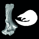
Palaeotis weigelti restudied : a small middle Eocene Ostrich (Aves : Struthioniformes)Peter Houde
Published online: 20/06/1987 |
|

|
The lower Miocene artiodactyls of Tagai bay, Olhon island, lake Baikal (Russia)Inesa Vislobokova
Published online: 20/05/1994 |
|

|
Préface au mémoire jubilaire en hommage à René LavocatJacques MichauxPublished online: 01/10/1980Keywords: Editorial https://doi.org/10.18563/pv.9.ext.1-13 Abstract Monsieur René Lavocat, Directeur du Laboratoire de Paléontologie des Vertébrés de la troisième section de l'Ecole Pratique des Hautes Etudes, quittait le service actif en l'année 1979. View editorial Published in Vol. 9, Ext (1980) |
|

|
Les mammifères post-glaciaires de Corse. Etude Archéozoologique.Jacques MichauxPublished online: 15/09/1989Keywords: Book review https://doi.org/10.18563/pv.19.1.45-46 Abstract Les Mammifères post-glaciaires de Corse de Jean-Denis Vigne, étudie l'évolution des mammifères en Corse depuis 7000 av. J.-C. jusqu'à aujourd'hui, en explorant leur adaptation insulaire, l'impact de l'homme sur leur extinction ou leur introduction, et les pratiques de chasse et d'élevage à travers l'analyse des ossements. PV article infos Published in Vol. 19, Fasc. 1 (1989) |
|

|
The stratigraphic sequence of North American rodent faunasRobert W. WilsonPublished online: 01/10/1980Keywords: North America; Rodents; Stratigraphic sequence https://doi.org/10.18563/pv.9.ext.273-283 Abstract Rodents first appear in the latest Paleocene or earliest Eocene as very fragmentary specimens (Family Paramyidae) known largely from a single locality. After this sparse beginning, rodents are usually abundant in the North American record if proper recovery methods are used. Utilization of rodents for biostratigraphic purposes depends on 1/ extinction, and 2/ replacement by evolution of endemic groups and/or incursions of Old World rodents, and rarely and late by South American kinds. These incursions are separated by relatively long periods of isolation in the Paleogene, but more episodic in the Neogene. At least 10 rodent zones can be characterized by major distinctions, and these zones can be amplified into as many as 16 with little trouble. In general, rodent genera permit as refined a zonation as do genera of large mammals. Distinction at a specific level has not been attempted herein except in the Blancan and Post-Blancan. PV article infos Published in Vol. 9, Ext (1980) |
|

|
Contributions à l'étude du gisement Miocène supérieur de Montredon (Hérault). Les grands mammifères. 3 - Les artiodactyles ruminantsLéonard Ginsburg and Herbert ThomasPublished online: 15/11/1988Keywords: Artiodactyla; France; Montredon; Ruminentia; Upper Miocene https://doi.org/10.18563/pv.18.ext.43-56 Abstract The remains of Ruminantia are very rare at Montredon. No specific determination was possible. We have only one Micromeryx, one small cervid, one tragocere and one (?) gazella. The faunal spectrum is in good correlation with the general retreat of the cervids in the European upper Miocene, but in contrast it is not convenient for the bovids, which are in expansion at the same time. For them, we have to invoke the local ecological conditions. PV article infos Published in Vol. 18, Ext (1988) |
|
|
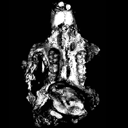
A pangolin (Manidae, Pholidota, Mammalia) from the French Quercy phosphorites (Pech du Fraysse, Saint-Projet, Tarn-et-Garonne, late Oligocene, MP 28)Jean-Yves Crochet, Lionel Hautier
Published online: 14/09/2015 |
|

|
|
|