
Abstract book of the 18th Conference of the EAVP
Pterosaurs from Coahuila
Pliocene-Pleistocene large mammals from Le Riège and Saint-Palais
Les sélaciens du Miocène de la région de Montpellier
Muridae du Pliocène supérieur d'Espagne et du midi de la France.
Contribution à l'étude des genres Gliravus et Microparamys.
Eocene (57) , Quercy Phosphorites (38) , Systematics (32) , Rodents (29) , Mammalia (27) , Rodentia (25) , Miocene (24)

|
The late Miocene percrocutas (Carnivora,Mammalia) of Madedonia, Greece.George D. KoufosPublished online: 14/06/1995Keywords: Biochronology; Carnivora; Comparisons; Dinocrocuta; Greece; Late Miocene; Mammalia Abstract Some new material of percrocutas from the late Miocene of Axios valley (Macedonia, Greece) is studied. They have been found in the locality of "Pentalophos 1" (PNT). The material has been described and compared with the known late Miocene percrocutas of Eurasia. This comparison indicates that it can be identified as Dinocrocuta gigantea (SCHLOSSER, 1903). A maxilla of a percrocuta, named ”Hyaena" salonicae, was found in the same area (Andrews, 1918). "Hyaena" salonicae is smaller than the PNT material. It is also compared with other material from Eurasia while its taxonomic and age problems are discussed. It belongs to Dinocrocuta and shows close relationships with D. robusta and D. senyureki; its age can be considered as late Vallesian-early Turolian. The age of the locality PNT is also discussed and a possible Vallesian age is proposed for it. PV article infos Published in Vol. 24, Fasc. 1-2 (1995) |
|
|
A new species of hippopotamine (Cetartiodactyla, Hippopotamidae) from the late Miocene Baynunah Formation, Abu Dhabi, United Arab EmiratesJean-Renaud Boisserie
Published online: 07/04/2017 |
S.I. Data |

|
Sur le remplissage des "poches à phosphorite" d'Aubrelong (commune de Bach, Lot)Bernard GèzePublished online: 15/11/1974Keywords: Aubrelong; Quercy Phosphorites https://doi.org/10.18563/pv.6.1-2.21-25 Abstract L’article analyse les poches à phosphorite d’Aubrelong (Quercy), comparant les fouilles de 1937 (Éocène supérieur) et celles des années 1967-1970 (Oligocène inférieur), révélant une stratification contrastée : argiles brunes pauvres en fossiles en surface, et argiles rouges riches en phosphates et faune en profondeur. Les analyses géochimiques confirment une origine latéritique sous climat tropical humide, avec des remplissages tantôt stratifiés, tantôt chaotiques, remettant en cause l’hypothèse d’une progression chronologique uniforme des dépôts PV article infos Published in Vol. 06, Fasc. 1-2 (1974) |
|

|
Neue Beobachtungen zum Schädel-und Gebiss-Bau der Paulchoffatiidae (Multituberculata,Ober-Jura).Gerhard HahnPublished online: 15/12/1987Keywords: Dentition; Paulchoffatiidae; Portugal; Skull structure; Upper Jurassic https://doi.org/10.18563/pv.17.4.155-196 Abstract The ventral face of the Paulchoffatiinae skull (Multituberculata, Lower Kimmeridgian, Portugal) is new reconstructed. Some details hitherto unknown are added, as the presence of jugals, the structure of the palatine and the extension of the pterygoids. The situation of the m2/ is discussed. Kielanodon hopsoni n. g., n. sp. is erected, known by its upper p3-5/. From Guimarotodon leiriensis the mandible with its dentition is made known. New informations concerning the milk-dentition and the replacement of teeth are also added. PV article infos Published in Vol. 17, Fasc. 4 (1987) |
|

|
Contribution à l'étude des genres Gliravus et Microparamys (Rodentia) de l'Eocène d'Europe.Jean-Louis HartenbergerPublished online: 15/03/1971Keywords: Eocene; Gliravus; Microparamys; Rodentia https://doi.org/10.18563/pv.4.4.97-135 Abstract Based on material found in about 15 localities the relationships of the genera Microparamys and Glirarus have been studied. One new genus, two subgenera and three species [Microparamys (Sparnacomys) chandoni n. subgen. and n. sp., Microparamys (Pantrogna) russelli n. subgen., Eoglirarus wildi n. gen. and n. sp., Gliravus meridionalis n. sp.] as well as the publication PV article infos Published in Vol. 04, Fasc. 4 (1971) |
|

|
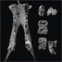
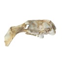
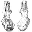
Etude du crâne de Pachynolophus lavocati n. sp. (Perissodactyla, Palaeotheriidae) des Phosphorites du QuercyJean-Albert RemyPublished online: 15/02/1972Keywords: Perissodactyla; Quercy Phosphorites https://doi.org/10.18563/pv.5.2.45-78 Abstract The genus Pachynolophus, one of the poorest known of the Palaeotheriidae, includes the brachyodont forms with reduced and non-molariform premolars and with upper molars lacking a mesostyle. Quantitative characters (divers surface indications and elongation of the teeth), while demonstrating a close relationship to Hyracotherium, permit a better differentiation of the genus, confirm its specific splitting, and permit the distinction of three lineages. The skull from Memerlein is taken as the type of a new species, P. Iavocati, of which the dentition is extremely characterized by its lophiodonty, the strong reduction of the premolars and the reduction of the cingula. This characterization testifies to a late age which extends the existence of the genus quite near to the Eocene-Oligocene limit. Compared with the only two skulls known of related species (Hyracotheríum vulpiceps and Pachynolophus Iivinierensis), that from Memerlein is distinguished by progressive characters affecting diferent regions but most particularly the braincase; it is not possible, however, to isolate within this evolution the part which leads to a systematic differentiation. Modernization is translated by a considerable increase in size of the braincase, principally in the frontal region, a development of the facial region with anterior displacement of the dental series and a greater specialization of the masticatory apparatus. This evolution parallels the history of the Equidae of the North American early Tertiary, but certain particularities, the form of the alisphenoid, the presence of an anterior frontal foramen, and the structure of the paroccipital apophysis, testifies to the independance of the European forms. PV article infos Published in Vol. 05, Fasc. 2 (1972) |
|

|
Les Affinités de Nyctereutes megamastoides (Pomel), canidé du gisement Villafranchien de Saint-Vallier (Drôme, France).R. MartinPublished online: 31/01/1971Keywords: Canidae; Nyctereutes; Villafranchian https://doi.org/10.18563/pv.4.2.39-58 Abstract Nyctereutes megamastoides (Pomel) from the Villafranchian of the Auvergne and from Saint-Vallier presents cranial and dental characters sufficiently close to those of the late Pliocene canid from Perpignan (Roussillon), described by Depéret under the specific name of Canis donnezani belonging to the same genus Nyctereutes. The extinction of the European "Nyctereutes" group seems due to the too great alimentary specialization of this canid, whereas the Asiatic lineage represented in the Villafranchian by Nyctereutes sinensis Schlosser and at present by Nyctereutes procyonider Gray was able to maintain itself probably by means of a profound change in its alimentary regime. PV article infos Published in Vol. 04, Fasc. 2 (1971) |
|

|
Analyse d'ouvrage : Koobi Fora Research Project, volume 3. The fossil ungulates: geology, fossil artiodactyls, and palaeoenvironments, édité par John Michael HARRIS, 1991. Clarendon Press, Oxford, xvi + 384 p. ISBN 0-19-857399-5.Jean SudrePublished online: 11/02/1993Keywords: Artiodactyls; palaeovenvironments; Ungulates https://doi.org/10.18563/pv.22.2-3.138-139 Abstract Avec ce volume se clôture l'étude géologique et paléontologique des sites à hominidés de Koobi Fora. Il fait suite aux deux précédents ouvrages, parus respectivement en 1978 et 1983, consacrés également à l'étude des faunes recueillies sur ces gisements dans le cadre du Koobi Fora Research Project. PV article infos Published in Vol. 22, Fasc. 2-3 (1993) |
|

|
Eurodexeinae, eine neue unterfamilie der Artiodactyla (Mammalia) aus dem unter- und mitteleozän europasJorg Erfurt and Jean SudrePublished online: 16/12/1996Keywords: Artiodactyls; Eocene; evolution; Germany; Lutetian; new genus; New subfamily Abstract Dichobunoid artiodactyls are described in this paper from the middle Eocene Geiseltal lignite deposits near Halle (Sachsen-Anhalt, Germany). The genera Eurodexis and Parahexacodus are established based on odontologica1 studies. The type-species are E. ceciliensis (FRANZEN & KRUMBIEGEL, 1981) and P. germanicus n. sp. from the "obere Mittelkohle" (see text), MP 13. The genera are referred to the new subfamily Eurodexeinae (Dichobunidae, Artiodactyla, Mammalia), which also contains the monospecific genus Eygalayodon SUDRE & MARANDAT, 1993. These new genera show many features similar to North American homacodontids and antiacodontids. They exemplify a high degree of diversity in European dichobunids. Moreover, the family Dichobunidae includes the Dichobuninae and the Hyperdichobuninae. The connection of the posthypocristid with the postentocristid, the shift of a medial to a lingual position of the hypoconulid on the lower molars and the development of a large, caniniform P1 are regarded as the principal tendencies of the eurodexeines. The entoconid is larger and more mesially situated compared to that of Diacodexis. The hypoconulid has a lingual position on a broad postcingulid. The elongation of the premolars and the presence of diastemata are considered associated with extension of the muzzle. This and the acute tubercles of the molars could indicate a limited degree of insectivory. Protodichobune cf. oweni and Diacodexis cf. varleli, first reported here from the "untere Unterkohle" (MP 11) of the Geiseltal, demonstrate the existence of forms in the basal part of the middle Eocene, that are direct descendants of lower Eocene ones. Eurodexis russelli n. sp. from MP 10 of Premontre is the most primitive representative of Eurodexis. A further part of this lineage could be Messelobunodon sp. from MP 11 of Messel (Germany). Another form from Premontre, referred to Eurodexeinae indet., represents the origin of the lineage to Parahexacodus germanicus n. sp. from the Geiseltal (MP 13). The identification of several lineages in MP 10 is evidence of an adaptive radiation during this time. Unfortunately the history of the earlier species of Diacodexis is not known well enough to exclude earlier diversifications. Our study shows the independent development of artiodactyls at the end of the lower Eocene in Europe, North America (and maybe in Asia) with much convergence. The question of the geographic origin of the genus Diacodexis is still open. PV article infos Published in Vol. 25, Fasc. 2-4 (1996) |
|

|
Insectivores pliocènes du Sud de la France (Languedoc-Roussillon) et du Nord-Est de l'Espagne.Jean-Yves CrochetPublished online: 31/10/1986Keywords: Biostratigraphy; Insectivora; Languedoc; Pliocene; Spain; Systematics https://doi.org/10.18563/pv.16.3.145-171 Abstract The first lists of Insectivores (Erinaceidae, Talpidae and Soricidae) from the Pliocene beds of Southern France and North-East Spain are given in this paper. The material from twelve localities is studied. These localities are geographically situated in Languedoc (Celleneuve, Vendargues, Nîmes, Sète, Balaruc 2 and Seynes), in Roussillon (Terrats, Serrats-d'en-Vacquer, Château d'eau and Mont-Hélène) and in North-East Spain (Layna, Medas Islands and Puebla de Valverde). These faunas correspond to the Early, Middle and Late Pliocene. 1 to 8 taxa are identified in these localities and 14 specific taxa are presently listed for this period in this area. Two new specific taxa are described as Galerix depereti nov. sp. from all the Early Pliocene localities in the North-Pyrenean area and as Desmanella gardiolensis nov. sp. from Balaruc 2. For this small mammals, two faunal assemblages are recognized. The first one is dated from the Early Pliocene (F 1, 2 and 3 zones in Aguilar et Michaux) and is characterized by Galerix depereti and rare and little diversified Soricids. The second one is Late Pliocene in age (zones G 2 and G 3). The fossils of the genus Talpa are relatively abundant and the Soricids are diversified and very abundant. The Middle Pliocene (zone G 1) is a transitional period. ln these faunas, most of the insectivore genera are known from the European Late Miocene beds (8 on 10). This fact demonstrates a relative continuity between the invectivore faunas from the Late Miocene to the Early Pliocene. In conclusion, somme paleoecological considerations are suggested. PV article infos Published in Vol. 16, Fasc. 3 (1986) |
|

|
La poche à phosphate de Ste-Néboule (Lot) et sa faune de vertebres du Ludien supérieur. 12- Fissipèdes (Carnivores)Louis de Bonis
Published online: 25/09/1978 |
|
|
A partial skeleton of Metaxytherium medium from the middle Miocene of La Morfassière quarry (Indre-et-Loire, France)Stéphane Ducrocq
Published online: 16/01/2025 |
|

|
Physogaleus hemmooriensis (Carcharhinidae, Elasmobranchii), a new shark species from the early to middle Miocene of the north sea basin.Thomas Reinecke and Kristiaan HoedemakersPublished online: 15/10/2006Keywords: Carcharhinidae; Early Miocene; Elasmobranchii; Hemmoorian; new species; North Sea Basin; Physogaleus https://doi.org/10.18563/pv.34.e14 Abstract A new carcharhinid shark species, Physogaleus hemmooriensis sp. nov., is described from the Lower Hemmoorian (Behrendorfian, late Burdigalian, early Miocene) of Werder, Lower Saxony, Germany. P. hemmooriensis also occurs in the Edegem and Antwerpen Sands Members of the Berchem Formation, Belgium, and in the Miste Bed, Aalten Member of the Breda Formation, The Netherlands, which have an early to middle Miocene age. In the Western Atlantic region, the taxon is present in the early Miocene Calvert Formation of Delaware, U.S.A, which is largely contemporaneous with the Hemmoorian. PV article infos Published in Vol. 34, Fasc. 1-2 (2006) |
|

|
L'occlusion dentaire chez Peradectes, Amphiperatherium et Peratherium, Marsupiaux du tertiaire d'Europe.Jean-Yves CrochetPublished online: 01/10/1980Keywords: Didelphidae; Eocene; Mastication; Oligocene; Wear facets https://doi.org/10.18563/pv.9.ext.79-89 Abstract The general principles guiding the study of wear facets which develop during mastication in mammals possessing tribosphenic molars are named. The application of this method of study to the molars of European Tertiary Didelphidae shows that the lineage of this family as represented by the species Peratherium cuvieri (Upper Eocene), P. elegans (Lower-middle Oligocene) and P. antiquum (Upper Oligocene) has propessively evolved toward a more carnivorous diet. PV article infos Published in Vol. 9, Ext (1980) |
|

|
Observations sur l'anatomie crânienne du genre Palaeotherium (Perissodactyla, Mammalia): mise en évidence d'un nouveau sous-genre, FranzenitheriumJean-Albert RemyPublished online: 01/12/1992Keywords: Palaeotherium; Paléogène; Perissodactyla; skull anatomy; Systematics https://doi.org/10.18563/pv.21.3-4.203-224 Abstract The skull remains referred to the genus Palaeotherium are recorded and described. Biometrical tests are made to elucidate intrageneric allometric relationships and to allow comparisons with various other perissodactyls. Apart from the well known shortness of post canine diastems and deepness of the narial opening, the genus is characterized by a great lengthening of the splanchnocranium, owing to a spreading of the post-orbital facial region, by a reduced area of the eye-socket and by the prevalence of the temporal muscle with regard to the masseter; this original shape of the masticatory apparatus needs to be related to the morphology of the jugal teeth and particularly to their asymmetrical semi-hypsodonty. PV article infos Published in Vol. 21, Fasc. 3-4 (1992) |
|

|
La poche à phosphate de Ste-Néboule (Lot) et sa faune de vertebres du Ludien supérieur. 8- Insectivores et Chiroptères.Bernard SigéPublished online: 25/09/1978Keywords: Eocene; Quercy Phosphorites https://doi.org/10.18563/pv.8.2-4.243-268 Abstract The small insectivorous placental mammals from Ste-Néboule are poorly varied. Two orders of insectivores are documented : the Proteutheria by Pseudorhyncocyon cayluxi (leptictids), the Lipotyphla by Saturninia gracilis and S. beata (nyctitheriids). The bats are only rhinolophoids and mostly hipposiderids. Sufficient populations allow us to restrict the original definitions of three previous species (Hipposideros schlosseri, Palaeophyllophora quercyi, P. oltina). From now, these species can be regarded as monophyletíc. PV article infos Published in Vol. 08, Fasc. 2-4 (1978) |
|
|
Rongeurs Miocènes dans le valles-Penedes 1 : Les rongeurs de Can Ponsic 1Jean-Louis Hartenberger and Miquel Crusafont i PairóPublished online: 20/04/1979Keywords: Can Ponsic 1; Miocene; Rodents; Valles-Penedes https://doi.org/10.18563/pv.9.1.1-15 Abstract The rodents from the spanish locality of Can Ponsic 1 bring new data about some rodents species of the beginning of the Upper Miocene in South-West Europe. The criticims made by Mein and Freudenthal about the validity of the species Hispanomys thaleri from Can Llobateres are not justiíied. The study of the anatomy of the skull of Rotundomys from Can Ponsic 1 gives accurate information about the affinity of this genus with Cricetulus, and shows that the hypothesis, according to which Rotundomys is an ancestral form of the Arvicolids, is unlikely. The systematics of Heteroxerus and the phylogeny of the mio-pliocene Muscardinus species are also discussed. The Can Ponsic 1 locality is a little older than Can Llobateres. PV article infos Published in Vol. 09, Fasc. 1 (1979) |
|

|
Première occurrence d'un mégachiroptère ptéropodidé dans le Miocène moyen d'Europe (Gisement de Lo Fournas-II, Pyrénées-Orientales, France).Jean-Pierre Aguilar, Marc Calvet
Published online: 31/10/1986 |
|

|
|
|

|
Nouveaux Mammifères Eocènes du Sahara OccidentalJean SudrePublished online: 01/11/1979Keywords: Eocene; Mammals; Occidental Sahara https://doi.org/10.18563/pv.9.3.83-115 Abstract The fossil mammals collected from the Eocene of Hammada du Dra (northwest Sahara. Algeria) and two fragmentary teeth from the Lutetian of M'Bodione Dadere (Senegal) are described. PV article infos Published in Vol. 09, Fasc. 3 (1979) |
|